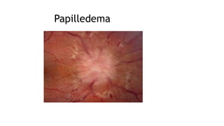
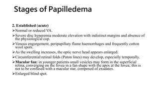
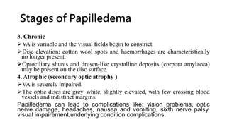
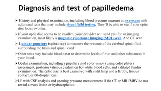
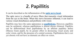
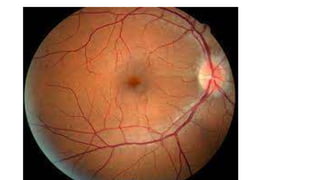
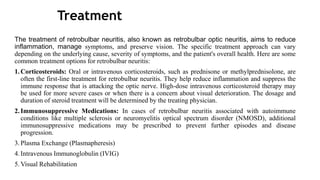
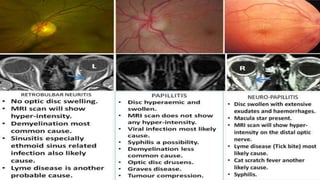
SCHOOL OF HEALTH SCIENCES
OPHTHALMOLOGY DEPARTMENT
YEAR OF STUDY: LEVEL II
Module name: OCULAR EMERGENCY
ACADEMIC YEAR: 2023/2024
GROUP I
Done at Kigali, 30th July 2023.
UNIVERSITY OF RWANDA
College of Medicine and Health Sciences
PRESENTATION ON PAPILLEDEMA, PAPILLITIS AND RETROBULBAR
NEURITIS.
Group Members
Presentation outline:
1. Papilledema
2. Papillitis
3. Retrobulbar neuritis
1. PAPILLEDEMA
Papilledema refers to the swelling of both optic discs in your eyes due to
increased intracranial pressure (intracranial hypertension).
Intracranial hypertension usually results in bilateral papilledema (disc swelling
of both optic discs). Swelling of the optic disc in only one eye (unilateral) is
usually not the result of intracranial hypertension. Because of this, its name is
optic disc edema.
Papilledema can be considered a medical emergency because intracranial
hypertension can be serious, even potentially life-threatening.
Papilledema
Etiology
 Primary and metastatic intracranial tumors.
 Hydrocephalus.
 Idiopathic intracranial hypertension: Often occurs in young, overweight females.
 Subdural and epidural hematomas.
 Subarachnoid hemorrhage: Severe headache, may have preretinal hemorrhages (Terson’s syndrome)
 Arteriovenous malformation.
 Brain abscess: Often produces high fever and mental status changes.
 Meningitis: Fever, stiff neck, headache (e.g., syphilis, tuberculosis, Lyme disease, bacterial, inflammatory,
neoplastic).
 Encephalitis: Often produces mental status abnormalities
 Cerebral venous sinus thrombosis
NOTE: Optic disc swelling in a patient with leukemia is often a sign of leukemic optic nerve infiltration.
Immediate radiation therapy is usually required to preserve vision.
CLINICAL MANIFESTATIONS
I. Symptoms
Transient visual obscurations: These events are periods of about five to 15 seconds
where your vision gets blurry, goes gray, or blacks out. An obscuration is similar to what
happens during a total eclipse when the moon blocks the sun from your sight, often
bilateral visual loss (lasting seconds) often precipitated after rising from a lying or sitting
position (altering intracranial pressure)
Headache
Double vision
Nausea
Vomiting
Neurological symptoms: These may include problems with movement or thinking.
Decrease in visual acuity (a mild decrease in visual acuity can occur in the acute setting if
associated with a macular disturbance). Visual field defects and severe loss of central
visual acuity occur more often with chronic papilledema.
II. Signs
Critical: Bilaterally swollen, hyperemic discs (in early papilledema, disc swelling
may be asymmetric) with nerve fiber layer edema causing blurring of the disc
margin, often obscuring the blood vessels.
Other: Papillary or peripapillary retinal hemorrhages (often flame shaped)
Loss of venous pulsations (20% of the normal population do not have venous
pulsations)
Dilated, tortuous retinal veins.
Normal pupillary response and color vision
An enlarged physiologic blind spot or other visual field defects by formal visual
field testing. In chronic papilledema, the hemorrhages and cotton-wool spots
resolve, disc hyperemia disappears and the disc becomes gray in color,
peripapillary gliosis and narrowing of the peripapillary retinal vessels occur, and
optociliary shunt vessels may develop on the disc. Loss of color vision, central
visual acuity, and visual fi eld defects, especially inferonasal, also occur.
Differential Diagnosis of Papilledema
Pseudo papilledema
Optic neuritis
Papillitis
Ischemic optic neuropathy
Chronic kidney disease
Malignant hypertension
Idiopathic intracranial hypertension
Space occupying lesions
Central retinal artery occlusion
Meningitis
NOTE: Unilateral or bilateral sixth nerve palsy also may result from increased intracranial
pressure.
Grades of Papilledema
Papilledema, also known as optic disc swelling, is graded based on its
severity. The most commonly used grading system is the Frisén Scale, which
has five grades:
Grade 0: No swelling of the optic disc.
Grade 1: Mild swelling with a blurred margin of the optic disc.
Grade 2: Moderate swelling with elevation of the optic disc and
obscuration of the vessels.
Grade 3: Marked swelling with a severely elevated optic disc and
obliteration of the vessels.
Grade 4: Severe swelling with a completely elevated optic disc and loss of
visibility of the vessels.
Stages of Papilledema
Papilledema is nearly always bilateral, but may be asymmetrical and it
may have different stages.
1. Early
Mild disc hyperemia with preservation of the optic cup.
Indistinct peripapillary retinal nerve striations and disc margins.
SVP is absent in about 20% of normal individuals and may be difficult
to identify even when present. An identifiable venous pulsation in at
least one eye means that the ICP is normal at that point in time,
bearing in mind that diurnal fluctuation can occur
Stages of Papilledema
2. Established (acute)
Normal or reduced VA.
Severe disc hyperemia moderate elevation with indistinct margins and absence of
the physiological cup.
Venous engorgement, peripapillary flame haemorrhages and frequently cotton
wool spots.
As the swelling increases, the optic nerve head appears enlarged.
Circumferential retinal folds (Paton lines) may develop, especially temporally.
Macular fan: in younger patients small vesicles may form in the superficial
retina, converging on the fovea in a fan shape with the apex at the fovea; this is
not to be confused with a macular star, composed of exudates.
Enlarged blind spot.
Stages of Papilledema
3. Chronic
VA is variable and the visual fields begin to constrict.
Disc elevation; cotton wool spots and haemorrhages are characteristically
no longer present.
Optociliary shunts and drusen-like crystalline deposits (corpora amylacea)
may be present on the disc surface.
4. Atrophic (secondary optic atrophy )
VA is severely impaired.
The optic discs are grey–white, slightly elevated, with few crossing blood
vessels and indistinct margins.
Papilledema can lead to complications like: vision problems, optic
nerve damage, headaches, nausea and vomiting, sixth nerve palsy,
visual impairement,underlying condition complications.
Diagnosis and test of papilledema
 History and physical examination, including blood pressure measure an eye exam with
additional tests that may include visual field testing. They’ll be able to see if your optic
disc looks swollen.
If your optic disc seems to be swollen, your provider will send you for an imaging
examination, most likely a magnetic resonance imaging (MRI) scan. And CT scan.
 Lumbar puncture (spinal tap) to measure the pressure of the cerebral spinal fluid
surrounding the brain and spinal cord
Other tests may include blood tests to determine levels of iron and other substances in
your blood.
Ocular examination, including a pupillary and color vision (using color plates)
assessment, posterior vitreous evaluation for white blood cells, and a dilated fundus
examination. The optic disc is best examined with a slit lamp and a Hruby, fundus
contact, or 60-diopter lens.
LP with CSF analysis and opening pressure measurement if the CT or MRI/MRV do not
reveal a mass lesion or hydrocephalus.
Treatment
For papilledema due to idiopathic intracranial hypertension you may prescribe a carbonic
anhydrase inhibitor such as acetazolamide. They may also work with you to achieve and
maintain a healthy weight as a long-term strategy.
Optic Nerve Sheath Fenestration: In cases where intracranial pressure cannot be
adequately controlled with medications, a surgical procedure called optic nerve sheath
fenestration may be considered. This procedure involves creating a small opening in the
protective sheath around the optic nerve to relieve
Pressure Shunting Procedures: In some cases, a shunt may be surgically placed to divert
excess cerebrospinal fluid away from the brain, reducing intracranial pressure.
For papilledema due to other cause treat the underlying cause.
It’s important to treat papilledema because untreated papilledema can lead to partial or
complete blindness in one or both eyes.
Lifestyle Modifications: Lifestyle changes may be recommended to help manage
intracranial pressure, such as maintaining a healthy weight, avoiding activities that increase
intracranial pressure (like straining or heavy lifting), and managing underlying conditions
like sleep apnea.
2. Papillitis
It can be described as the inflammation of the optic nerve head.
The optic nerve is a bundle of nerve fibers that transmits visual information
from the eye to the brain. When this nerve becomes inflamed, it can lead to
various visual disturbances and problems with vision.
Papillitis may have the same appearance as papilledema. However, papillitis
may be unilateral, whereas papilledema is almost always bilateral. Papillitis
can be differentiated from papilledema by an afferent pupillary defect
(Marcus Gunn pupil), by its greater effect in decreasing visual acuity and
color vision, and by the presence of a central scotoma. Papilledema that is not
yet chronic will not have as dramatic an effect on vision.
Etiology
1.Autoimmune Disorders
2. Multiple Sclerosis (MS)
3. Infections
4. Medications and Vaccines
5. Systemic Inflammatory Disorders: Inflammatory conditions like
sarcoidosis, systemic lupus erythematosus (SLE), and others can affect the
optic nerve and cause papillitis.
6. Toxic Exposure
7. Nutritional Deficiencies
8. Idiopathic: In some instances, the exact cause of papillitis cannot be
identified, and it is classified as idiopathic.
Signs
1. Visual acuity is usually reduced markedly.
2. Colour vision is often severely impaired.
3. Pupil shows ill-sustained constriction to light. Marcus Gunn pupil which
indicates relative afferent pupillary defect (RAPD) is a diagnostic sign. It is
detected by the swinging flash light test .
4. Ophthalmoscopic features. Papillitis is characterised by hyperaemia of
the disc and blurring of the margins. Disc becomes oedematous and
physiological cup is obliterated. Retinal veins are congested and tortuous.
Splinter haemorrhages and fine exudates may be seen on the disc. Slit-lamp
examination may reveal inflammatory cells in the vitreous. Inflammatory
signs may also be present in the surrounding retina when papillitis is
associated with macular star formation and the condition is labelled as
‘neuroretinitis’
Symptoms of papillitis can include:
1.Sudden loss of vision in one eye, which may worsen over a period of a
few hours to a few days.
2.Pain or discomfort in the affected eye, especially with eye movement.
3.Blurred or dimmed vision.
4.Changes in color perception.
5.The appearance of flashing lights or other visual disturbances
Differential diagnosis
1. Optic neuritis
2. Infectious optic neuritis
3. Systemic inflammatory disorders
4. Neurological conditions
5. Ischemic optic neuropathy
6. Posterior uveitis
• The diagnosis of papillitis involves a comprehensive evaluation that
includes a combination of clinical examination, imaging studies and
laboratory tests.
Here are the diagnostic process for papillitis
1. Ophthalmic examination
2. Visual field testing
3. Imaging studies
4. Lumbar puncture(spinal tap)
5. Blood test
Complications
Color Vision abnormalities
Partial or complete vision loss
Pain and discomfort
Visual field defect
Recurrence
Multiple sclerosis
TREATMENTS OF PAPILLITIS
Pappillitis can be treated by
Corticosteroids: like oral or intravenous corticosteroids such as
predinisolone
Immunomodulatory therapy
Pain management: pain relievers like acetaminophen or nonsteroids
anti-inflammatory drugs(NSAIDS)
Treatment of underlying cause
Observation and monitoring
3. RETROBULBAR NEURITIS
Retrobulbar neuritis, also known as retrobulbar optic neuritis, is a condition characterized by
inflammation of the optic nerve behind the eye. Unlike papillitis, which involves inflammation at the
optic nerve head (where the nerve fibers exit the eye), retrobulbar neuritis affects the optic nerve
further back along its course.
it is a form of Optic neuritis which includes inflammatory and demyelinating disorders of the optic
nerve.
Etiology
1. Idiopathic. In a large proportion of cases the underlying cause is unidentifiable.
2. Hereditary optic neuritis (Leber’s disease)
3. Demyelinating disorders are by far the most common cause of optic neuritis. These include
multiple sclerosis, neuromyelitis optica (Devic’s disease) and diffuse periaxial encephalitis of
Schilder. About 70% cases of established multiple sclerosis may develop optic neuritis.
4. Parainfectious optic neuritis is associated with various viral infections such as measles, mumps,
chickenpox, whooping cough and glandular fever. It may also occur following immunization.
Etiology cont’
5. Infectious optic neuritis may be sinus related (with acute ethmoiditis) or
associated with cat scratch fever, syphilis (during primary or secondary stage), lyme
disease and cryptococcal meningitis in patients with AIDS.
6. Toxic optic neuritis (see toxic amblyopias).
7. Nutritional deficiencies: In rare cases, vitamin deficiencies, especially vitamin
B12, have been implicated in the development of optic neuritis.
8. Autoimmune disorders: Retrobulbar neuritis can be associated with autoimmune
diseases, such as multiple sclerosis (MS). In MS, the body's immune system
mistakenly attacks the protective myelin sheath that covers nerve fibers, including
those in the optic nerve. This demyelination disrupts nerve signal transmission and
leads to inflammation.
Differential diagnosis
1.Papillitis
2. Anterior Ischemic Optic Neuropathy (AION): AION is a condition in which there is decreased blood flow to the
optic nerve, resulting in sudden vision loss. It may be caused by arteritis or non-arteritic factors.
3. Central Retinal Artery Occlusion (CRAO) or Central Retinal Vein Occlusion (CRVO)
4. Multiple Sclerosis (MS)
5. Neuromyelitis Optica Spectrum Disorder (NMOSD)
6. Toxic Optic Neuropathy
7. compressive Optic Neuropathy: Tumors or other masses pressing on the optic nerve can result in vision changes
similar to optic neuritis.
8.Traumatic Optic Neuropathy: Head trauma or injury to the eye can cause optic nerve damage and vision loss.
9.Ischemic Optic Neuropathy: This condition involves reduced blood flow to the optic nerve, leading to vision
loss
10.Acute papilledema
11.Systemic sereve hypertension
12.Optical tumor compressing optic nerve
Clinical manifestation
1.Vision loss: Sudden, painless loss of vision is the hallmark symptom of
retrobulbar neuritis. The vision loss is usually unilateral (affecting one eye)
but can, in some cases, be bilateral (affecting both eyes). The severity of
vision loss can range from mild blurriness to significant visual impairment.
2.Visual disturbances: Apart from vision loss, patients may experience other
visual disturbances, such as dimming of vision, reduced color perception,
and difficulty seeing in low light conditions.
3.Pain or discomfort: Unlike typical optic neuritis (which occurs in the optic
nerve head and can cause eye pain), retrobulbar neuritis is characterized by
painless vision loss. However, some individuals with retrobulbar neuritis
may experience mild pain or discomfort during eye movement.
Clinical manifestation
4.Visual field defects: Retrobulbar neuritis can lead to specific patterns of visual
field loss. Most commonly, central or cecocentral scotomas (areas of reduced or
absent vision) are observed. Patients may have difficulty seeing objects in the center
of their visual field or may experience a blind spot in the center of their vision.
5.Contrast sensitivity reduction: Retrobulbar neuritis can affect contrast
sensitivity, making it difficult to distinguish between objects of similar brightness or
color.
6.Optic disc appearance: The optic disc, where the optic nerve exits the eye, may
appear normal during the early stages of retrobulbar neuritis. Unlike in papillitis
(inflammation at the optic nerve head), there may be no obvious swelling or disc
edema.
Retrobulbar neuritis has complications like: vision loss, color vision deficits and
impaired contrast sensitivity.
DIAGNOSIS
1.Patient History: The healthcare provider will begin by taking a detailed
history of the patient's symptoms, including the onset, duration, and
progression of vision changes. They will inquire about any pain or
discomfort in the eyes and whether there have been any recent infections or
systemic illnesses. It is important to identify any potential triggers or
underlying conditions that could be associated with retrobulbar neuritis.
2.Visual Acuity Test: This test assesses how well a person can see at
different distances using an eye chart. The healthcare provider will ask the
patient to read letters or numbers from the chart to determine the extent of
visual impairment.
3.Visual Field Testing: Visual field testing evaluates a person's peripheral
vision. Specific patterns of visual field loss, such as central or cecocentral
scotomas, may indicate retrobulbar neuritis.
DIAGNOSIS
4.Pupillary Light Reflex: The provider will examine the pupil's
response to light, which can help assess the function of the optic
nerve and identify any abnormalities.
5.Ophthalmoscopy: In this examination, the provider uses an
ophthalmoscope to look at the back of the eye, including the optic
disc. In retrobulbar neuritis, the optic disc may appear normal or only
show subtle changes compared to other forms of optic neuritis.
6.Color Vision Testing: Color vision assessment can detect any
abnormalities in color perception, which may be affected in cases of
retrobulbar neuritis.
7.Contrast Sensitivity Test: This test evaluates the ability to
distinguish between objects of similar brightness or color, which may
be reduced in retrobulbar neuritis.
8.Optical Coherence Tomography (OCT):
Treatment
The treatment of retrobulbar neuritis, also known as retrobulbar optic neuritis, aims to reduce
inflammation, manage symptoms, and preserve vision. The specific treatment approach can vary
depending on the underlying cause, severity of symptoms, and the patient's overall health. Here are some
common treatment options for retrobulbar neuritis:
1.Corticosteroids: Oral or intravenous corticosteroids, such as prednisone or methylprednisolone, are
often the first-line treatment for retrobulbar neuritis. They help reduce inflammation and suppress the
immune response that is attacking the optic nerve. High-dose intravenous corticosteroid therapy may
be used for more severe cases or when there is a concern about visual deterioration. The dosage and
duration of steroid treatment will be determined by the treating physician.
2.Immunosuppressive Medications: In cases of retrobulbar neuritis associated with autoimmune
conditions like multiple sclerosis or neuromyelitis optical spectrum disorder (NMOSD), additional
immunosuppressive medications may be prescribed to prevent further episodes and disease
progression.
3.Plasma Exchange (Plasmapheresis)
4.Intravenous Immunoglobulin (IVIG)
5.Visual Rehabilitation
References
1.kanski’s ophthalmology 8th edition book
2. The Wills Eye Manual_ Office and Emergency Room Diagnosis and
Treatment of Eye
3. A.K. Khurana-Ophthalmology
4. https://www.mayoclinic.org/diseases-conditions/optic-
neuritis/symptoms-causes/syc-20354953

GROUP B PAPILLEDEMA.pptx

  • 1.
    SCHOOL OF HEALTHSCIENCES OPHTHALMOLOGY DEPARTMENT YEAR OF STUDY: LEVEL II Module name: OCULAR EMERGENCY ACADEMIC YEAR: 2023/2024 GROUP I Done at Kigali, 30th July 2023. UNIVERSITY OF RWANDA College of Medicine and Health Sciences PRESENTATION ON PAPILLEDEMA, PAPILLITIS AND RETROBULBAR NEURITIS.
  • 2.
  • 3.
    Presentation outline: 1. Papilledema 2.Papillitis 3. Retrobulbar neuritis
  • 4.
    1. PAPILLEDEMA Papilledema refersto the swelling of both optic discs in your eyes due to increased intracranial pressure (intracranial hypertension). Intracranial hypertension usually results in bilateral papilledema (disc swelling of both optic discs). Swelling of the optic disc in only one eye (unilateral) is usually not the result of intracranial hypertension. Because of this, its name is optic disc edema. Papilledema can be considered a medical emergency because intracranial hypertension can be serious, even potentially life-threatening.
  • 5.
  • 6.
    Etiology  Primary andmetastatic intracranial tumors.  Hydrocephalus.  Idiopathic intracranial hypertension: Often occurs in young, overweight females.  Subdural and epidural hematomas.  Subarachnoid hemorrhage: Severe headache, may have preretinal hemorrhages (Terson’s syndrome)  Arteriovenous malformation.  Brain abscess: Often produces high fever and mental status changes.  Meningitis: Fever, stiff neck, headache (e.g., syphilis, tuberculosis, Lyme disease, bacterial, inflammatory, neoplastic).  Encephalitis: Often produces mental status abnormalities  Cerebral venous sinus thrombosis NOTE: Optic disc swelling in a patient with leukemia is often a sign of leukemic optic nerve infiltration. Immediate radiation therapy is usually required to preserve vision.
  • 7.
    CLINICAL MANIFESTATIONS I. Symptoms Transientvisual obscurations: These events are periods of about five to 15 seconds where your vision gets blurry, goes gray, or blacks out. An obscuration is similar to what happens during a total eclipse when the moon blocks the sun from your sight, often bilateral visual loss (lasting seconds) often precipitated after rising from a lying or sitting position (altering intracranial pressure) Headache Double vision Nausea Vomiting Neurological symptoms: These may include problems with movement or thinking. Decrease in visual acuity (a mild decrease in visual acuity can occur in the acute setting if associated with a macular disturbance). Visual field defects and severe loss of central visual acuity occur more often with chronic papilledema.
  • 8.
    II. Signs Critical: Bilaterallyswollen, hyperemic discs (in early papilledema, disc swelling may be asymmetric) with nerve fiber layer edema causing blurring of the disc margin, often obscuring the blood vessels. Other: Papillary or peripapillary retinal hemorrhages (often flame shaped) Loss of venous pulsations (20% of the normal population do not have venous pulsations) Dilated, tortuous retinal veins. Normal pupillary response and color vision An enlarged physiologic blind spot or other visual field defects by formal visual field testing. In chronic papilledema, the hemorrhages and cotton-wool spots resolve, disc hyperemia disappears and the disc becomes gray in color, peripapillary gliosis and narrowing of the peripapillary retinal vessels occur, and optociliary shunt vessels may develop on the disc. Loss of color vision, central visual acuity, and visual fi eld defects, especially inferonasal, also occur.
  • 9.
    Differential Diagnosis ofPapilledema Pseudo papilledema Optic neuritis Papillitis Ischemic optic neuropathy Chronic kidney disease Malignant hypertension Idiopathic intracranial hypertension Space occupying lesions Central retinal artery occlusion Meningitis NOTE: Unilateral or bilateral sixth nerve palsy also may result from increased intracranial pressure.
  • 10.
    Grades of Papilledema Papilledema,also known as optic disc swelling, is graded based on its severity. The most commonly used grading system is the Frisén Scale, which has five grades: Grade 0: No swelling of the optic disc. Grade 1: Mild swelling with a blurred margin of the optic disc. Grade 2: Moderate swelling with elevation of the optic disc and obscuration of the vessels. Grade 3: Marked swelling with a severely elevated optic disc and obliteration of the vessels. Grade 4: Severe swelling with a completely elevated optic disc and loss of visibility of the vessels.
  • 11.
    Stages of Papilledema Papilledemais nearly always bilateral, but may be asymmetrical and it may have different stages. 1. Early Mild disc hyperemia with preservation of the optic cup. Indistinct peripapillary retinal nerve striations and disc margins. SVP is absent in about 20% of normal individuals and may be difficult to identify even when present. An identifiable venous pulsation in at least one eye means that the ICP is normal at that point in time, bearing in mind that diurnal fluctuation can occur
  • 12.
    Stages of Papilledema 2.Established (acute) Normal or reduced VA. Severe disc hyperemia moderate elevation with indistinct margins and absence of the physiological cup. Venous engorgement, peripapillary flame haemorrhages and frequently cotton wool spots. As the swelling increases, the optic nerve head appears enlarged. Circumferential retinal folds (Paton lines) may develop, especially temporally. Macular fan: in younger patients small vesicles may form in the superficial retina, converging on the fovea in a fan shape with the apex at the fovea; this is not to be confused with a macular star, composed of exudates. Enlarged blind spot.
  • 13.
    Stages of Papilledema 3.Chronic VA is variable and the visual fields begin to constrict. Disc elevation; cotton wool spots and haemorrhages are characteristically no longer present. Optociliary shunts and drusen-like crystalline deposits (corpora amylacea) may be present on the disc surface. 4. Atrophic (secondary optic atrophy ) VA is severely impaired. The optic discs are grey–white, slightly elevated, with few crossing blood vessels and indistinct margins. Papilledema can lead to complications like: vision problems, optic nerve damage, headaches, nausea and vomiting, sixth nerve palsy, visual impairement,underlying condition complications.
  • 14.
    Diagnosis and testof papilledema  History and physical examination, including blood pressure measure an eye exam with additional tests that may include visual field testing. They’ll be able to see if your optic disc looks swollen. If your optic disc seems to be swollen, your provider will send you for an imaging examination, most likely a magnetic resonance imaging (MRI) scan. And CT scan.  Lumbar puncture (spinal tap) to measure the pressure of the cerebral spinal fluid surrounding the brain and spinal cord Other tests may include blood tests to determine levels of iron and other substances in your blood. Ocular examination, including a pupillary and color vision (using color plates) assessment, posterior vitreous evaluation for white blood cells, and a dilated fundus examination. The optic disc is best examined with a slit lamp and a Hruby, fundus contact, or 60-diopter lens. LP with CSF analysis and opening pressure measurement if the CT or MRI/MRV do not reveal a mass lesion or hydrocephalus.
  • 15.
    Treatment For papilledema dueto idiopathic intracranial hypertension you may prescribe a carbonic anhydrase inhibitor such as acetazolamide. They may also work with you to achieve and maintain a healthy weight as a long-term strategy. Optic Nerve Sheath Fenestration: In cases where intracranial pressure cannot be adequately controlled with medications, a surgical procedure called optic nerve sheath fenestration may be considered. This procedure involves creating a small opening in the protective sheath around the optic nerve to relieve Pressure Shunting Procedures: In some cases, a shunt may be surgically placed to divert excess cerebrospinal fluid away from the brain, reducing intracranial pressure. For papilledema due to other cause treat the underlying cause. It’s important to treat papilledema because untreated papilledema can lead to partial or complete blindness in one or both eyes. Lifestyle Modifications: Lifestyle changes may be recommended to help manage intracranial pressure, such as maintaining a healthy weight, avoiding activities that increase intracranial pressure (like straining or heavy lifting), and managing underlying conditions like sleep apnea.
  • 16.
    2. Papillitis It canbe described as the inflammation of the optic nerve head. The optic nerve is a bundle of nerve fibers that transmits visual information from the eye to the brain. When this nerve becomes inflamed, it can lead to various visual disturbances and problems with vision. Papillitis may have the same appearance as papilledema. However, papillitis may be unilateral, whereas papilledema is almost always bilateral. Papillitis can be differentiated from papilledema by an afferent pupillary defect (Marcus Gunn pupil), by its greater effect in decreasing visual acuity and color vision, and by the presence of a central scotoma. Papilledema that is not yet chronic will not have as dramatic an effect on vision.
  • 18.
    Etiology 1.Autoimmune Disorders 2. MultipleSclerosis (MS) 3. Infections 4. Medications and Vaccines 5. Systemic Inflammatory Disorders: Inflammatory conditions like sarcoidosis, systemic lupus erythematosus (SLE), and others can affect the optic nerve and cause papillitis. 6. Toxic Exposure 7. Nutritional Deficiencies 8. Idiopathic: In some instances, the exact cause of papillitis cannot be identified, and it is classified as idiopathic.
  • 19.
    Signs 1. Visual acuityis usually reduced markedly. 2. Colour vision is often severely impaired. 3. Pupil shows ill-sustained constriction to light. Marcus Gunn pupil which indicates relative afferent pupillary defect (RAPD) is a diagnostic sign. It is detected by the swinging flash light test . 4. Ophthalmoscopic features. Papillitis is characterised by hyperaemia of the disc and blurring of the margins. Disc becomes oedematous and physiological cup is obliterated. Retinal veins are congested and tortuous. Splinter haemorrhages and fine exudates may be seen on the disc. Slit-lamp examination may reveal inflammatory cells in the vitreous. Inflammatory signs may also be present in the surrounding retina when papillitis is associated with macular star formation and the condition is labelled as ‘neuroretinitis’
  • 20.
    Symptoms of papillitiscan include: 1.Sudden loss of vision in one eye, which may worsen over a period of a few hours to a few days. 2.Pain or discomfort in the affected eye, especially with eye movement. 3.Blurred or dimmed vision. 4.Changes in color perception. 5.The appearance of flashing lights or other visual disturbances
  • 21.
    Differential diagnosis 1. Opticneuritis 2. Infectious optic neuritis 3. Systemic inflammatory disorders 4. Neurological conditions 5. Ischemic optic neuropathy 6. Posterior uveitis
  • 22.
    • The diagnosisof papillitis involves a comprehensive evaluation that includes a combination of clinical examination, imaging studies and laboratory tests. Here are the diagnostic process for papillitis 1. Ophthalmic examination 2. Visual field testing 3. Imaging studies 4. Lumbar puncture(spinal tap) 5. Blood test
  • 23.
    Complications Color Vision abnormalities Partialor complete vision loss Pain and discomfort Visual field defect Recurrence Multiple sclerosis
  • 24.
    TREATMENTS OF PAPILLITIS Pappillitiscan be treated by Corticosteroids: like oral or intravenous corticosteroids such as predinisolone Immunomodulatory therapy Pain management: pain relievers like acetaminophen or nonsteroids anti-inflammatory drugs(NSAIDS) Treatment of underlying cause Observation and monitoring
  • 25.
    3. RETROBULBAR NEURITIS Retrobulbarneuritis, also known as retrobulbar optic neuritis, is a condition characterized by inflammation of the optic nerve behind the eye. Unlike papillitis, which involves inflammation at the optic nerve head (where the nerve fibers exit the eye), retrobulbar neuritis affects the optic nerve further back along its course. it is a form of Optic neuritis which includes inflammatory and demyelinating disorders of the optic nerve. Etiology 1. Idiopathic. In a large proportion of cases the underlying cause is unidentifiable. 2. Hereditary optic neuritis (Leber’s disease) 3. Demyelinating disorders are by far the most common cause of optic neuritis. These include multiple sclerosis, neuromyelitis optica (Devic’s disease) and diffuse periaxial encephalitis of Schilder. About 70% cases of established multiple sclerosis may develop optic neuritis. 4. Parainfectious optic neuritis is associated with various viral infections such as measles, mumps, chickenpox, whooping cough and glandular fever. It may also occur following immunization.
  • 26.
    Etiology cont’ 5. Infectiousoptic neuritis may be sinus related (with acute ethmoiditis) or associated with cat scratch fever, syphilis (during primary or secondary stage), lyme disease and cryptococcal meningitis in patients with AIDS. 6. Toxic optic neuritis (see toxic amblyopias). 7. Nutritional deficiencies: In rare cases, vitamin deficiencies, especially vitamin B12, have been implicated in the development of optic neuritis. 8. Autoimmune disorders: Retrobulbar neuritis can be associated with autoimmune diseases, such as multiple sclerosis (MS). In MS, the body's immune system mistakenly attacks the protective myelin sheath that covers nerve fibers, including those in the optic nerve. This demyelination disrupts nerve signal transmission and leads to inflammation.
  • 28.
    Differential diagnosis 1.Papillitis 2. AnteriorIschemic Optic Neuropathy (AION): AION is a condition in which there is decreased blood flow to the optic nerve, resulting in sudden vision loss. It may be caused by arteritis or non-arteritic factors. 3. Central Retinal Artery Occlusion (CRAO) or Central Retinal Vein Occlusion (CRVO) 4. Multiple Sclerosis (MS) 5. Neuromyelitis Optica Spectrum Disorder (NMOSD) 6. Toxic Optic Neuropathy 7. compressive Optic Neuropathy: Tumors or other masses pressing on the optic nerve can result in vision changes similar to optic neuritis. 8.Traumatic Optic Neuropathy: Head trauma or injury to the eye can cause optic nerve damage and vision loss. 9.Ischemic Optic Neuropathy: This condition involves reduced blood flow to the optic nerve, leading to vision loss 10.Acute papilledema 11.Systemic sereve hypertension 12.Optical tumor compressing optic nerve
  • 29.
    Clinical manifestation 1.Vision loss:Sudden, painless loss of vision is the hallmark symptom of retrobulbar neuritis. The vision loss is usually unilateral (affecting one eye) but can, in some cases, be bilateral (affecting both eyes). The severity of vision loss can range from mild blurriness to significant visual impairment. 2.Visual disturbances: Apart from vision loss, patients may experience other visual disturbances, such as dimming of vision, reduced color perception, and difficulty seeing in low light conditions. 3.Pain or discomfort: Unlike typical optic neuritis (which occurs in the optic nerve head and can cause eye pain), retrobulbar neuritis is characterized by painless vision loss. However, some individuals with retrobulbar neuritis may experience mild pain or discomfort during eye movement.
  • 30.
    Clinical manifestation 4.Visual fielddefects: Retrobulbar neuritis can lead to specific patterns of visual field loss. Most commonly, central or cecocentral scotomas (areas of reduced or absent vision) are observed. Patients may have difficulty seeing objects in the center of their visual field or may experience a blind spot in the center of their vision. 5.Contrast sensitivity reduction: Retrobulbar neuritis can affect contrast sensitivity, making it difficult to distinguish between objects of similar brightness or color. 6.Optic disc appearance: The optic disc, where the optic nerve exits the eye, may appear normal during the early stages of retrobulbar neuritis. Unlike in papillitis (inflammation at the optic nerve head), there may be no obvious swelling or disc edema. Retrobulbar neuritis has complications like: vision loss, color vision deficits and impaired contrast sensitivity.
  • 31.
    DIAGNOSIS 1.Patient History: Thehealthcare provider will begin by taking a detailed history of the patient's symptoms, including the onset, duration, and progression of vision changes. They will inquire about any pain or discomfort in the eyes and whether there have been any recent infections or systemic illnesses. It is important to identify any potential triggers or underlying conditions that could be associated with retrobulbar neuritis. 2.Visual Acuity Test: This test assesses how well a person can see at different distances using an eye chart. The healthcare provider will ask the patient to read letters or numbers from the chart to determine the extent of visual impairment. 3.Visual Field Testing: Visual field testing evaluates a person's peripheral vision. Specific patterns of visual field loss, such as central or cecocentral scotomas, may indicate retrobulbar neuritis.
  • 32.
    DIAGNOSIS 4.Pupillary Light Reflex:The provider will examine the pupil's response to light, which can help assess the function of the optic nerve and identify any abnormalities. 5.Ophthalmoscopy: In this examination, the provider uses an ophthalmoscope to look at the back of the eye, including the optic disc. In retrobulbar neuritis, the optic disc may appear normal or only show subtle changes compared to other forms of optic neuritis. 6.Color Vision Testing: Color vision assessment can detect any abnormalities in color perception, which may be affected in cases of retrobulbar neuritis. 7.Contrast Sensitivity Test: This test evaluates the ability to distinguish between objects of similar brightness or color, which may be reduced in retrobulbar neuritis. 8.Optical Coherence Tomography (OCT):
  • 33.
    Treatment The treatment ofretrobulbar neuritis, also known as retrobulbar optic neuritis, aims to reduce inflammation, manage symptoms, and preserve vision. The specific treatment approach can vary depending on the underlying cause, severity of symptoms, and the patient's overall health. Here are some common treatment options for retrobulbar neuritis: 1.Corticosteroids: Oral or intravenous corticosteroids, such as prednisone or methylprednisolone, are often the first-line treatment for retrobulbar neuritis. They help reduce inflammation and suppress the immune response that is attacking the optic nerve. High-dose intravenous corticosteroid therapy may be used for more severe cases or when there is a concern about visual deterioration. The dosage and duration of steroid treatment will be determined by the treating physician. 2.Immunosuppressive Medications: In cases of retrobulbar neuritis associated with autoimmune conditions like multiple sclerosis or neuromyelitis optical spectrum disorder (NMOSD), additional immunosuppressive medications may be prescribed to prevent further episodes and disease progression. 3.Plasma Exchange (Plasmapheresis) 4.Intravenous Immunoglobulin (IVIG) 5.Visual Rehabilitation
  • 35.
    References 1.kanski’s ophthalmology 8thedition book 2. The Wills Eye Manual_ Office and Emergency Room Diagnosis and Treatment of Eye 3. A.K. Khurana-Ophthalmology 4. https://www.mayoclinic.org/diseases-conditions/optic- neuritis/symptoms-causes/syc-20354953