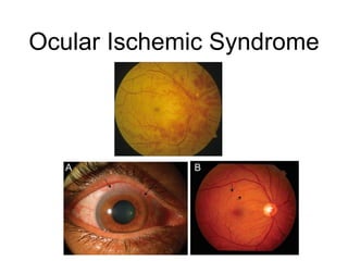
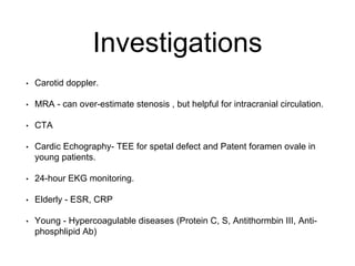
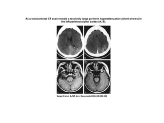
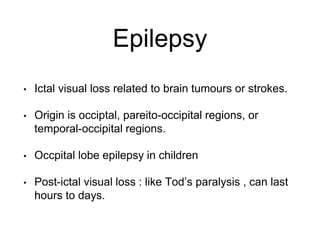
Transient Visual Loss
Raed Behbehani , MD FRCSC
TVL
• Common complaint
• History is critical (onset , mono-ocular or binocular,
duartion , precipitating factors , associated
symptoms)
• Examination is often normal
TVL
• Ocular vs non-ocular
• Ischemic , non-ischmic , migraine
Non-ischemic Causes
• Usually reduced quality of vision “blurr” , “fog” rather
than “black outs”.
• Symptoms often age
• Can be accompanied by ocular discomfort or pain
(PDS, Microhyhema, Dry Eyes, intermittent angle
closure).
Ischemic Causes
• Sudden in onset and slow recovery in central or
peripheral visual field.
• Deficit can respect vertical or horizontal meridian
“Curtain coming down” , “half of my vision”.
• Associated symptoms ( Pain , headache, jaw
claudication, neurologic signs)
Migraine
• Stereotyped pattern ( 15 min - 1 hour )
• Young patient , with associated migraine symptoms
(nausea, vomiting , photosensitivity , phonophbia)
• Binocular (patient may notice temporal field changes only)
• Scilntillating pattern, colored lights, (positive symptoms).
• Can be followed by headache but can be isolated
(acephalgic migraine)
• Migraine trigger (stress , hunger , food )
Migraine
Other causes
• Uhthohff’s phenomenon (in any optic neuropathy)
• Monocular TVL associated with exercise .
• Changes in posture (Transient visual obscurrations
in papilledema)
• Gaze-evoked amaurosis (orbital intraconal mass ,
optic nerve sheath meningioma)
Clinical Approach
• Careful History is crucial !
• Inquire about vascular risk factors , history of
cardiac disease , arrhythmias and hyperoculable
disease.
• Careful slit lamp exam : tear film , angles
(gonioscopy), microhyphemua , Iris TI defects .
• Fundus : disc , cotton wool spots , haemorrhages ,
emboli
Ocular Causes
• Check tear film (blurring of vision with reading )
• Blepharitis , flourescin staining , meibomitis.
• Guttatae ( blurring of vision in the morning) ,
examine patient and corneal thickness AM.
Ocular Causes
• Intermittent angle-closure (pain with halos).
• Microhyphema ( juvenile XG, malpositioned IOL).
• One piece IOL in ciliary sulcus will lead iris chaffing
and pigment release.
• Lenticular swelling from hyperglycemia.
Ocular Causes
PG
Pigment Dispersion
Syndrome
•Pigment is lost from the posterior surface of the iris.
•Concave posterior iris surface mechanically rubs against
anteriorly oriented zonular fibers.
•Reverve Pupillary block.
•Bilateral but can be asymmetric.
•Young myopic males , visual blurring with exercise .
• Glaucoma.
Optic Disc
Ischemic Causes
• Interruption of blood supply to retina , optic nerve or
visual pathways (monocaulr)
• Cortical (binocular)
• Carotid artery disease ( plaque, thrombus , or
vasculitis , Fibromscular dysplasia)
• Risk of stroke from carotid-artery TVL is much lower
than TIA (2% vs 5-8% from hemispheric TIA).
Ischemic TVL
• Abrupt
• Painless.
• Lasts 1-5 minutes
• Can respect vertical or horizontal meridien
• Resolves ove few minute
Monoocular vs Binocular
• Monocular : patient usually can read or continue
activity.
• Binocular : patient is more symptomatic and is
unable to read and has to stop activity.
• Neurologic signs (hemiparesis , hemisensory loss
,or hemianopia) in MCA territory , (vertigo , ataxia,
diplopia) in Vertebrobasilar distribution.
• Pain , jaw claudication and TVL - rule out GCA !
Ischemic TVL
• Transient monocular blindness upon exposure to
light ( bright light-induced amaurosis).
• Impaired retinal circulation to outer retina.
• Differential from delayed light-adaptation (cone
disease)
Neuroophthalmic Signs
Ocular Ischemic Syndrome
• Severe ocular ischemia from carotid disease
• Episcleral and conjunctival injection, corneal edema,
cells and flare in the AC.
• Iris neovasculrization and glaucoma.
• Venous stasis retinopathy (Mid-peripher) , unilateral.
• Disc edema in late stages.
Ocular Ischemic Syndrome
Investigations
• Carotid doppler.
• MRA - can over-estimate stenosis , but helpful for intracranial circulation.
• CTA
• Cardic Echography- TEE for spetal defect and Patent foramen ovale in
young patients.
• 24-hour EKG monitoring.
• Elderly - ESR, CRP
• Young - Hypercoagulable diseases (Protein C, S, Antithormbin III, Anti-
phosphlipid Ab)
Treatment
• Aspirin or clopidogrel in all patients.
• Warfarin or heparin in non-surgical patients resistant
to anti-platelet therapy.
Surgical Treatment
• Carotid endarterectomy : (NASCET study) compared to
hemiaspheric TIA patients who will benefit if they have 3 or
more of the following :
• 1) > 75 years
• 2) make
• 3) History of hemispheric TIA or stroke
• 4) History of intermittant claudications
• 5) Caortid stenosis 80%-94% and no collateral
Surgery
• Complication rate for major post-operative stroke
was 2.1 % in NASCET.
• Asymptomatic carotid stenosis and carotid stenosis
less then 70% with monocular TVL is probably best
managed medically.
• Percutaneous trans-luminal angioplasty - for poor
surgical candidates.
Retinal Vasospasm
• Very rare.
• Diagnosis of exclusion.
• Young patients with repeated monocular visual loss.
• Has been associated with exercise and cocaine.
• Calcium channel blockers can help.
Retinal Vasospasm
Always R/O GCA !
Transient Cortical Blindness
Following Contrast Media
• Usually following hyperosmolar iodinated contrast.
• Transient cortical edema due to break down of
blood-brain barrier.
• Following cerebral or coronary angiography.
• Vision often returns in hours-3 days.
• Patients with hypertension more at risk.
FLAIR MR images show hyperintensities (arrows) in occipital cortices bilaterally (A, B).
Saigal G et al. AJNR Am J Neuroradiol 2004;25:252-256
Axial noncontrast CT scan reveals a relatively large gyriform hyperattenuation (short arrows) in
the left parietooccipital cortex (A, B).
Saigal G et al. AJNR Am J Neuroradiol 2004;25:252-256
Epilepsy
• Ictal visual loss related to brain tumours or strokes.
• Origin is occiptal, pareito-occipital regions, or
temporal-occipital regions.
• Occpital lobe epilepsy in children
• Post-ictal visual loss : like Tod’s paralysis , can last
hours to days.

Transient visual loss

  • 1.
    Transient Visual Loss RaedBehbehani , MD FRCSC
  • 2.
    TVL • Common complaint •History is critical (onset , mono-ocular or binocular, duartion , precipitating factors , associated symptoms) • Examination is often normal
  • 3.
    TVL • Ocular vsnon-ocular • Ischemic , non-ischmic , migraine
  • 4.
    Non-ischemic Causes • Usuallyreduced quality of vision “blurr” , “fog” rather than “black outs”. • Symptoms often age • Can be accompanied by ocular discomfort or pain (PDS, Microhyhema, Dry Eyes, intermittent angle closure).
  • 5.
    Ischemic Causes • Suddenin onset and slow recovery in central or peripheral visual field. • Deficit can respect vertical or horizontal meridian “Curtain coming down” , “half of my vision”. • Associated symptoms ( Pain , headache, jaw claudication, neurologic signs)
  • 6.
    Migraine • Stereotyped pattern( 15 min - 1 hour ) • Young patient , with associated migraine symptoms (nausea, vomiting , photosensitivity , phonophbia) • Binocular (patient may notice temporal field changes only) • Scilntillating pattern, colored lights, (positive symptoms). • Can be followed by headache but can be isolated (acephalgic migraine) • Migraine trigger (stress , hunger , food )
  • 7.
  • 8.
    Other causes • Uhthohff’sphenomenon (in any optic neuropathy) • Monocular TVL associated with exercise . • Changes in posture (Transient visual obscurrations in papilledema) • Gaze-evoked amaurosis (orbital intraconal mass , optic nerve sheath meningioma)
  • 9.
    Clinical Approach • CarefulHistory is crucial ! • Inquire about vascular risk factors , history of cardiac disease , arrhythmias and hyperoculable disease. • Careful slit lamp exam : tear film , angles (gonioscopy), microhyphemua , Iris TI defects . • Fundus : disc , cotton wool spots , haemorrhages , emboli
  • 10.
    Ocular Causes • Checktear film (blurring of vision with reading ) • Blepharitis , flourescin staining , meibomitis. • Guttatae ( blurring of vision in the morning) , examine patient and corneal thickness AM.
  • 11.
    Ocular Causes • Intermittentangle-closure (pain with halos). • Microhyphema ( juvenile XG, malpositioned IOL). • One piece IOL in ciliary sulcus will lead iris chaffing and pigment release. • Lenticular swelling from hyperglycemia.
  • 12.
  • 13.
  • 14.
    Pigment Dispersion Syndrome •Pigment islost from the posterior surface of the iris. •Concave posterior iris surface mechanically rubs against anteriorly oriented zonular fibers. •Reverve Pupillary block. •Bilateral but can be asymmetric. •Young myopic males , visual blurring with exercise . • Glaucoma.
  • 15.
  • 16.
    Ischemic Causes • Interruptionof blood supply to retina , optic nerve or visual pathways (monocaulr) • Cortical (binocular) • Carotid artery disease ( plaque, thrombus , or vasculitis , Fibromscular dysplasia) • Risk of stroke from carotid-artery TVL is much lower than TIA (2% vs 5-8% from hemispheric TIA).
  • 17.
    Ischemic TVL • Abrupt •Painless. • Lasts 1-5 minutes • Can respect vertical or horizontal meridien • Resolves ove few minute
  • 18.
    Monoocular vs Binocular •Monocular : patient usually can read or continue activity. • Binocular : patient is more symptomatic and is unable to read and has to stop activity. • Neurologic signs (hemiparesis , hemisensory loss ,or hemianopia) in MCA territory , (vertigo , ataxia, diplopia) in Vertebrobasilar distribution. • Pain , jaw claudication and TVL - rule out GCA !
  • 19.
    Ischemic TVL • Transientmonocular blindness upon exposure to light ( bright light-induced amaurosis). • Impaired retinal circulation to outer retina. • Differential from delayed light-adaptation (cone disease)
  • 20.
  • 21.
    Ocular Ischemic Syndrome •Severe ocular ischemia from carotid disease • Episcleral and conjunctival injection, corneal edema, cells and flare in the AC. • Iris neovasculrization and glaucoma. • Venous stasis retinopathy (Mid-peripher) , unilateral. • Disc edema in late stages.
  • 22.
  • 23.
    Investigations • Carotid doppler. •MRA - can over-estimate stenosis , but helpful for intracranial circulation. • CTA • Cardic Echography- TEE for spetal defect and Patent foramen ovale in young patients. • 24-hour EKG monitoring. • Elderly - ESR, CRP • Young - Hypercoagulable diseases (Protein C, S, Antithormbin III, Anti- phosphlipid Ab)
  • 24.
    Treatment • Aspirin orclopidogrel in all patients. • Warfarin or heparin in non-surgical patients resistant to anti-platelet therapy.
  • 25.
    Surgical Treatment • Carotidendarterectomy : (NASCET study) compared to hemiaspheric TIA patients who will benefit if they have 3 or more of the following : • 1) > 75 years • 2) make • 3) History of hemispheric TIA or stroke • 4) History of intermittant claudications • 5) Caortid stenosis 80%-94% and no collateral
  • 26.
    Surgery • Complication ratefor major post-operative stroke was 2.1 % in NASCET. • Asymptomatic carotid stenosis and carotid stenosis less then 70% with monocular TVL is probably best managed medically. • Percutaneous trans-luminal angioplasty - for poor surgical candidates.
  • 27.
    Retinal Vasospasm • Veryrare. • Diagnosis of exclusion. • Young patients with repeated monocular visual loss. • Has been associated with exercise and cocaine. • Calcium channel blockers can help.
  • 28.
  • 29.
  • 30.
    Transient Cortical Blindness FollowingContrast Media • Usually following hyperosmolar iodinated contrast. • Transient cortical edema due to break down of blood-brain barrier. • Following cerebral or coronary angiography. • Vision often returns in hours-3 days. • Patients with hypertension more at risk.
  • 31.
    FLAIR MR imagesshow hyperintensities (arrows) in occipital cortices bilaterally (A, B). Saigal G et al. AJNR Am J Neuroradiol 2004;25:252-256
  • 32.
    Axial noncontrast CTscan reveals a relatively large gyriform hyperattenuation (short arrows) in the left parietooccipital cortex (A, B). Saigal G et al. AJNR Am J Neuroradiol 2004;25:252-256
  • 33.
    Epilepsy • Ictal visualloss related to brain tumours or strokes. • Origin is occiptal, pareito-occipital regions, or temporal-occipital regions. • Occpital lobe epilepsy in children • Post-ictal visual loss : like Tod’s paralysis , can last hours to days.