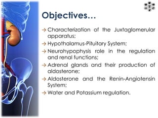
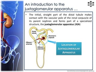
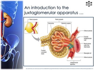
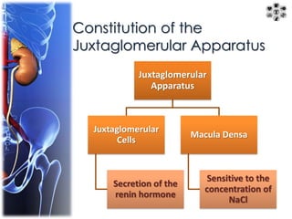
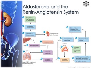
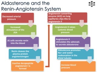
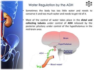
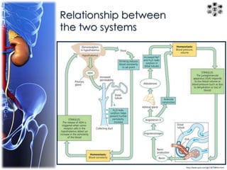
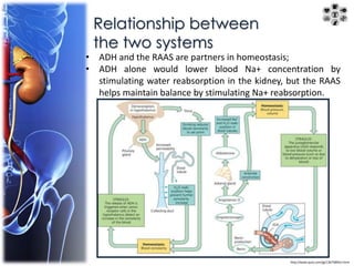
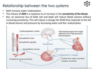
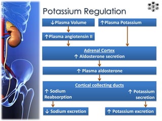
Functions of the 
juxtaglomerular 
apparatus and 
hormonal control in 
the urinary system
Objectives… 
→ Characterization of the Juxtaglomerular 
apparatus; 
→ Hypothalamus-Pituitary System; 
→ Neurohypophysis role in the regulation 
and renal functions; 
→ Adrenal glands and their production of 
aldosterone; 
→ Aldosterone and the Renin-Angiotensin 
System; 
→Water and Potassium regulation.
An introduction to the 
juxtaglomerular apparatus … 
• The initial, straight part of the distal tubule makes 
contact with the vascular pole of the renal corpuscle of 
its parent nephron and forms part of a specialized 
structure, the juxtaglomerular apparatus (JGA) 
LOCATION OF 
JUXTAGLOMERULAR 
APPARATUS 
http://withfriendship.com/user/neeraj/juxtaglomerular_apparatus.php
An introduction to the 
juxtaglomerular apparatus … 
http://apbrwww5.apsu.edu/thompsonj/Anatomy%20&%20Physiology/2020/2020%20Exam%20Reviews/Exam%204/CH25%20Nephron%20III%20-%20Blood%20Supply%20&JGA.htm
Constitution of the 
Juxtaglomerular Apparatus 
Juxtaglomerular 
Apparatus 
Juxtaglomerular 
Cells 
Secretion of the 
renin hormone 
Macula Densa 
Sensitive to the 
concentration of 
NaCl
Constitution of the Juxtaglomerular 
Apparatus 
• JGA forms at the point of contact between a nephron's distal tubule (D) and the 
vascular pole of its glomerulus (G). 
• At that point cells of the distal tubule become columnar as a thickened region 
called the macula densa (MD).
Constitution of the Juxtaglomerular 
Apparatus 
• Smooth muscle cells of the afferent arteriole's (AA) tunica media are converted 
from a contractile to a secretory morphology as juxtaglomerular granule cells (JG). 
• Also present are lacis cells (L), which are extraglomerular mesangial cells adjacent 
to the macula densa, the afferent arteriole, and the efferent arteriole (EA).
Constitution of the Juxtaglomerular 
Apparatus 
• A plastic section through an afferent 
arteriole of a JGA shows the JG cells 
(arrowheads) with secretory granules 
containing renin. 
• Activities of the macula densa and 
renin released by JG cells produce an 
incompletely understood 
autoregulatory tubuloglomerular 
feedback loop: 
- To control arterial blood pressure, 
- To maintain a relatively constant rate 
of glomerular filtration despite 
changes in blood pressure.
Hypothalamus-Pituitary System 
• The pituitary gland is composed of an anterior part and a posterior part, 
directly attached to the hypothalamus region of the brain by a stalk called the 
infundibulum. 
• The gland occupies a fossa of the sphenoid bone called the sella turcica .
Hypothalamus-Pituitary System 
The hypothalamo-hypophyseal portal system and hormone release 
in the pituitary.
Posterior Pituitary 
Oxytocin ADH 
- Uterine 
contractions; 
- Lactation. 
- Stimulates 
water 
retention; 
- Raises blood 
pressure by 
contracting 
arterioles; 
- Induces male 
aggression.
Posterior Pituitary 
Pars Nervosa 
• Modified neural tissues containing 
unmyelinated axons supported and 
ensheathed by glia cells - pituicytes 
(P); 
• The axons run from the supraoptic 
and paraventricular hypothalamic 
nuclei and have swellings called 
neurosecretory (Herring) bodies 
(NB) 
- from which oxytocin and 
vasopressin are released upon 
neural stimulation. 
• The released hormones are picked 
up by capillaries (C) for distribution 
throughout the body.
Adrenal Glands 
http://academic.kellogg.cc.mi.us/herbrandsonc/bio201_mckinley/http://drmichaelroth.wordpress.com/tag/adrenals/ endocrine%20system.htm
Aldosterone and the 
Renin-Angiotensin System 
http://antranik.org/the-renin-angiotensin-aldosterone-reflex/
Aldosterone and the 
Renin-Angiotensin System 
Decreased arterial 
pressure 
Increased 
stimulation of the 
JGA 
JG cells secrete renin 
into the blood 
Renin cleaves the 
plasma protein 
angiotensinogen 
Inactive decapeptide 
angiotensin I is 
formed 
Angiotensin converting 
enzyme (ACE) on lung 
capillaries clips this to 
angiotensin II 
Angiotensin II raises 
systemic blood 
pressure 
Angiotensin II 
stimulates the adrenals 
to secrete aldosterone 
Aldosterone promotes 
Na+ and water 
reabsorption in the 
distal tubules 
Increase blood 
pressure
Water Regulation by the ADH 
• Sometimes the body has too little water and needs to 
conserve it and too much water and needs to get rid of it. 
• Most of the control of water takes place in the distal and 
collecting tubules under control of ADH released by the 
posterior pituitary under control of the hypothalamus in the 
mid-brain area.
Water Regulation by the ADH 
• ADH travels in the blood stream to the peritubular capillaries 
and binds to receptors on the distal and collecting tubules 
which causes water channels to open in the tubule walls. 
• This allows water to diffuse through the tubule walls into the 
interstitial fluid where it is collected by the peritubular 
capillaries.
Water Regulation by the ADH 
↓ Plasma Volume 
↓ Venous, atrial and arterial 
pressures 
Posterior Pituitary 
↑ Vasopressin Secretion 
↑ Plasma Vasopressin 
Collecting Ducts 
↑ Tubular Permeability to H2O 
↑ H2O Reabsorption 
↓ H2O excretion 
Excess H2O ingested 
↓ Body-fluid osmolarity 
(↑ H2O concentration) 
↓ Firing by the hypothalamic 
osmoreceptors 
Posterior Pituitary 
↓ Vasopressin Secretion 
↓ Plasma Vasopressin 
Collecting Ducts 
↓ Tubular Permeability to H2O 
↓ H2O Reabsorption 
↑ H2O excretion
Water Regulation by the ADH 
• Over 99% of the filtrate produced each day can be 
reabsorbed. 
• The amount of water reabsorbed from the filtrate back 
into the blood depends on the water situation in the 
body. 
• When the body is dehydrated, most of the filtrate is 
reabsorbed.
Relationship between 
the two systems 
http://www.quia.com/jg/1367589list.html
Relationship between 
the two systems 
• ADH and the RAAS are partners in homeostasis; 
• ADH alone would lower blood Na+ concentration by 
stimulating water reabsorption in the kidney, but the RAAS 
helps maintain balance by stimulating Na+ reabsorption. 
http://www.quia.com/jg/1367589list.html
Relationship between the two systems 
• Both increase water reabsorption. 
• The release of ADH is a response to an increase in the osmolarity of the blood. 
• But, an excessive loss of both salt and body will reduce blood volume without 
increasing osmolarity. This will induce a change the RAAS that responds to the fall 
in blood volume and pressure by increasing water and Na+ reabsorption. 
http://www.samsca.com/pathophysiology-heart-failure.aspx
Potassium Regulation 
↓Plasma Volume 
↑Plasma angiotensin II 
↑Plasma Potassium 
Adrenal Cortex 
↑ Aldosterone secretion 
↑ Plasma aldosterone 
Cortical collecting ducts 
↑ Sodium 
Reabsorption 
↑ Potassium 
secretion 
↓ Sodium excretion ↑ Potassium excretion
Thank you for 
your attention 
Follow Us: 
http://biomedeeks.blogspot.pt/

Juxtaglomerular Apparatus

  • 1.
    Functions of the juxtaglomerular apparatus and hormonal control in the urinary system
  • 2.
    Objectives… → Characterizationof the Juxtaglomerular apparatus; → Hypothalamus-Pituitary System; → Neurohypophysis role in the regulation and renal functions; → Adrenal glands and their production of aldosterone; → Aldosterone and the Renin-Angiotensin System; →Water and Potassium regulation.
  • 3.
    An introduction tothe juxtaglomerular apparatus … • The initial, straight part of the distal tubule makes contact with the vascular pole of the renal corpuscle of its parent nephron and forms part of a specialized structure, the juxtaglomerular apparatus (JGA) LOCATION OF JUXTAGLOMERULAR APPARATUS http://withfriendship.com/user/neeraj/juxtaglomerular_apparatus.php
  • 4.
    An introduction tothe juxtaglomerular apparatus … http://apbrwww5.apsu.edu/thompsonj/Anatomy%20&%20Physiology/2020/2020%20Exam%20Reviews/Exam%204/CH25%20Nephron%20III%20-%20Blood%20Supply%20&JGA.htm
  • 5.
    Constitution of the Juxtaglomerular Apparatus Juxtaglomerular Apparatus Juxtaglomerular Cells Secretion of the renin hormone Macula Densa Sensitive to the concentration of NaCl
  • 6.
    Constitution of theJuxtaglomerular Apparatus • JGA forms at the point of contact between a nephron's distal tubule (D) and the vascular pole of its glomerulus (G). • At that point cells of the distal tubule become columnar as a thickened region called the macula densa (MD).
  • 7.
    Constitution of theJuxtaglomerular Apparatus • Smooth muscle cells of the afferent arteriole's (AA) tunica media are converted from a contractile to a secretory morphology as juxtaglomerular granule cells (JG). • Also present are lacis cells (L), which are extraglomerular mesangial cells adjacent to the macula densa, the afferent arteriole, and the efferent arteriole (EA).
  • 8.
    Constitution of theJuxtaglomerular Apparatus • A plastic section through an afferent arteriole of a JGA shows the JG cells (arrowheads) with secretory granules containing renin. • Activities of the macula densa and renin released by JG cells produce an incompletely understood autoregulatory tubuloglomerular feedback loop: - To control arterial blood pressure, - To maintain a relatively constant rate of glomerular filtration despite changes in blood pressure.
  • 9.
    Hypothalamus-Pituitary System •The pituitary gland is composed of an anterior part and a posterior part, directly attached to the hypothalamus region of the brain by a stalk called the infundibulum. • The gland occupies a fossa of the sphenoid bone called the sella turcica .
  • 10.
    Hypothalamus-Pituitary System Thehypothalamo-hypophyseal portal system and hormone release in the pituitary.
  • 11.
    Posterior Pituitary OxytocinADH - Uterine contractions; - Lactation. - Stimulates water retention; - Raises blood pressure by contracting arterioles; - Induces male aggression.
  • 12.
    Posterior Pituitary ParsNervosa • Modified neural tissues containing unmyelinated axons supported and ensheathed by glia cells - pituicytes (P); • The axons run from the supraoptic and paraventricular hypothalamic nuclei and have swellings called neurosecretory (Herring) bodies (NB) - from which oxytocin and vasopressin are released upon neural stimulation. • The released hormones are picked up by capillaries (C) for distribution throughout the body.
  • 13.
  • 14.
    Aldosterone and the Renin-Angiotensin System http://antranik.org/the-renin-angiotensin-aldosterone-reflex/
  • 15.
    Aldosterone and the Renin-Angiotensin System Decreased arterial pressure Increased stimulation of the JGA JG cells secrete renin into the blood Renin cleaves the plasma protein angiotensinogen Inactive decapeptide angiotensin I is formed Angiotensin converting enzyme (ACE) on lung capillaries clips this to angiotensin II Angiotensin II raises systemic blood pressure Angiotensin II stimulates the adrenals to secrete aldosterone Aldosterone promotes Na+ and water reabsorption in the distal tubules Increase blood pressure
  • 16.
    Water Regulation bythe ADH • Sometimes the body has too little water and needs to conserve it and too much water and needs to get rid of it. • Most of the control of water takes place in the distal and collecting tubules under control of ADH released by the posterior pituitary under control of the hypothalamus in the mid-brain area.
  • 17.
    Water Regulation bythe ADH • ADH travels in the blood stream to the peritubular capillaries and binds to receptors on the distal and collecting tubules which causes water channels to open in the tubule walls. • This allows water to diffuse through the tubule walls into the interstitial fluid where it is collected by the peritubular capillaries.
  • 18.
    Water Regulation bythe ADH ↓ Plasma Volume ↓ Venous, atrial and arterial pressures Posterior Pituitary ↑ Vasopressin Secretion ↑ Plasma Vasopressin Collecting Ducts ↑ Tubular Permeability to H2O ↑ H2O Reabsorption ↓ H2O excretion Excess H2O ingested ↓ Body-fluid osmolarity (↑ H2O concentration) ↓ Firing by the hypothalamic osmoreceptors Posterior Pituitary ↓ Vasopressin Secretion ↓ Plasma Vasopressin Collecting Ducts ↓ Tubular Permeability to H2O ↓ H2O Reabsorption ↑ H2O excretion
  • 19.
    Water Regulation bythe ADH • Over 99% of the filtrate produced each day can be reabsorbed. • The amount of water reabsorbed from the filtrate back into the blood depends on the water situation in the body. • When the body is dehydrated, most of the filtrate is reabsorbed.
  • 20.
    Relationship between thetwo systems http://www.quia.com/jg/1367589list.html
  • 21.
    Relationship between thetwo systems • ADH and the RAAS are partners in homeostasis; • ADH alone would lower blood Na+ concentration by stimulating water reabsorption in the kidney, but the RAAS helps maintain balance by stimulating Na+ reabsorption. http://www.quia.com/jg/1367589list.html
  • 22.
    Relationship between thetwo systems • Both increase water reabsorption. • The release of ADH is a response to an increase in the osmolarity of the blood. • But, an excessive loss of both salt and body will reduce blood volume without increasing osmolarity. This will induce a change the RAAS that responds to the fall in blood volume and pressure by increasing water and Na+ reabsorption. http://www.samsca.com/pathophysiology-heart-failure.aspx
  • 23.
    Potassium Regulation ↓PlasmaVolume ↑Plasma angiotensin II ↑Plasma Potassium Adrenal Cortex ↑ Aldosterone secretion ↑ Plasma aldosterone Cortical collecting ducts ↑ Sodium Reabsorption ↑ Potassium secretion ↓ Sodium excretion ↑ Potassium excretion
  • 24.
    Thank you for your attention Follow Us: http://biomedeeks.blogspot.pt/