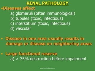
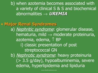
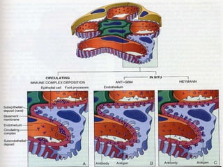
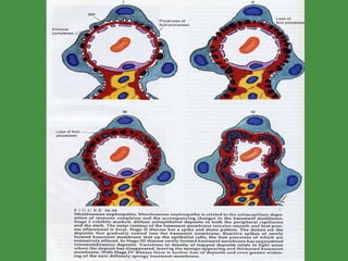
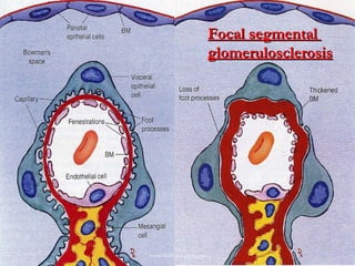
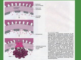
RENAL PATHOLOGY Diseases affect a) glomeruli (often immunological) b) tubules  (toxic, infectious) c) interstitium (toxic, infectious) d) vascular Disease in one area usually results in  damage or disease on neighboring areas Large functional reserve a) > 75% destruction before impairment www.freelivedoctor.com
Congenital Anomalies Review page 961 ,   “Robbins and Cotran”  PATHOLOGIC BASIS OF DISEASE . 7 TH  ed. Understand and   describe the following congenital anomalies of the kidney a) agenesis b) hypoplasia c) ectopic kidney d) horseshoe kidney www.freelivedoctor.com
GLOMERULI Network of capillaries a) lined by fenestrated endothelium b) basement membrane c) podocytes (“foot processes”) Glomeruli capillary wall a) lined with fenestrated endothelium ( 70-   100 nm) b) glomerular basement membrane  (GBM) i) consist of collagen (type IV),    heparan sulfate, laminin,    glycoproteins www.freelivedoctor.com
-  Type IV collagen forms network to    which glycoprotein's attach c) visceral epithelial cells (podocytes; “foot    processes”) i)   composed of interdigitating    processes embedded to basement    membrane ii)  adjacent foot processes are    separated by 20-30 nm filtration slits   bridged by thin diaphragm (nephrin) d) entire glomerulus is supported by      mesangial cells i) lying between capillaries  www.freelivedoctor.com
ii) phagocytic, contractile, proliferate,    secretion of biologically active    mediators - modified smooth muscle cells iii) involved in many types of GN Glomeruli a) very permeable to H 2 O and small solutes b) impermeable to proteins (~ 70 kDa or    larger; i.e., albumin) c) “glomerular barrier function” i) selective permeability based on: - size - charge: cationic more permeable www.freelivedoctor.com
ii) podocytes important in maintaining    this “function” - slit diaphragm maintain size-   selectivity by specific proteins 1.-  NEPHRIN : extend towards each    other from neighboring podocytes   comprising the slit diaphragm !! 2.-  PODOCIN : intracellular (podocyte)    protein where nephrin attaches - mutations in genes encoding    these proteins give rise to    nephrotic syndrome (i.e.,    glomerular disease) www.freelivedoctor.com
www.freelivedoctor.com
Clinical Manifestations Termimology a)  Azotemia :    BUN and     creatinine i) related to    GFR - prerenal azotemia:    RBF,    hypoperfusion w/out    parenchymal damage  - postrenal azotemia: obstruction    of urine flow below level of    kidney www.freelivedoctor.com
b) when azotemia becomes associated with    a variety of clinical S & S and biochemical    abnormalities     UREMIA   Major Renal Syndromes   a)  Nephritic syndrome :   glomerular disease,    hematuria, mild    moderate proteinuria,    azotemia, edema,    BP i) classic presentation of post    streptococcal GN b)  Nephrotic syndrome : heavy proteinuria    (> 3.5 g/day), hypoalbuminemia, severe    edema, hyperlipidemia and lipiduria www.freelivedoctor.com
c)  Acute renal failure : oliguria/anuria, recent    onset of azotemia, can result from GN,    tubular or interstitial disease d)  Nephroliathiasis : renal stones, renal colic,    hematuria, recurrent stone formation e)  Chronic renal failure : 4 stages i)    renal reserve:  GFR ~ 50% normal    BUN & creatinine normal, pt.    asymptomatic, more susceptible to    develop azotemia ii)  renal insufficiency:  GFR 20-50% of    normal, azotemia, anemia,    BP,    polyuria/nocturia (via       concentrating ability) www.freelivedoctor.com
iii)  renal failure : GFR less than 20-25%   kidneys cannot regulate volume,    ions: edema, hypocalcemia,    metabolic acidosis, uremia with    neurological, CV and GI    complications iv)  end stage renal disease : GFR < 5%    of normal, terminal stage of uremia www.freelivedoctor.com
www.freelivedoctor.com
Glomerular Disease Chronic GN one of most common causes of chronic renal failure Glomerular disease often associated with systemic disorders such as: a) diabetes mellitus b) SLE c) amyloidosis d)  vasculitis - pts. with manifestations of glomerular    disease should be considered for    these systemic syndromes, etc. www.freelivedoctor.com
GN characterized by one or more of the following (inflammatory diseases of glomerulus)  a)  hypercellularity :  i) cell proliferation of mesangial cells or    endothelial cells ii) leukocyte infiltration (neutrophils,    monocytes and sometimes    lymphocytes) iii) formation of crescents - epithelial cell proliferation (from    immune/inflammatory injury) - fibrin thought to elicit this injury   (TNF, IL-1, IFN-   are others) www.freelivedoctor.com
b)  basement membrane thickening i) deposition of immune complexes on    either the endothelial or epithelial    side of GBM or w/in GBM itself ii) thickening of GBM proper as with    diabetes mellitus (diabetic    glomerulosclerosis) c)  hyalinization (hyalinosis) and sclerosis   i) accumulation of material that is    eosinophilic and homogeneous - obliterates capillary lumen of    glomerulus (sclerotic feature) www.freelivedoctor.com
- result of capillary or endothelial    injury. Usually end result of    various forms of glomerular    damage (intraglomerular    thromboses, accumulation of    other metabolic materials) Since etiology of primary GN is unknown,  classification is based on histology.  Subdivided: a) diffuse (all glomeruli) b) global (entire glomerulus) c) focal (portion of glomeruli) d) segmental (part of each glomerulus) e) mesangial (affecting mesangial region) www.freelivedoctor.com
Pathogenesis of Glomerular Disease/Injury Little is known regarding etiology or triggering Immune mechanisms underlie most cases of primary GN and many of the secondary cases   a) 2 forms of Ab-associated injury i) injury resulting from soluble Ag-Ab    deposits in glomerulus ii) injury from Ab reacting in-situ with    glomerulus - insoluble fixed glomerular Ag - molecules planted w/in    glomerulus www.freelivedoctor.com
Examples In Situ Immune Complex Deposition a)  Ab act directly with intrinsic tissue Ag   “ planted” in the glomerulus from the    circulation b)  2 forms of Ab-mediated glomerular      injury i)   anti-GBM Ab-induced nephritis -  Ab directed against fixed Ag in    GBM -  in humans spontaneous AGBM    nephritis is  autoimmune disease www.freelivedoctor.com
- Ab bind along GBM forming a    “linear pattern” - sometimes AGBM Ab cross react      with BM of lung       GOODPASTURE  SYNDROME - < 1% of GN cases - some cases show severe    glomerular damage and rapidly    progressive crescentic GN ii)  Heymann nephritis - a form of membranous GN - Ab bind along GBM in “granular    pattern” www.freelivedoctor.com
c) Trigger for induction of autoimmune Ab is   unclear i) ETX ii) mercuric chloride iii) graft-vs.-host reaction Ab can react with “planted” Ag in GBM a) cationic Ag binding to anionic GBM      sites b) bacterial byproducts c) IgG deposition in mesangium www.freelivedoctor.com
www.freelivedoctor.com
re: Ab-mediated injury    Ag-Ab deposition in GBM is major pathway of glomerular injury !! a)  largest proportion of cases of GN are      granular immune pattern along the GBM    or mesangium Cell mediated immune GN a)  sensitized T cells can cause glomerular    injury, in absence of immune deposits i) may occur in some forms of rapidly    progressive GN  www.freelivedoctor.com
www.freelivedoctor.com
Mediators of immune injury seen as loss of glomerular barrier function a) proteinuria b)    GFR in some instances Complement-leukocyte mechanism a) well established i) activated complement (C5a)     neutrophils and monocytes - release proteases    degrade    GBM ii) ROS iii) some are neutrophil-independent - C5-C9 (lytic component;    membrane attack complex) www.freelivedoctor.com
- Membrane attack complex stimulate    growth factors (TGF)     GBM    thickening iv) direct cytotoxicity Other mechanisms of glomerular injury a) epithelial cell injury i) can be induced by Ab to visceral    epithelial cell Ag ii) toxins iii) cytokines iv) loss of foot processes - caused by alterations in nephrin www.freelivedoctor.com
b) renal ablation GN i) any renal disease       GFR (30-50%    of normal)  - lead to end stage renal failure ii) patients develop proteinuria and    diffuse glomerulosclerosis - initiated by unaffected glomeruli      hypertrophy to maintain    function    single nephron    hypertension    damage www.freelivedoctor.com
Glomerular Syndromes and Disorders Nephrotic Syndrome a)  massive proteinuria (> 3.5 g/day) b)  hypoalbuminemia c)  generalized edema d)  hyperlipidemia and lipiduria Initial event is derangement of GBM   increasing permeability and progressive loss of plasma proteins   hypoalbuminemia    decrease in plasma colloid osmotic pressure    edema      plasma volume       aldosterone www.freelivedoctor.com
   ANP, GFR       water and solute retention by kidney    exacerbation of edema (anasarca; massive amounts of edematous fluid);  hypoalbuminemia       lipoprotein production by the liver In children < 15 yrs, nephrotic syndrome almost always caused by primary renal  disease (~ 98 %) In adults nephrotic syndrome may often be associated with secondary renal disease   www.freelivedoctor.com
www.freelivedoctor.com
GLOMERULAR DISEASES ( NONINFLAMMATORY ) Membranous Glomerulopathy  (epithelial cell and BM disease)   a)  most common cause of nephrotic      syndrome in adults (C5-C9 cytotoxic) b) diffuse thickening of glomerular        capillary wall !! c) most cases are idiopathic  i)  most believed to be autoimmune d) most cells are normocellular or only      mildly hypercellular www.freelivedoctor.com
e)  believed to be caused by   i) deposition of immune complexes    w/in  capillary wall - IgG and C3 ii) formation of  in situ  immune    complexes iii) refer to Heymans nephritis and    Goodpasture syndrome iv) classified as non inflammatory since    there is NO cellular proliferation f)  In adults, a frequent association is    with carcinoma !! (i.e., melanoma,    lung and colon) www.freelivedoctor.com
g) associated with systemic infections i) HBV ii) SLE h) associated with certain drug treatments i) gold, penicillamine, NSAID Clinical a) variable i) spontaneous remission or renal    failure w/in 10-15 yrs b) persistent proteinuria w/ normal function c) better response to corticosteroids in      children vs. adults www.freelivedoctor.com
d) Progression of disease i)  stage I : small granular subepithelial    deposits ii)  stage II : “spikes” of BM protrude    between deposits of electron dense    material (e.g., IgG, C3) iii)  stage III : deposits of electron dense    material are incorporated into GBM iv)  stage iv : GBM very distorted and    damaged www.freelivedoctor.com
www.freelivedoctor.com
Minimal change disease (Lipoid nephrosis;  (Epithelial cell disease)) a) major cause of nephrotic syndrome in  b)    children < 15 yrs; peak 2-6 yrs  i) also in adults with nephrotic    syndrome (~ 20 %) c)  effacement  of “foot” processes d) glomeruli show only “minimal” changes e) most patients are boys who usually    present prior to 6 yrs of age i) selective proteinuria (albumin) ii) history of recent Ag exposure ?? f) idiopathic (sometimes follows respiratory    infection or routine immunizations) www.freelivedoctor.com
Minimal Change Disease www.freelivedoctor.com
www.freelivedoctor.com
g) T cell involvement suggested i) HL patients present w/ similar S & S ii) epithelial cell diseases have altered    T ell function h) loss of lipoproteins through the glomeruli      accumulates lipids in proximal tubule    cells    foamy cytoplasm.  Together with    lipids in the urine    LIPOID NEPHROSIS i) remission w/in 8 weeks with use of      corticosteroid use (very dramatic response    is one hallmark of this disease) j) relapses do not tend to progress to      chronic renal failure www.freelivedoctor.com
k) development of azotemia should suggest    incorrect diagnosis of minimal change    disease l) in absence of complications, outcome of    patients with epithelial cell disease is      same as general population Focal segmental glomerulosclerosis   (epithelial cell and BM disease) a) some glomeruli exhibit segmental areas    of sclerosis whereas others are normal b) nephrotic syndrome   www.freelivedoctor.com
c) occurs in the following setting: i) associated with other conditions - HIV - heroin addiction - sickle cell disease - morbid obesity ii) secondary event - IgA nephropathy iii) adaptive process to loss of kidney - renal ablation - advanced stages of other renal    diseases (e.g., hypertension) iv) primary disease (e.g., idiopathic    focal segmental glomerulosclerosis) www.freelivedoctor.com
d) most common cause of nephrotic      syndrome in USA i) Hispanics and African Americans e) differs from minimal change disease i) higher incidence of hematuria,    reduced GFR,  and hypertension ii) poor response to corticosteroids iii) proteinuria is non selective iv) progression to chronic    glomerulosclerosis v) IgM and C3 trapping on sclerotic    segments www.freelivedoctor.com
vi) whether this is a specific disease or   is an evolution of minimal change    disease is unresolved !! - degeneration of visceral    epithelial cells hallmark of FSGN - similar cell damage as seen in    minimal change disease vii) genetic basis - NPHS1 gene    encodes nephrin - several mutations of this gene    give rise to congenital nephrotic    syndrome of the Finnish type    (CNF) www.freelivedoctor.com
Focal segmental  glomerulosclerosis www.freelivedoctor.com
- NPHS2 gene - encodes to podocin - mutations give rise to    steroid resistant nephrotic    syndrome in children Paraproteinemic nephropathies a) abnormally elevated immunoglobulin or   immunoglobulin fragments (i.e.,        paraproteins) may cause renal disease i) present in blood and/or urine ii) renal diseases include - multiple myeloma - Waldenstrom macroglobulinemia - cryoglobulinemia www.freelivedoctor.com
Multiple myeloma a) neoplasm of Ig producing plasma cells b) occurs in 5 th  decade c)  more than 50% of all patients with    multiple myeloma have renal      involvement   i) hypercalcemia ii) hyperuricemia iii) renal infections iv) renal lesions are of three types - tubular and interstitial lesions - amyloidosis  - light chain deposition disease www.freelivedoctor.com
2.   Waldenstrom macroglobulinemia   a)    serum IgM b)    blood viscosity c) renal involvement results in partial or      total occlusion of glomerular capillaries 3.  Cryoglobulinemia   a) IgM or IgG b) defined by their capacity to precipitate at    4   C c) renal disease is often immune complex    mediated d) microthrombi in glomeruli e) mesangial & endothelial cell proliferation www.freelivedoctor.com
Hereditary nephritis (Alport syndrome) a) most often present as recurrent        hematuria b) structural defects in GBM i) specific molecular defect affecting    type IV collagen c) usually does not present with nephrotic    syndrome and proteinuria  d) more severe in men i) die by age 40 e) progressive hearing loss (high        frequencies) f) ocular defects most often    the lens www.freelivedoctor.com
Benign familial hematuria (thin GBM  disease) a) presents as recurrent hematuria in      childhood or young adults (similar to      Alport syndrome) b) no progression to renal failure (unlike    Alport syndrome) c) reduced thickness of GBM (capillary site) d) one of most important causes of      asymptomatic hematuria e) IgA nephropathy and this disease are two    most major diagnostic considerations of    asymptomatic hematuria www.freelivedoctor.com
GLOMERULAR LESIONS ASSOCIATED WITH SYSTEMIS DISEASES Diabetic glomerulosclerosis  (Kimmelstiel- Wilson Disease) a) diabetic microangiopathy i) small arteries, arterioles and    capillaries) - hyaline arteriosclerosis in    diabetics involves both afferent    and efferent arterioles www.freelivedoctor.com
ii) occur by progressive accumulation    of GBM material - severity and duration of    hyperglycemia !!  ?? b) etiology of proteinuria not known i) non-nephrotic proteinuria ii) nephrotic  c) earliest lesion is thickening of    GBM d) followed by glomerular enlargement e) “diffuse glomerulosclerosis” refers to    enlarged glomeruli w/expanded      mesangium and diffusely thickened GBM www.freelivedoctor.com
f) nodular glomerulosclerosis (i.e.,    Kimmelstiel-Wilson Disease) – highly      specific for diabetes  g) one of leading causes of chronic renal    failure in USA h) 2 processes play role in diabetic        glomerular lesions i) metabolic defect (i.e., glycosylation    end products that: -    GBM thickening -    mesangial matrix ii) hemodynamic effects: - glomerular hypertrophy (   GFR) - develop of glomerulosclerosis www.freelivedoctor.com
Diabetic  glomerulosclerosis www.freelivedoctor.com
i) Clinical i) proteinuria usually mild ii) nephrotic syndrome present    renal    failure w/in 6 yrs. - severe proteinuria usually    associated with other signs of    advanced diabetes (i.e.,    retinopathy) Amyloidosis a) deposits of amyloid w/in glomeruli i) mostly light chain AA or AL type ii) Congo red amyloid positive deposits    mainly in mesangium and capillaries www.freelivedoctor.com
Amyloid nephropathy www.freelivedoctor.com
b) clinical i) nephrotic syndrome ii) progressive glomerular destruction    leads to death from uremia Henoch-Schönlein Purpura a) purpuric skin lesions on legs, arms and    buttock b) abdominal pain, vomiting, intestinal      bleeding, arthralgias and renal        abnormalities (hematuria, proteinuria,    nephrotic syndrome)  c) not all these S & S need to be present www.freelivedoctor.com
d)  disease most common in children i) 3-8 yrs e) does occur in adults where disease is      more severe i) may develop rapidly progressive form    of glomerulornephritis with many    crescents f) onset often follows upper respiratory      infection i) IgA deposits in mesangium which    has led to concept that IgA    nephropathy (purely a renal disease)    and Henoch-Schönlein Purpura are    spectra of same disease www.freelivedoctor.com
Glomerular Diseases   (INFLAMMATORY) (“GLOMERULONEPHRITIS”) Inflammatory lesions of glomerulus characterized by hypercellularity which may be diffuse or focal + proliferation a) Diffuse types of  glomerulornephritis i) post infectious ii) membranoproliferative iii) some lupus forms  b) Focal types of  glomerulornephritis i) IgA ii) other lupus forms www.freelivedoctor.com
c) glomerular injury may be either global    (entire glomerulus) or segmental d) glomerulornephritis clinically        characterized by the  NEPHRITIC      SYNDROME  i) may present with only portions of    these S & S (i.e., hematuria) ii) on occasion, proteinuria may    predominate e) different forms of GN are differentiated    via microscopy www.freelivedoctor.com
f) neutrophils contribute to    cellularity,      particularly in children i) many neutrophils in glomeruli    referred to as “exudative” g) proliferation of podocytes and Bowman’s    capsule (visceral and parietal epithelium,    respectively)  i) leads to formation of “crescents” - highly cellular lesions - extend from Bowman’s capsule    into glomerulus having shape of    crescents.  These are severe    lesions (associated with    necrosis, early and fibrosis, late) www.freelivedoctor.com
Pathogenesis of inflammatory GN Immunologic injury a) trapping of circulating immune complexes b)  in situ  immune complex formation c) activation of alternative complement  d) cell mediated processes  Circulating immune complex nephritis a) glomerular trapping of circulating Ag-Ab v  complexes.  i) glomerulus as “innocent bystander”  - penetrate GBM than are   - trapped in GBM www.freelivedoctor.com
b) confirmed by EM i) presence of subepithelial “humps” ii) peripheral granular staining directed - IgG - C3 c) alternatively, circulating immune complex    do not penetrate GBM but localize to i) mesangium or subendothelium d) trapping is affected by: i) size and charge of aggregates ii) glomerular hemodynamics iii) presence of vasoactive substances   www.freelivedoctor.com
e) Ag may be exogenous or endogenous i) exogenous - bacterial Ag induced by 1. Strep infections 2. bacterial endocarditis 3. viruses (HBV) ii) endogenous - DNA in pathogenesis of lupus 2.  In Situ  immune complex formation a) Goodpasture Syndrome b) Ag (endogenous) already embedded in    GBM i) Ab binds !! ii) linear localization of IgG along GBM www.freelivedoctor.com
c) Ag-Ab interaction activates complement ! i) rapidly progressive GN occurs 3.  Alternative complement activation a) focal glomerulornephritis i) caused by IgA b) form of membranoproliferative GN 4.  Cell-mediated Immunity a) no direct evidence for any specific GN    caused by cell mediated processes\ i) indirect evidence for a delayed (type    iv) cell type GN - lymphocytes from some patients    with GN react in vitro with a    glomerular Ag www.freelivedoctor.com
once immune complexes localized w/in GBM other secondary affects cause immune mediated injury    mediators: a) complement (C5b-C9; cytotoxic) b) neutrophils (chemotaxis via C5a) c) monocytes & macrophages d) coagulation system NEPHRITIC SYNDROME hematuria oliguria    BUN and creatinine hypertension proteinuria (< 3.5 g/day); ± edema www.freelivedoctor.com
www.freelivedoctor.com
Glomerulornephritis - - INFLAMMATORY Acute GN (post infectious GN) a) sudden onset of nephritic syndrome b) diffuse hypercellularity og glomeruli c) most often associated with i) group A  β -hemolytic streptococci  - S. pyogenes ii) others less frequently - staph - spirochetes - viruses d) most often affect children i) one of most common renal diseases www.freelivedoctor.com
e) latent period of ~ 10-14 days f) diffuse enlargement and hypercellularity    of glomeruli, hypercellularity due to:  i) proliferation of endothelial and    mesangial cells and infiltration of    neutrophils and monocytes g) characteristics: i) subepithelial “humps” of GBM ii) granular IgG and C3  along GBM   in association with “humps” h) Clinical: i) most resolve but in rare occasions    can progress to develop many    crescents and renal failure www.freelivedoctor.com
Acute GN  (post infectious GN) www.freelivedoctor.com
ii) primary infection in pharynx or skin iii) nephritic syndrome (abrupt) - hematuria - oliguria - facial edema - hypertension iv)    serum C3 Membranoproliferative GN a) characterized by GBM thickening (i.e.,    “membrano”) + mesangial cell    proliferation (“proliferative ”) www.freelivedoctor.com
b) two major groups: Types I and II (+ III) i)  Type I:   majority of cases are    idiopathic. Associations with - HBV - HCV - bacterial endocarditis - strep infections - granular deposition of Ig (IgG,    IgM) and complement (C3) and    C1q and C4 ii) type II (and III) - circulating C3 Ab (C3 nephritic    factor)       C3    (hypocomplementemia) www.freelivedoctor.com
- characteristic “ribbon-like” zone of    cellularity on thickened GBM (“dense deposit disease”) c) Clinical: i) occurs primarily in older children and    young adults ii) nephritic or nephrotic syndrome iii) low levels of C3 iv) do not have postinfectious GN v) no systemic inflammatory condition vi) most progress to end-stage renal    failure, regardless of treatment !! www.freelivedoctor.com
Membranoproliferative GN Type I www.freelivedoctor.com
SLE (immune complex disease); “Lupus Nephritis” a) chronic autoimmune disorder b) affects 1   young women c) > 70% will develop renal disease d) circulating anti DNA Ab and    C3, etc. i) T cell function is decreased ii) trapping of immune complexes    cause of the renal damage ( in situ ) e) nephritic or nephrotic syndrome f) dysfunction of renal tubules (usually    accompanies GN) g) cellular proliferation is mesangial,      subepithelial and subendothelial cells www.freelivedoctor.com
i) involves the glomeruli w/more severe    inflammation h) hematoxylin bodies    only light        microscopic feature of tissue damage i) episodic inflammation – usually present    with old lesions  j) IgG most common.  IgA and IgM also      present.  Complement present i) IgG, IgA, IgM, C3, C4 and C1q    present in same glomerulus “FULL    HOUSE” www.freelivedoctor.com
k) 5 classes based on WHO classification i) Class I – histologically normal ii) Class II – pure mesangial lesion iii) Class III – focal and segmental GN iv) Class IV – diffuse proliferative GN v) Class V – diffuse membranous - Class II and V have more benign    course relative to Class III and    IV   l) renal disease    major consequence of    SLE i) renal failure    cause of death in ~    33 % of patients with SLE www.freelivedoctor.com
Focal GN a) only some of the glomeruli are involved i) or to segments of the glomerulus b) different from focal & segmental    glomerulosclerosis which is a        noninflammatory disease i) glomeruli essentially normocellular c) many conditions produce this defect i) primary renal disease or systemic    diseases such as IgA nephropathy    and Henoch-Schönlein GN (see table) www.freelivedoctor.com
d)  IgA nephropathy (Berger Disease) i) association with chronic liver disease - impaired capacity to remove    circulating immune complexes ii) IgA and fibronectin found in > 70 %    of IgA nephropathy patients.  iii) Ag involve bacterial, viral and    dietary - infectious agents is suggested    from data showing hematuria    following upper respiratory or GI    infection !! - dietary agents    milk proteins I   in mesangium; gluten-sensitivity www.freelivedoctor.com
iv) C3 and properdin (via activation of    alternate pathway) usually present   together with IgA in mesangium - C1q and C4 (classic pathway    activation) are typically absent v) IgA nephropathy is a mesangial    proliferative lesion (granular    deposits) vi) clinical - common in young men (15-30) - presents with hematuria - nephrotic type proteinuria is    uncommon (may indicate more    severe glomerular damage) www.freelivedoctor.com
- ~ 20 % of IgA nephropathy    patients progress to end-stage    renal failure !! - most common type of 1   GN in    several parts of the world    (France, Italy, Japan, Singapore    and Austria) -- ~ 20 %.  In USA    is responsible for ~ 3-10 % of 1      GN  www.freelivedoctor.com
IgA nephropathy  (Berger Disease) www.freelivedoctor.com
e)  Henoch-Schönlein (HS) Purpura i) close relationship with IgA    nephropathy  - differentiate: IgA purely renal;    HS is a systemic disease, etc. Crescentic GN a) ominous morphological pattern i) majority of glomeruli are surrounded   by accumulation of cells in Bowman’s    capsule (parietal epithelial cells) ii) indicative of fulminant glomerular    damage and always leaves scarring  www.freelivedoctor.com
iii) does not denote a specific etiologic    form of GN b) most patients with substantial (~ 80%)    crescents progress to renal failure c) Fibrin in Bowman’s capsule is important    for the formation of glomerular crescents i) Tx with anticoagulants d) associated with areas of segmental      necrosis within glomeruli e)  Types: i)  Type I  – anti-GBM antibody disease    (GOODPASTURE SYNDROME) or    idiopathic www.freelivedoctor.com
- plasmapheresis to remove    circulating Ab  is helpful  in this    type of RPGN (i.e., crescentic) - etiology unknown ii)  Type II  – immune-complex mediated    disease - can be complication of any of    the immune complex nephritides  SLE, IgA nephropathy,  HS Purpura    all these show granular      pattern (characteristic of    immune complex) -  not helped  with plasmapheresis  www.freelivedoctor.com
iii)  Type III  – pauci-immune type - lack of anti-GBM Ab or immune    complexes - patients do have ANCA (~90%)      either c or p patterns   } in some cases, is a    component of    vasculitides (i.e.,  Wegener Granulomatosis) f) clinical: i) hematuria with red cell cast in urine ii) transplant or chronic dialysis in most    patients   www.freelivedoctor.com
Crescentic GN www.freelivedoctor.com
TUBULOINTERSTITIAL DISEASE Most tubular diseases involve the interstitium 2 distinct types of diseases a) ischemic or toxic tubular injury   i) ATN ii) acute renal failure b) inflammatory diseases i)  “tubulointerstitial nephritis” ATN   (Clinical entity) Destruction of tubular epithelial cells Acute suppression of renal function www.freelivedoctor.com
  Most common cause of acute renal failure: a) oliguria (, 400 ml/day) b) severe glomerular disease (RPGN c) acute thrombotic angioplasties d) diffuse renal vascular disease        (Polyarteritis nodosa) e) diffuse cortical necrosis f)  interstitial nephritis (acute drug-       induced) g)  acute papillary necrosis www.freelivedoctor.com
Is reversible and arise from: a) severe trauma b) septicemia (shock and hypotension)  c) ATN associated with shock – “ischemic” -  d) mismatched blood transfusion and other    hemodynamic problems as well as      myoglobinuria    all reversible  ischemic   ATN e)  nephrotoxic ATN  – variety of poisons i) - heavy metals (Hg) ii) - CCl4 iii) - etc. www.freelivedoctor.com
Occurs frequently a) since it is reversible, proper    management means difference between    recovery and death 2 major problems are: a) - tubular injuries b) - blood flow disturbances Major disturbances: a) Change charge in tubules (mainly -)  i) Na+ - K+ - ATPase cause   less Na+ reabsorption and traps    Na+, within tubule with more distal    tube delivery of Na+ which causes    vasoconstriction (feedback) www.freelivedoctor.com
Treatment protocol 1) - initiating phase 2) - maintenance phase 3) - recovery phase Initiating phase Last about 36 hours. Incited by: a) medical, surgical, obstetric event i) slight oliguria (transient decrease in    blood flow) ii) rise in BUN www.freelivedoctor.com
Maintenance phase Anywhere from 2-6 days a) sharp decline in urine output (50-400    ml/day) i) may last few days to 3 weeks b) fluid overload, uremia c) may die from poor management Recovery phase Steady increase in urine output (up to 3L/day) Electrolyte imbalances may continue Increased vulnerability to infection Because of these, about 25% patients die in this phase www.freelivedoctor.com
Tubulointerstitial Nephritis  (TIN) Inflammatory disease of Interstitium/tubules Glomerulus not involved at all or only late in  disease Infections induced TIN – “pyelonephritis” Non infection – interstitial nephritis a) Caused by: i) drugs ii) metabolic disorders (hypokalemia) iii radiation injury iv) immune reactions www.freelivedoctor.com
TIN divided into 2 categories, regardless of etiology a) - acute b) - chronic Acute Pyelonephritis Kidney/renal pelvis (distal to collecting ducts) Caused by bacterial infections (lower UTI) –  cystitis, urethritis and prostatitis  or upper UTI – (pyelonephritis) or both tracts Principle causative bacteria are gram - rods a) E. coli (most common), Proteus,          enterobacter, Klebsiella www.freelivedoctor.com
2 routes bacteria can reach kidney  a) blood stream (not very common) b) lower urinary tract (ascending infections) i) - catheterization ii) - cystoscopy Most commonly affect females (in absence of instrumentation) a) close proximity to rectum b) shorter urethra  Urine sterile, flushing keeps bladder sterile a) Obstruction increased incidence of UTI i) prostate hypertrophy ii) uterine prolapse iii) UT obstructions www.freelivedoctor.com
Incompetent vesicoureteral orifice a) one way valve (at level of bladder) b) incompetence – reflux of urine into      ureters – vesicoureteral reflux (VUR) –    usually congenital defect – 30-50% of    young children with UTI c) - usually congenital defect d) spinal cord injury can produce a flaccid    bladder (residual volume remain in      urinary tract) – favors bacterial growth www.freelivedoctor.com
e) Diabetes increases risk of serious      complications i) septicemia ii) recurrence of infection iii) diabetic neuropathy – dysfunction    of bladder f) pregnancy i) 6% develop pyelonephritis; 40- 60%    develop UTI if not treated Chronic pyelonephritis and reflux nephropathy Interstitial inflammation with scarring of renal parenchyma Important cause of chronic renal failure  www.freelivedoctor.com
Two forms: a) - Chronic obstructive pyelonephritis b) - Chronic reflux-associated pyelonephritis Chronic obstructive pyelonephritis Can be bilateral (congenital disease) Obstruction predisposes kidney to infection recurrent infections on obstructive foci causes scarring – chronic pyelonephritis!  www.freelivedoctor.com
chronic reflux-associated pyelonephritis (reflux nephropathy) More common form of chronic pyelonephritis scarring Occurs from superimposed of a UTI on vesiculouretheral and intrarenal reflux a) reflux may be bi- or unilateral i) unilateral causes atrophy ii) bilateral can cause chronic renal    insufficiency iii) diffuse or patchy - Unclear if sterile vesiculouretheral disease causes renal damage www.freelivedoctor.com
Hallmark is scarring involving pelvis/calyces, leading to papillary blunting and deformities Renal papilla – area of kidney where opening from collecting ducts enters renal pelvis Kidneys are asymmetrically contracted Signs and Symptoms: a) hypertension b) seen following normal physical exam c) slowly progressive    late in disease d) can cause loss of concentrating        mechanisms (if bilateral and progressive) i) - polyuria ii) - nocturia  www.freelivedoctor.com
Drug-induced interstitial nephritis Acute TIN – seen with synthetic penicillins, diuretics (thiazides), NSAI a) disease begins ~15 days (2-40 range) i) fever ii) rash (25% cases) iii) renal findings: hematuria, leukouria iv) increased serum creatinine or acute    renal failure with oliguria (50% of    cases) Immune mechanism is indicated (suggested) a) IgE increased (hypersensitivity – Type I)   Injury produced by IgE and cell-mediated      immune reactions www.freelivedoctor.com
Analgesic Nephropathy Patients who consume large quantities of analgesics may develop  chronic interstitial  nephritis , often associated with  renal papillary  necrosis Usually result from consumption of a mixture for long periods of time: a) - aspirin b) - caffeine c) - acetaminophen d) - codeine e) - phenacetin  www.freelivedoctor.com
Primary pathogenesis is a) papillary necrosis followed by b) interstitial nephritis is secondary c) acetaminophen – oxidative damage d) aspirin inhibits prostaglandins –        vasoconstriction e) all the above leads to papillary ischemia Chronic renal failure, hypertension and anemia Complications may be incidence of “transitional cell carcinoma” of renal pelvis or bladder. www.freelivedoctor.com
Diseases of Blood Vessels Nearly all diseases of kidney involve blood vessels. Kidneys involved in pathogenesis of essential and secondary hypertension Systemic vascular disease (i.e. arteritis) also involve kidney  www.freelivedoctor.com
Benign nephrosclerosis Renal changes associated with benign hypertension a) always associated with  hyaline     arteriosclerosis Kidneys are atrophic Many renal diseases cause hypertension which in turn may lead to benign nephrosclerosis. Therefore this disease seen simultaneously with other diseases of kidney www.freelivedoctor.com
This disease by itself usually does  not  cause severe damage a) mild oliguria b) loss (slight) of concentrating mechanism c) decreases GFR d) mild degree of proteinuria is a constant    finding These patients usually die from hypertensive heart disease or cerebrovascular disease rather than from renal disease www.freelivedoctor.com
Malignant hypertension Less common than benign May arise de novo (without preexisting  hypertension) or may arise suddenly in patient with mild hypertension Factors: a) initial event – some form of vascular damage to kidney b) result is increased permeability of small      blood vessels to fibrinogen and other        plasma proteins, endothelial injury    and platelet deposits www.freelivedoctor.com
c) This leads to appearance of fibroid      necrosis in small arteries and arterioles    and intravascular thrombosis d) platelets (platelet derived growth factors)    and plasma cause intimal hyperplasia of    vessels resulting in hyperplastic        arteriosclerosis, which is typical of      malignant hypertension e) narrowing of renal afferent arteriole      stimulates angiotensin II production      (ischemic-induced) with aldosterone      secretion increases www.freelivedoctor.com
Diastolic pressure > 120 mmHg, papilledema,  encephalopathy, CV disorders, renal failure 90% deaths due to uremia 10% deaths due to CV or cerebral disorders (hemorrhage) www.freelivedoctor.com
Thrombotic Microangiopathies Clinical syndromes Widespread thrombosis in microcirculation (a/C) Damage to endothelial cells !! Diseases: a) childhood hemolytic-uremia syndrome (HUS) b) Thrombotic thrombocytopenic purpura Most follow intestinal infection (E. Coli) Disease is one of main causes of acute renal failure in children Vasoconstriction (decreased NO, increased endothelium, decreased PGI 2 ) www.freelivedoctor.com
Although the various diseases have diverse etiologies, 2 predominant factors   a) endothelial injury and activation, leading    to vascular  thrombosis  and, b) platelet aggregation c) both of these causing vascular        obstruction and vasoconstriction 1.-   Endothelial injury activation can be initiated by a variety of agents, while some remain elusive  a) denuding the endothelial cells, exposes    vascular to thrombogenic subendothelium i)    NO, PGI 2 , enhance platelet    aggregation and vasoconstriction www.freelivedoctor.com
ii) vasoconstriction also initiated via    endothelial derived endothelin-1 iii) activation of endothelial cells  increases adhesiveness to platelets, etc iv) endothelial cells elaborate large    multimers of vW factor    platelet    aggregation 2.- Platelet Aggregation serum factors causing platelet aggregation a) large multimers of vW factor (secreted by    endothelial cells)  i) usually cleaved by ADAMTS-13 (vW    factor-cleaving metalloprotease www.freelivedoctor.com
HUS/TTP 1.- Classic childhood HUS  (> 75%) bloody diarrhea    intestinal infection a) verocytotoxin-releasing bacteria i) Verocytotoxin-producing strains    of E. coli (eg 0157:H7 or 0103); ii) Similar to Shigella toxin. iii) undercooked hamburger iv) “petting” zoos  www.freelivedoctor.com
characterized: a) sudden onset (post GI or influenza      infection)  b) hematemesis c) melena d) severe oliguria e) hematuria f) hemolytic anemia (microangiopathic) g) hypertension in > 50% of cases www.freelivedoctor.com
Pathogenesis a) related to Shigella toxin i) affects endothelium -    adhesion of leukocytes -    endothelin and    NO - endothelial lysis (inpresence of    cytokines such as TNF ii) these changes favor thrombosis and    vasoconstriction iii) verocytotoxin can directly bind to    platelets and cause activation most patients recover in few weeks, with proper care (i.e., dialysis, etc); < 5% lethality www.freelivedoctor.com
2.- Adult HUS associated with: a) infection i) typhoid fever ii) E. coli septicemia iii) etx or shiga toxin iv) viral infections b) antiphospholipid syndrome i) SLE ii) similar to membranoproliferative GN    but w/out immune complex deposits c) complication of pregnancy (“postpartum    renal failure” www.freelivedoctor.com
d) vascular renal disease i) systemic sclerosis ii) malignant hypertension e) chemotherapeutic and          immunosuppressive drugs i) mitomycin ii) cyclosporine iii) bleomycin iv) cisplatin v) radiation Tx 3.- Familial HUS recurrent thromboses (~ 50 lethality) deficit of complement regulatory protein  a) Factor H www.freelivedoctor.com
4.- Idiopathic Thrombotic Thrombocytopenic Purpura Manifested by: a) thrombi in glomeruli b) fever c) hemolytic anemia d) neurologic symptoms e) thrombocytopenic purpura defect in ADAMTS-13 (acquired or inherited) a) normally cleaves large vW multimers i) large vW factors promote platelet    aggregation more common in women most patients < 40 years www.freelivedoctor.com
neurologic involvement is dominant feature renal involvement in ~ 50% of patients a) eosinophilic thrombi in glomerular      capillaries, interlobular artery and      afferent arterioles b) similar changes as with HUS exchange transfusion and steroid Tx   mortality rate to < 50% www.freelivedoctor.com
Cystic Diseases Common and difficult to diagnose In adult polycystic disease – major cause of chronic renal failure Confused with malignant tumors Simple cyst a) Innocuous lesion b) Occur as single or multiple cysts c) Usually 1-5 cm diameter d) Clear fluid, smooth membrane, gray glistening www.freelivedoctor.com
e) Single layer of cuboidal cells   f) Usually confined to cortex   g) No clinical significance Importance to differentiate from tumors a) are fluid filled rather than solid b) have smooth contours c) almost always avascular  Occur in patients with end-stage renal disease who have undergone long term dialysis Occasionally, renal adenomas or adenosarcoma arise from these cysts www.freelivedoctor.com
Adult polycystic kidney disease  (autosomal dominant) Multiple expanding cysts of both kidneys that eventually destroy parenchyma of kidney Accounts for 10% of chronic renal failure In 90% of families, PKD1 (defective gene) is located on chromosome #16 a) encodes for protein (polycystin-1),      extracellular and is a cell membrane      associated protein b) how mutations in this gene cause cysts    formation is unclear www.freelivedoctor.com
Polycystin 2 (PKD2 gene) mutations also cause cyst formation No  symptoms until 4th decade a) by then, kidneys are very large b) common complaint is “flank pain” c) hematuria d) most important complications i) hypertension (~75% patients) ii) UTI iii) aneurysms in circle of Willis (10-   30%) and risk for subarachnoid    hemorrhage www.freelivedoctor.com
iv) Asymptomatic liver cysts in ~30-   40% v) fatal disease (uremia or      hypertension) vi) progresses very slowly viii) Treatment with renal    transplantation Childhood polycystic kidney disease  (autosomal recessive) Rare Serious manifestations at birth and young infants may die quickly a) pulmonary failure b) renal failure www.freelivedoctor.com
Numerous small cysts in cortex and medulla Bilateral disease Many epithelial cysts in liver Patients who survive infancy develop liver cirrhosis (congenital hepatic cirrhosis) Unidentified gene location on chromosome 6p Urinary Outflow-Obstruction Renal Stones Urolithiasis:  Calculus formation at any level in urine collecting system, most often arise in kidney  www.freelivedoctor.com
Occur frequently (!1% of all autopsies) More common in males Familial tendency ~75% of renal stone a) calcium oxalate b) calcium phosphate 15% composed of magnesium ammonium phosphate 10% uric acid or cystine stones All stones composed of mucoprotein  www.freelivedoctor.com
Cause of stones is obscure a) Supersaturation in urine of stones      constituents (exceeds solubility) b) 50% of patients forming “calcium stones”    do not have increased plasma Ca ++  but    do have high urine Ca ++ i) most Ca ++  absorbed from gut in    large amounts (absorptive    hypercalciuria) ii) only 5-10% has associated    hypercalcemia  www.freelivedoctor.com
- hyperparathyroidism - Vit D intoxication - Sarcoidosis (autoimmune    disease, bacterial  - productions of Vit D (toxic)  c) Magnesium ammonium Phosphate stones i) almost always occur in patients with    alkaline urine due to UTI ii) proteus vulgaris and staph split urea    in kidney and therefore predispose    patient to urolithiasis www.freelivedoctor.com
Gout and diseases involved with rapid cell turnover (e.g. leukemia) lead to high uric acid levels in urine and possibility of uric acid stones Unlike magnesium ammonium phosphate stone, both uric acid and cystine stones are more likely to form when urine is  acidic  (pH < 5.5) Stone formation 80% unilateral Hematuria and predispose to infection www.freelivedoctor.com
Hydronephrosis Dilation of renal pelvis and calyces with atrophy of parenchyma caused by obstruction of outflow of urine Most common causes: a) congenital i) atresia of the urethra (absence of a    normal body passage or opening from    an organ to other parts of the body) www.freelivedoctor.com
b) acquired i) stones ii) tumors iii) inflammation iv) spinal cord damage with paralysis of    bladder v) normal pregnancy Bilateral  nephrons only if blockage is below level or ureters Major problems are tubular with impaired concentration mechanisms Obstruction leads to inflammatory response  a) interstitial fibrosis Complicating pyelonephritis is common  www.freelivedoctor.com
Tumors Most common malignant tumor is: a) renal cell carcinoma (80-85% of all 1° Ca      in kidney) b) nephroblastoma (Wilms tumor) c) calyces and pelvis Tumor of lower urinary tract are 2x as common as renal cell Cancer 1.- Renal cell Ca Derived from renal tubular epithelial cells a) located primarily in cortex 2-3% of all cell Ca in adults (~30,000 cases/yr) 6th to 7th decades in life  www.freelivedoctor.com
Higher risk in smokers and occupational exposure to cadmium 30 fold increase in susceptibility in patients with polycystic disease Classification: a)  clear cell Cancer i) most common (70-80% renal ca) b) most are sporadic c) familial links (von Hippel-Lindau [VHL]) i) autosomal dominant disease ii) predispose to a variety of CA –    hemangioblastoma of cerebellum    and retina www.freelivedoctor.com
iii) genetic abnormality chromosome 3    (loss of tumor suppressor gene) –    clear cell CA 2.- Papillary renal cell Ca 10-15% of all renal CA Multifocal and bilateral Both sporadic and familial forms No genetic abnormalities in chromosome 3 a) Protooncogene on chromosome 7 www.freelivedoctor.com
3- Chromophobe Renal Carcinoma Least common (~5% of all renal cell CA) Cortical collecting ducts or their collated cells Stain more darkly than clear cell CA Lack of a lot of chromosomes (1,2,6,10,17,&21) Have good prognosis Renal cell CA are difficult to diagnose! a) Present with hematuria in ~50% of cases 4. Wilms tumor Occurs infrequently in adults 1/3 most common organ cancer in children <10  years, therefore, one of major cancers in children Sporadic or familial in nature a) autosomal dominant  www.freelivedoctor.com
Urinary bladder and collecting system tumors (Renal pelvis to urethra) Tumors in collecting system above bladder are uncommon Bladder Cancer more frequent cause of death than are kidney tumors a) Bladder tumors i) small benign papillomas (rare)  ii) large invasive CA iii) most recur after removal and kill by    infiltrative obstruction of ureters      rather than by metastasizing iv) Shallow lesion have good prognosis www.freelivedoctor.com
v) deep invasive Cancer, survival (5yr)    is <20% with overall 5 yr survival at    50-60% b) - Cancer of ureters is very rare i) 5 yr survival <10% www.freelivedoctor.com
RENAL PATHOLOGY Dr. Richard M Raymond Diseases affect a)  glomeruli (often immunological) b)  tubules  (toxic, infectious) c)  interstitium (toxic, infectious) d)  vascular Disease in one area usually results in  damage or disease on neighboring areas Large functional reserve a)  > 75% destruction before impairment www.freelivedoctor.com
Congenital Anomalies Review page 961 ,   “Robbins and Cotran”  PATHOLOGIC BASIS OF DISEASE . 7 TH  ed. Understand and   describe the following congenital anomalies of the kidney a)  agenesis b)  hypoplasia c)  ectopic kidney d)  horseshoe kidney www.freelivedoctor.com
GLOMERULI Network of capillaries a)  lined by fenestrated endothelium b)  basement membrane c)  podocytes (“foot processes”) Glomeruli capillary wall a)  lined with fenestrated endothelium ( 70-   100 nm) b)  glomerular basement membrane  (GBM) i)  consist of collagen (type IV),    heparan sulfate, laminin,    glycoproteins www.freelivedoctor.com
-  Type IV collagen forms network to    which glycoprotein's attach c)  visceral epithelial cells (podocytes; “foot    processes”) i)   composed of interdigitating    processes embedded to basement    membrane ii)  adjacent foot processes are    separated by 20-30 nm filtration slits   bridged by thin diaphragm (nephrin) d)  entire glomerulus is supported by      mesangial cells i)  lying between capillaries  www.freelivedoctor.com
ii)  phagocytic, contractile, proliferate,    secretion of biologically active    mediators -  modified smooth muscle cells iii)  involved in many types of GN Glomeruli a)  very permeable to H 2 O and small solutes b)  impermeable to proteins (~ 70 kDa or    larger; i.e., albumin) c)  “glomerular barrier function” i)  selective permeability based on: -  size -  charge: cationic more permeable www.freelivedoctor.com
ii)  podocytes important in maintaining    this “function” -  slit diaphragm maintain size-   selectivity by specific proteins 1.-  NEPHRIN : extend towards each    other from neighboring podocytes   comprising the slit diaphragm !! 2.-  PODOCIN : intracellular (podocyte)    protein where nephrin attaches -  mutations in genes encoding    these proteins give rise to    nephrotic syndrome (i.e.,    glomerular disease) www.freelivedoctor.com
www.freelivedoctor.com
Clinical Manifestations Termimology a)   Azotemia :    BUN and     creatinine i)  related to    GFR -  prerenal azotemia:    RBF,    hypoperfusion w/out    parenchymal damage  -  postrenal azotemia: obstruction    of urine flow below level of    kidney www.freelivedoctor.com
b)  when azotemia becomes associated with    a variety of clinical S & S and biochemical    abnormalities     UREMIA   Major Renal Syndromes   a)   Nephritic syndrome :   glomerular disease,    hematuria, mild    moderate proteinuria,    azotemia, edema,    BP i)  classic presentation of post    streptococcal GN b)   Nephrotic syndrome : heavy proteinuria    (> 3.5 g/day), hypoalbuminemia, severe    edema, hyperlipidemia and lipiduria www.freelivedoctor.com
c)   Acute renal failure : oliguria/anuria, recent    onset of azotemia, can result from GN,    tubular or interstitial disease d)   Nephroliathiasis : renal stones, renal colic,    hematuria, recurrent stone formation e)   Chronic renal failure : 4 stages i)     renal reserve:  GFR ~ 50% normal    BUN & creatinine normal, pt.    asymptomatic, more susceptible to    develop azotemia ii)   renal insufficiency:  GFR 20-50% of    normal, azotemia, anemia,    BP,    polyuria/nocturia (via       concentrating ability) www.freelivedoctor.com
iii)   renal failure : GFR less than 20-25%   kidneys cannot regulate volume,    ions: edema, hypocalcemia,    metabolic acidosis, uremia with    neurological, CV and GI    complications iv)   end stage renal disease : GFR < 5%    of normal, terminal stage of uremia www.freelivedoctor.com
www.freelivedoctor.com
Glomerular Disease Chronic GN one of most common causes of chronic renal failure Glomerular disease often associated with systemic disorders such as: a)  diabetes mellitus b)  SLE c)  amyloidosis d)   vasculitis -  pts. with manifestations of glomerular    disease should be considered for    these systemic syndromes, etc. www.freelivedoctor.com
GN characterized by one or more of the following (inflammatory diseases of glomerulus)  a)   hypercellularity :  i)  cell proliferation of mesangial cells or    endothelial cells ii)  leukocyte infiltration (neutrophils,    monocytes and sometimes    lymphocytes) iii)  formation of crescents -  epithelial cell proliferation (from    immune/inflammatory injury) -  fibrin thought to elicit this injury   (TNF, IL-1, IFN-   are others) www.freelivedoctor.com
b)   basement membrane thickening i)  deposition of immune complexes on    either the endothelial or epithelial    side of GBM or w/in GBM itself ii)  thickening of GBM proper as with    diabetes mellitus (diabetic    glomerulosclerosis) c)   hyalinization (hyalinosis) and sclerosis   i)  accumulation of material that is    eosinophilic and homogeneous -  obliterates capillary lumen of    glomerulus (sclerotic feature) www.freelivedoctor.com
-  result of capillary or endothelial    injury. Usually end result of    various forms of glomerular    damage (intraglomerular    thromboses, accumulation of    other metabolic materials) Since etiology of primary GN is unknown,  classification is based on histology.  Subdivided: a)  diffuse (all glomeruli) b)  global (entire glomerulus) c)  focal (portion of glomeruli) d)  segmental (part of each glomerulus) e)  mesangial (affecting mesangial region) www.freelivedoctor.com
Pathogenesis of Glomerular Disease/Injury Little is known regarding etiology or triggering Immune mechanisms underlie most cases of primary GN and many of the secondary cases   a)  2 forms of Ab-associated injury i)  injury resulting from soluble Ag-Ab    deposits in glomerulus ii)  injury from Ab reacting in-situ with    glomerulus -  insoluble fixed glomerular Ag -  molecules planted w/in    glomerulus www.freelivedoctor.com
Examples In Situ Immune Complex Deposition a)  Ab act directly with intrinsic tissue Ag   “ planted” in the glomerulus from the    circulation b)  2 forms of Ab-mediated glomerular      injury i)   anti-GBM Ab-induced nephritis -  Ab directed against fixed Ag in    GBM -  in humans spontaneous AGBM    nephritis is  autoimmune disease www.freelivedoctor.com
-  Ab bind along GBM forming a    “linear pattern” -  sometimes AGBM Ab cross react      with BM of lung       GOODPASTURE  SYNDROME -  < 1% of GN cases -  some cases show severe    glomerular damage and rapidly    progressive crescentic GN ii)   Heymann nephritis -  a form of membranous GN -  Ab bind along GBM in “granular    pattern” www.freelivedoctor.com
c)  Trigger for induction of autoimmune Ab is   unclear i)  ETX ii)  mercuric chloride iii)  graft-vs.-host reaction Ab can react with “planted” Ag in GBM a)  cationic Ag binding to anionic GBM      sites b)  bacterial byproducts c)  IgG deposition in mesangium www.freelivedoctor.com
www.freelivedoctor.com
re: Ab-mediated injury    Ag-Ab deposition in GBM is major pathway of glomerular injury !! a)  largest proportion of cases of GN are      granular immune pattern along the GBM    or mesangium Cell mediated immune GN a)  sensitized T cells can cause glomerular    injury, in absence of immune deposits i)  may occur in some forms of rapidly    progressive GN  www.freelivedoctor.com
www.freelivedoctor.com
Mediators of immune injury seen as loss of glomerular barrier function a)  proteinuria b)     GFR in some instances Complement-leukocyte mechanism a)  well established i)  activated complement (C5a)     neutrophils and monocytes -  release proteases    degrade    GBM ii)  ROS iii)  some are neutrophil-independent -  C5-C9 (lytic component;    membrane attack complex) www.freelivedoctor.com
-  Membrane attack complex    stimulate growth factors (TGF)       GBM thickening iv)  direct cytotoxicity Other mechanisms of glomerular injury a)  epithelial cell injury i)  can be induced by Ab to visceral    epithelial cell Ag ii)  toxins iii)  cytokines iv)  loss of foot processes -  caused by alterations in nephrin www.freelivedoctor.com
b)  renal ablation GN i)  any renal disease       GFR (30-50%    of normal)  - lead to end stage renal failure ii)  patients develop proteinuria and    diffuse glomerulosclerosis -  initiated by unaffected glomeruli      hypertrophy to maintain    function    single nephron      hypertension    damage www.freelivedoctor.com
Glomerular Syndromes and Disorders Nephrotic Syndrome a)  massive proteinuria (> 3.5 g/day) b)  hypoalbuminemia c)  generalized edema d)  hyperlipidemia and lipiduria Initial event is derangement of GBM   increasing permeability and progressive loss of plasma proteins   hypoalbuminemia    decrease in plasma colloid osmotic pressure    edema      plasma volume       aldosterone www.freelivedoctor.com
   ANP, GFR       water and solute retention by kidney    exacerbation of edema (anasarca; massive amounts of edematous fluid);  hypoalbuminemia       lipoprotein production by the liver In children < 15 yrs, nephrotic syndrome almost always caused by primary renal  disease (~ 98 %) In adults nephrotic syndrome may often be associated with secondary renal disease   www.freelivedoctor.com
www.freelivedoctor.com
GLOMERULAR DISEASES ( NONINFLAMMATORY ) Membranous Glomerulopathy  (epithelial cell and BM disease)   a)   most common cause of nephrotic      syndrome in adults (C5-C9 cytotoxic) b)  diffuse thickening of glomerular        capillary wall !! c)  most cases are idiopathic  i)  most believed to be autoimmune d)  most cells are normocellular or only      mildly hypercellular www.freelivedoctor.com
e)   believed to be caused by   i)  deposition of immune complexes    w/in  capillary wall - IgG and C3 ii)  formation of  in situ  immune    complexes iii)  refer to Heymans nephritis and    Goodpasture syndrome iv)  classified as non inflammatory since    there is NO cellular proliferation f)   In adults, a frequent association is    with carcinoma !! (i.e., melanoma,    lung and colon) www.freelivedoctor.com
g)  associated with systemic infections i)  HBV ii)  SLE h)  associated with certain drug treatments i)  gold, penicillamine, NSAID Clinical a)  variable i)  spontaneous remission or renal    failure w/in 10-15 yrs b)  persistent proteinuria w/ normal function c)  better response to corticosteroids in      children vs. adults www.freelivedoctor.com
d)  Progression of disease i)   stage I : small granular subepithelial    deposits ii)   stage II : “spikes” of BM protrude    between deposits of electron dense    material (e.g., IgG, C3) iii)   stage III : deposits of electron dense    material are incorporated into GBM iv)   stage iv : GBM very distorted and    damaged www.freelivedoctor.com
www.freelivedoctor.com
Minimal change disease (Lipoid nephrosis;  (Epithelial cell disease)) a)  major cause of nephrotic syndrome in  b)     children < 15 yrs; peak 2-6 yrs  i)  also in adults with nephrotic    syndrome (~ 20 %) c)   effacement  of “foot” processes d)  glomeruli show only “minimal” changes e)  most patients are boys who usually    present prior to 6 yrs of age i)  selective proteinuria (albumin) ii)  history of recent Ag exposure ?? f)  idiopathic (sometimes follows respiratory    infection or routine immunizations) www.freelivedoctor.com
Minimal Change Disease www.freelivedoctor.com
www.freelivedoctor.com
g)  T cell involvement suggested i)  HL patients present w/ similar S & S ii)  epithelial cell diseases have altered    T ell function h)  loss of lipoproteins through the glomeruli      accumulates lipids in proximal tubule    cells    foamy cytoplasm.  Together with    lipids in the urine    LIPOID NEPHROSIS i)  remission w/in 8 weeks with use of      corticosteroid use (very dramatic response    is one hallmark of this disease) j)  relapses do not tend to progress to      chronic renal failure www.freelivedoctor.com
k)  development of azotemia should suggest    incorrect diagnosis of minimal change    disease l)  in absence of complications, outcome of    patients with epithelial cell disease is      same as general population Focal segmental glomerulosclerosis   (epithelial cell and BM disease) a)  some glomeruli exhibit segmental areas    of sclerosis whereas others are normal b)  nephrotic syndrome   www.freelivedoctor.com
c)  occurs in the following setting: i)  associated with other conditions -  HIV -  heroin addiction -  sickle cell disease -  morbid obesity ii)  secondary event -  IgA nephropathy iii)  adaptive process to loss of kidney -  renal ablation -  advanced stages of other renal    diseases (e.g., hypertension) iv)  primary disease (e.g., idiopathic    focal segmental glomerulosclerosis) www.freelivedoctor.com
d)  most common cause of nephrotic      syndrome in USA i)  Hispanics and African Americans e)  differs from minimal change disease i)  higher incidence of hematuria,    reduced GFR,  and hypertension ii)  poor response to corticosteroids iii)  proteinuria is non selective iv)  progression to chronic    glomerulosclerosis v)  IgM and C3 trapping on sclerotic    segments www.freelivedoctor.com
vi)  whether this is a specific disease or   is an evolution of minimal change    disease is unresolved !! -  degeneration of visceral    epithelial cells hallmark of FSGS -  similar cell damage as seen in    minimal change disease vii)  genetic basis -  NPHS1 gene    encodes nephrin -  several mutations of this gene    give rise to congenital nephrotic    syndrome of the Finnish type    (CNF) www.freelivedoctor.com
Focal segmental  glomerulosclerosis www.freelivedoctor.com
-  NPHS2 gene -  encodes to podocin -  mutations give rise to    steroid resistant nephrotic    syndrome in children Paraproteinemic nephropathies a)  abnormally elevated immunoglobulin or   immunoglobulin fragments (i.e.,        paraproteins) may cause renal disease i)  present in blood and/or urine ii)  renal diseases include -  multiple myeloma -  Waldenstrom macroglobulinemia -  cryoglobulinemia www.freelivedoctor.com
Multiple myeloma a)  neoplasm of Ig producing plasma cells b)  occurs in 5 th  decade c)   more than 50% of all patients with    multiple myeloma have renal      involvement   i)  hypercalcemia ii)  hyperuricemia iii)  renal infections iv)  renal lesions are of three types -  tubular and interstitial lesions -  amyloidosis  -  light chain deposition disease www.freelivedoctor.com
2.   Waldenstrom macroglobulinemia   a)     serum IgM b)     blood viscosity c)  renal involvement results in partial or      total occlusion of glomerular capillaries 3.  Cryoglobulinemia   a)  IgM or IgG b)  defined by their capacity to precipitate at    4   C c)  renal disease is often immune complex    mediated d)  microthrombi in glomeruli e)  mesangial & endothelial cell proliferation www.freelivedoctor.com
Hereditary nephritis (Alport syndrome) a)  most often present as recurrent        hematuria b)  structural defects in GBM i)  specific molecular defect affecting    type IV collagen c)  usually does not present with nephrotic    syndrome and proteinuria  d)  more severe in men i)  die by age 40 e)  progressive hearing loss (high        frequencies) f)  ocular defects most often    the lens www.freelivedoctor.com
Benign familial hematuria (thin GBM  disease) a)  presents as recurrent hematuria in      childhood or young adults (similar to      Alport syndrome) b)  no progression to renal failure (unlike    Alport syndrome) c)  reduced thickness of GBM (capillary site) d)  one of most important causes of      asymptomatic hematuria e)  IgA nephropathy and this disease are two    most major diagnostic considerations of    asymptomatic hematuria www.freelivedoctor.com
GLOMERULAR LESIONS ASSOCIATED WITH SYSTEMIS DISEASES Diabetic glomerulosclerosis  (Kimmelstiel- Wilson Disease) a)  diabetic microangiopathy i)  small arteries, arterioles and    capillaries) -  hyaline arteriosclerosis in    diabetics involves both afferent    and efferent arterioles www.freelivedoctor.com
ii)  occur by progressive accumulation    of GBM material -  severity and duration of    hyperglycemia !!  ?? b)  etiology of proteinuria not known i)  non-nephrotic proteinuria ii)  nephrotic  c)  earliest lesion is thickening of    GBM d)  followed by glomerular enlargement e)  “diffuse glomerulosclerosis” refers to    enlarged glomeruli w/expanded      mesangium and diffusely thickened GBM www.freelivedoctor.com
f)  nodular glomerulosclerosis (i.e.,    Kimmelstiel-Wilson Disease) – highly      specific for diabetes  g)  one of leading causes of chronic renal    failure in USA h)  2 processes play role in diabetic        glomerular lesions i)  metabolic defect (i.e., glycosylation    end products that: -     GBM thickening -     mesangial matrix ii)  hemodynamic effects: -  glomerular hypertrophy (   GFR) -  develop of glomerulosclerosis www.freelivedoctor.com
Diabetic  glomerulosclerosis www.freelivedoctor.com
I)  Clinical i)  proteinuria usually mild ii)  nephrotic syndrome present    renal    failure w/in 6 yrs. -  severe proteinuria usually    associated with other signs of    advanced diabetes (i.e.,    retinopathy) Amyloidosis a)  deposits of amyloid w/in glomeruli i)  mostly light chain AA or AL type ii)  Congo red amyloid positive deposits    mainly in mesangium and capillaries www.freelivedoctor.com
Amyloid nephropathy www.freelivedoctor.com
b)  clinical i)  nephrotic syndrome ii)  progressive glomerular destruction    leads to death from uremia Henoch-Schönlein Purpura a)  purpuric skin lesions on legs, arms and    buttock b)  abdominal pain, vomiting, intestinal      bleeding, arthralgias and renal        abnormalities (hematuria, proteinuria,    nephrotic syndrome)  c)  not all these S & S need to be present www.freelivedoctor.com
d)   disease most common in children i)  3-8 yrs e)  does occur in adults where disease is      more severe i)  may develop rapidly progressive form    of glomerulornephritis with many    crescents f)  onset often follows upper respiratory      infection i)  IgA deposits in mesangium which    has led to concept that IgA    nephropathy (purely a renal disease)    and Henoch-Schönlein Purpura are    spectra of same disease www.freelivedoctor.com
Glomerular Diseases   (INFLAMMATORY) (“GLOMERULONEPHRITIS”) Inflammatory lesions of glomerulus characterized by hypercellularity which may be diffuse or focal + proliferation a)  Diffuse types of  glomerulornephritis i)  post infectious ii)  membranoproliferative iii)  some lupus forms  b)  Focal types of  glomerulornephritis i)  IgA ii)  other lupus forms www.freelivedoctor.com
c)  glomerular injury may be either global    (entire glomerulus) or segmental d)  glomerulornephritis clinically        characterized by the  NEPHRITIC      SYNDROME  i)  may present with only portions of    these S & S (i.e., hematuria) ii)  on occasion, proteinuria may    predominate e)  different forms of GN are differentiated    via microscopy www.freelivedoctor.com
f)  neutrophils contribute to    cellularity,      particularly in children i)  many neutrophils in glomeruli    referred to as “exudative” g)  proliferation of podocytes and Bowman’s    capsule (visceral and parietal epithelium,    respectively)  i)  leads to formation of “crescents” -  highly cellular lesions -  extend from Bowman’s capsule    into glomerulus having shape of    crescents.  These are severe    lesions (associated with    necrosis, early and late fibrosis) www.freelivedoctor.com
Pathogenesis of inflammatory GN Immunologic injury a)  trapping of circulating immune complexes b)   in situ  immune complex formation c)  activation of alternative complement  d)  cell mediated processes  Circulating immune complex nephritis a)  glomerular trapping of circulating Ag-Ab v  complexes.  i)  glomerulus as “innocent bystander”  -  penetrate GBM than are   -  trapped in GBM www.freelivedoctor.com
b)  confirmed by EM i)  presence of subepithelial “humps” ii)  peripheral granular staining directed -  IgG -  C3 c)  alternatively, circulating immune complex    do not penetrate GBM but localize to i)  mesangium or subendothelium d)  trapping is affected by: i)  size and charge of aggregates ii)  glomerular hemodynamics iii)  presence of vasoactive substances   www.freelivedoctor.com
e)  Ag may be exogenous or endogenous i)  exogenous -  bacterial Ag induced by 1. Strep infections 2. bacterial endocarditis 3. viruses (HBV) ii)  endogenous -  DNA in pathogenesis of lupus 2.  In Situ  immune complex formation a)  Goodpasture Syndrome b)  Ag (endogenous) already embedded in    GBM i)  Ab binds !! ii)  linear localization of IgG along GBM www.freelivedoctor.com
c)  Ag-Ab interaction activates complement ! i)  rapidly progressive GN occurs 3.  Alternative complement activation a)  focal glomerulornephritis i)  caused by IgA b)  form of membranoproliferative GN 4.  Cell-mediated Immunity a)  no direct evidence for any specific GN    caused by cell mediated processes\ i)  indirect evidence for a delayed (type    iv) cell type GN -  lymphocytes from some patients    with GN react in vitro with a    glomerular Ag www.freelivedoctor.com
once immune complexes localized w/in GBM other secondary affects cause immune mediated injury    mediators: a)  complement (C5b-C9; cytotoxic) b)  neutrophils (chemotaxis via C5a) c)  monocytes & macrophages d)  coagulation system NEPHRITIC SYNDROME hematuria oliguria    BUN and creatinine hypertension proteinuria (< 3.5 g/day); ± edema www.freelivedoctor.com
www.freelivedoctor.com
Glomerulornephritis - - INFLAMMATORY Acute GN (post infectious GN) a)  sudden onset of nephritic syndrome b)  diffuse hypercellularity of glomeruli c)  most often associated with i)  group A  β -hemolytic streptococci  -  S. pyogenes ii)  others less frequently -  staph -  spirochetes -  viruses d)  most often affect children i)  one of most common renal diseases www.freelivedoctor.com
e)  latent period of ~ 10-14 days f)  diffuse enlargement and hypercellularity    of glomeruli, hypercellularity due to:  i)  proliferation of endothelial and    mesangial cells and infiltration of    neutrophils and monocytes g)  characteristics: i)  subepithelial “humps” of GBM ii)  granular IgG and C3  along GBM   in association with “humps” h)  Clinical: i)  most resolve but in rare occasions    can progress to develop many    crescents and renal failure www.freelivedoctor.com
Acute GN  (post infectious GN) www.freelivedoctor.com
ii)  primary infection in pharynx or skin iii)  nephritic syndrome (abrupt) -  hematuria -  oliguria -  facial edema -  hypertension iv)     serum C3 Membranoproliferative GN a)  characterized by GBM thickening (i.e.,    “membrano”) + mesangial cell    proliferation (“proliferative ”) www.freelivedoctor.com
b)  two major groups: Types I and II (+ III) i)   Type I:   majority of cases are    idiopathic. Associations with -  HBV -  HCV -  bacterial endocarditis -  strep infections -  granular deposition of Ig (IgG,    IgM) and complement (C3) and    C1q and C4 ii)  type II (and III) -  circulating C3 Ab (C3 nephritic    factor)       C3    (hypocomplementemia) www.freelivedoctor.com
-  characteristic “ribbon-like”  zone of    cellularity on  thickened GBM (“dense  deposit disease”) c)  Clinical: i)  occurs primarily in older children and    young adults ii)  nephritic or nephrotic syndrome iii)  low levels of C3 iv)  do not have postinfectious GN v)  no systemic inflammatory condition vi)  most progress to end-stage renal    failure, regardless of treatment !! www.freelivedoctor.com
Membranoproliferative GN Type I www.freelivedoctor.com
SLE (immune complex disease); “Lupus Nephritis” a)  chronic autoimmune disorder b)  affects 1   young women c)  > 70% will develop renal disease d)  circulating anti DNA Ab and    C3, etc. i)  T cell function is decreased ii)  trapping of immune complexes    cause of the renal damage ( in situ ) e)  nephritic or nephrotic syndrome f)  dysfunction of renal tubules (usually    accompanies GN) g)  cellular proliferation is mesangial,      subepithelial and subendothelial cells www.freelivedoctor.com
i)  involves the glomeruli w/more severe    inflammation h)  hematoxylin bodies    only light        microscopic feature of tissue damage i)  episodic inflammation – usually present    with old lesions  j)  IgG most common.  IgA and IgM also      present.  Complement present i)  IgG, IgA, IgM, C3, C4 and C1q    present in same glomerulus “FULL    HOUSE” www.freelivedoctor.com
k)  5 classes based on WHO classification i)  Class I – histologically normal ii)  Class II – pure mesangial lesion iii)  Class III – focal and segmental GN iv)  Class IV – diffuse proliferative GN v)  Class V – diffuse membranous -  Class II and V have more benign    course relative to Class III and    IV   l)  renal disease    major consequence of    SLE i)  renal failure    cause of death in ~    33 % of patients with SLE www.freelivedoctor.com
Focal GN a)  only some of the glomeruli are involved i)  or to segments of the glomerulus b)  different from focal & segmental    glomerulosclerosis which is a        noninflammatory disease i)  glomeruli essentially normocellular c)  many conditions produce this defect i)  primary renal disease or systemic    diseases such as IgA nephropathy    and Henoch-Schönlein GN (see table) www.freelivedoctor.com
d)   IgA nephropathy (Berger Disease) i)  association with chronic liver disease -  impaired capacity to remove    circulating immune complexes ii)  IgA and fibronectin found in > 70 %    of IgA nephropathy patients.  iii)  Ag involve bacterial, viral and    dietary -  infectious agents is suggested    from data showing hematuria    following upper respiratory or GI    infection !! -  dietary agents    milk proteins I   in mesangium; gluten-sensitivity www.freelivedoctor.com
iv)  C3 and properdin (via activation of    alternate pathway) usually present   together with IgA in mesangium -  C1q and C4 (classic pathway    activation) are typically absent v)  IgA nephropathy is a mesangial    proliferative lesion (granular    deposits) vi)  clinical -  common in young men (15-30) -  presents with hematuria -  nephrotic type proteinuria is    uncommon (may indicate more    severe glomerular damage) www.freelivedoctor.com
-  ~ 20 % of IgA nephropathy    patients progress to end-stage    renal failure !! -  most common type of 1   GN in    several parts of the world    (France, Italy, Japan, Singapore    and Austria) -- ~ 20 %.  In USA    is responsible for ~ 3-10 % of 1      GN  www.freelivedoctor.com
IgA nephropathy  (Berger Disease) www.freelivedoctor.com
e)   Henoch-Schönlein (HS) Purpura i)  close relationship with IgA    nephropathy  -  differentiate: IgA purely renal;    HS is a systemic disease, etc. Crescentic GN a)  ominous morphological pattern i)  majority of glomeruli are surrounded   by accumulation of cells in Bowman’s    capsule (parietal epithelial cells) ii)  indicative of fulminant glomerular    damage and always leaves scarring  www.freelivedoctor.com
iii)  does not denote a specific etiologic    form of GN b)  most patients with substantial (~ 80%)    crescents progress to renal failure c)  Fibrin in Bowman’s capsule is important    for the formation of glomerular crescents i)  Tx with anticoagulants d)  associated with areas of segmental      necrosis within glomeruli e)   Types: i)   Type I  – anti-GBM antibody disease    (GOODPASTURE SYNDROME) or    idiopathic www.freelivedoctor.com
-  plasmapheresis to remove    circulating Ab  is helpful  in this    type of RPGN (i.e., crescentic) -  etiology unknown ii)   Type II  – immune-complex mediated    disease -  can be complication of any of    the immune complex nephritides  SLE, IgA nephropathy,  HS Purpura    all these show granular      pattern (characteristic of    immune complex) -   not helped  with plasmapheresis  www.freelivedoctor.com
iii)   Type III  – pauci-immune type -  lack of anti-GBM Ab or immune    complexes -  patients do have ANCA (~90%)      either c or p patterns   } in some cases, is a    component of    vasculitides (i.e.,  Wegener Granulomatosis) f)  clinical: i)  hematuria with red cell cast in urine ii)  transplant or chronic dialysis in most    patients   www.freelivedoctor.com
Crescentic GN www.freelivedoctor.com
TUBULOINTERSTITIAL DISEASE Most tubular diseases involve the interstitium 2 distinct types of diseases a)  ischemic or toxic tubular injury   i)  ATN ii)  acute renal failure b)  inflammatory diseases i)   “tubulointerstitial nephritis” ATN   (Clinical entity) Destruction of tubular epithelial cells Acute suppression of renal function www.freelivedoctor.com
  Most common cause of acute renal failure: a)  oliguria (< 400 ml/day) b)  severe glomerular disease (RPGN) c)  acute thrombotic angioplasties d)  diffuse renal vascular disease        (Polyarteritis nodosa) e)  diffuse cortical necrosis f)   interstitial nephritis (acute drug-     induced) g)   acute papillary necrosis www.freelivedoctor.com
Is reversible and arise from: a)  severe trauma b)  septicemia (shock and hypotension)  c)  ATN associated with shock – “ischemic” -  d)  mismatched blood transfusion and other    hemodynamic problems as well as      myoglobinuria    all reversible  ischemic   ATN e)   nephrotoxic ATN  – variety of poisons i)   heavy metals (Hg) ii)   CCl4 iii)  etc. www.freelivedoctor.com
Occurs frequently a)  since it is reversible, proper    management means difference between    recovery and death 2 major problems are: a)   tubular injuries b)   blood flow disturbances Major disturbances: a)  Change charge in tubules (mainly -)  i)  Na+ - K+ - ATPase cause     less Na+ reabsorption and traps    Na+, within tubule with more distal      tube delivery of Na+ which causes    vasoconstriction (feedback) www.freelivedoctor.com
Treatment protocol 1)  - initiating phase 2)  - maintenance phase 3)  - recovery phase Initiating phase Last about 36 hours. Incited by: a)  medical, surgical, obstetric event i)  slight oliguria (transient decrease in      blood flow) ii)  rise in BUN www.freelivedoctor.com
Maintenance phase Anywhere from 2-6 days a)  sharp decline in urine output (50-400      ml/day)   i)  may last few days to 3 weeks b)  fluid overload, uremia c)  may die from poor management Recovery phase Steady increase in urine output (up to 3L/day) Electrolyte imbalances may continue Increased vulnerability to infection Because of these, about 25% patients die in this phase www.freelivedoctor.com
Tubulointerstitial Nephritis  (TIN) Inflammatory disease of Interstitium/tubules Glomerulus not involved at all or only late in  disease Infections induced TIN – “pyelonephritis” Non infection – interstitial nephritis a)  Caused by: i)  drugs ii)  metabolic disorders (hypokalemia) iii)  radiation injury iv)  immune reactions www.freelivedoctor.com
TIN divided into 2 categories, regardless of  etiology a)   acute b)   chronic Acute Pyelonephritis Kidney/renal pelvis (distal to collecting ducts) Caused by bacterial infections (lower UTI) –  cystitis, urethritis and prostatitis  or upper UTI – (pyelonephritis) or both tracts Principle causative bacteria are gram - rods a)  E. coli (most common), Proteus,        enterobacter, Klebsiella www.freelivedoctor.com
2 routes bacteria can reach kidney  a)  blood stream (not very common) b)  lower urinary tract (ascending infections) i)   catheterization ii)  cystoscopy Most commonly affect females (in absence of instrumentation) a)  close proximity to rectum b)  shorter urethra  Urine sterile, flushing keeps bladder sterile a)  Obstruction increased incidence of UTI i)  prostate hypertrophy ii)  uterine prolapse iii)  UT obstructions www.freelivedoctor.com
Incompetent vesicoureteral orifice a)  one way valve (at level of bladder) b)  incompetence – reflux of urine into      ureters – vesicoureteral reflux (VUR) –    usually congenital defect – 30-50% of      young children with UTI c)   usually congenital defect d)  spinal cord injury can produce a flaccid    bladder (residual volume remain in      urinary tract) – favors bacterial growth www.freelivedoctor.com
e)  Diabetes increases risk of serious      complications i)  septicemia ii)  recurrence of infection iii)  diabetic neuropathy – dysfunction    of bladder f)  pregnancy i)  6% develop pyelonephritis; 40- 60%    develop UTI if not treated Chronic pyelonephritis and reflux nephropathy Interstitial inflammation with scarring of renal parenchyma Important cause of chronic renal failure  www.freelivedoctor.com
Two forms: a)   Chronic obstructive pyelonephritis b)   Chronic reflux-associated pyelonephritis Chronic obstructive pyelonephritis Can be bilateral (congenital disease) Obstruction predisposes kidney to infection recurrent infections on obstructive foci causes scarring – chronic pyelonephritis!  www.freelivedoctor.com
chronic reflux-associated pyelonephritis (reflux nephropathy) More common form of chronic pyelonephritis scarring Occurs from superimposed of a UTI on vesicoureteral and intrarenal reflux a)  reflux may be bi- or unilateral i)  unilateral causes atrophy ii)  bilateral can cause chronic renal    insufficiency iii)  diffuse or patchy Unclear if sterile vesicoureteral disease causes renal damage www.freelivedoctor.com
Hallmark is scarring involving pelvis/calyces, leading to papillary blunting and deformities Renal papilla – area of kidney where opening from collecting ducts enters renal pelvis Kidneys are asymmetrically contracted Signs and Symptoms: a)  hypertension b)  seen following normal physical exam c)  slowly progressive    late in disease d)  can cause loss of concentrating        mechanisms (if bilateral and progressive) i)   polyuria ii)   nocturia  www.freelivedoctor.com
Drug-induced interstitial nephritis Acute TIN – seen with synthetic penicillins, diuretics (thiazides), NSAI a)  disease begins ~15 days (2-40 range) i)  fever ii)  rash (25% cases) iii)  renal findings: hematuria, leukouria iv)  increased serum creatinine or acute    renal failure with oliguria (50% of    cases) Immune mechanism is indicated (suggested) a)  IgE increased (hypersensitivity – Type I)   Injury produced by IgE and cell-mediated      immune reactions www.freelivedoctor.com
Analgesic Nephropathy Patients who consume large quantities of analgesics may develop  chronic interstitial  nephritis , often associated with  renal papillary  necrosis Usually result from consumption of a mixture for long periods of time: a)   aspirin b)   caffeine c)   acetaminophen d)   codeine e)   phenacetin  www.freelivedoctor.com
Primary pathogenesis is a)  papillary necrosis followed by b)  interstitial nephritis is secondary c)  acetaminophen – oxidative damage d)  aspirin inhibits prostaglandins –        vasoconstriction e)  all the above leads to papillary ischemia Chronic renal failure, hypertension and anemia Complications may be incidence of “transitional cell carcinoma” of renal pelvis or bladder. www.freelivedoctor.com
Diseases of Blood Vessels Nearly all diseases of kidney involve blood vessels. Kidneys involved in pathogenesis of essential and secondary hypertension Systemic vascular disease (i.e. arteritis) also involve kidney  www.freelivedoctor.com
Benign Nephrosclerosis Sclerosis of renal arterioles and small arteries a)  some degree of ischemia i)  hypertension Renal changes associated with benign hypertension a)  always associated with  hyaline     arteriosclerosis i)  deposits in arterioles -  extravasation of plasma    proteins through injured    endothelial cells; also in BM www.freelivedoctor.com
ii)  results in narrowing of lumen no renal insufficiency nor uremia in  uncomplicated cases  a)  diabetes, blacks, more severe blood      pressure elevations    risk of renal      insufficiency Kidneys are atrophic Many renal diseases cause hypertension which in turn may lead to benign nephrosclerosis. Therefore this disease seen simultaneously with other diseases of kidney www.freelivedoctor.com
This disease by itself usually does  not  cause severe damage a)  mild oliguria b)  loss (slight) of concentrating mechanism c)  decreases GFR d)  mild degree of proteinuria is a constant    finding These patients usually die from hypertensive heart disease or cerebrovascular disease rather than from renal disease Pathogenesis a)  medial and intimal thickening. Due to: i)  age, hemodynamic changes, genetic    factors, etc. www.freelivedoctor.com
Malignant hypertension (malignant nephrosclerosis) malignant nephrosclerosis  is the f orm of renal disease associated with malignant hypertension  Less common than benign May arise de novo (without preexisting  hypertension) or may arise suddenly in      patient with mild hypertension (i.e., essential  benign hypertension) Frequent cause of death from uremia www.freelivedoctor.com
Factors: a)  initial event – some form of vascular damage to kidney b)  result is increased permeability of small    blood vessels to fibrinogen and other      plasma proteins, endothelial injury    and platelet deposits c)  This leads to appearance of fibrinoid      necrosis in small arteries and arterioles    and intravascular thrombosis www.freelivedoctor.com
d)  platelets (platelet derived growth factors)    and plasma cause intimal hyperplasia of    vessels resulting in hyperplastic        arteriosclerosis, which is typical of      malignant hypertension e)  narrowing of renal afferent arteriole      (i.e., progressive ischemia) stimulates    angiotensin II production (ischemic-     induced) with renin secretion        increased.  Aldosterone also increased. i)  self perpetuating cycle -  constriction (Angio II)       vasoconstriction    ischemia     renin    etc. www.freelivedoctor.com
Diastolic pressure > 130 mmHg, papilledema,  encephalopathy, CV disorders, renal failure a)  often, early symptoms due to       intracranial pressure i)  headache, nausea, vomiting, visual    impairments b)  hypertensive crisis i)  loss of consciousness, convulsions ii)  proteinuria and hematuria (micro- or    macro-)  90% deaths due to uremia 10% deaths due to CV or cerebral disorders (hemorrhage) www.freelivedoctor.com
This is a different kind of arteriosclerosis. This is hyperplastic arteriolosclerosis, which most often appears in the kidney in patients with malignant hypertension. The arteriolar wall is markedly thickened and the lumen is narrowed.  Arteriosclerosis, or &quot;hardening of the arteries&quot; is a generic term that includes atherosclerosis, arteriolosclerosis, and medial calcific sclerosis. www.freelivedoctor.com
Renal Artery Stenosis Unilateral is uncommon cause of hypertension a)  ~ 70 % due to atheromatous plaque at    origin of renal artery i)  second leading cause is    fibromuscular dysplasia of renal    artery (hyperplasia of all layers) -  More common in women -  Tend to occur in younger groups    (20-30 yrs) www.freelivedoctor.com
b)  potentially curable form of hypertension i)  related to degree of stenosis -  caused primarily by renin    secretion -  ACE inhibitors show marked    in    arterial blood pressure -  revascularization Ischemic kidney shows signs of diffuse ischemic atrophy Patients present usually resembling essential hypertension arteriography required to localize stenotic lesion 70-80 % cure rate  www.freelivedoctor.com
Thrombotic Microangiopathies Clinical syndromes Widespread thrombosis in microcirculation (a/c) Damage to endothelial cells !! Diseases: a)  childhood hemolytic-uremia syndrome (HUS) b)  Thrombotic thrombocytopenic purpura Most follow intestinal infection ( E. coli ) Disease is one of main causes of acute renal failure in children Vasoconstriction (decreased NO, increased endothelin-1, decreased PGI 2 ) www.freelivedoctor.com
Although the various diseases have diverse etiologies, 2 predominant factors   a)  endothelial injury and activation, leading    to vascular  thrombosis  and, b)  platelet aggregation c)  both of these causing vascular        obstruction and vasoconstriction 1.-   Endothelial injury activation can be initiated by a variety of agents, while some remain elusive  a)  denuding the endothelial cells, exposes    vascular to thrombogenic subendothelium i)     NO, PGI 2 , enhance platelet    aggregation and vasoconstriction www.freelivedoctor.com
ii)  vasoconstriction also initiated via    endothelial derived endothelin-1 iii)  activation of endothelial cells    increases adhesiveness to platelets,    etc. iv)  endothelial cells elaborate large    multimers of vW factor    platelet    aggregation 2.- Platelet Aggregation serum factors causing platelet aggregation a)  large multimers of vW factor (secreted by    endothelial cells)  i)  usually cleaved by ADAMTS-13 (vW    factor-cleaving metalloprotease) www.freelivedoctor.com
HUS/TTP 1.- Classic childhood HUS  (> 75% following infection) bloody diarrhea    intestinal infection a)  verocytotoxin-releasing bacteria i)  Verocytotoxin-producing strains    of E. coli (eg 0157:H7 or 0103); ii)  Similar to Shigella toxin. iii)  undercooked hamburger iv)  “petting” zoos  www.freelivedoctor.com
characterized: a)  sudden onset (post GI or influenza      infection)  b)  hematemesis c)  melena d)  severe oliguria e)  hematuria f)  hemolytic anemia (microangiopathic) g)  hypertension in > 50% of cases www.freelivedoctor.com
Pathogenesis a)  related to Shigella toxin i)  affects endothelium -     adhesion of leukocytes -     endothelin and    NO -  endothelial lysis (in presence of    cytokines such as TNF) ii)  these changes favor thrombosis and    vasoconstriction iii)  verocytotoxin can directly bind to    platelets and cause activation most patients recover in few weeks, with proper care (i.e., dialysis, etc); < 5% lethality www.freelivedoctor.com
2.- Adult HUS associated with: a)  infection i)  typhoid fever ii)  E. coli septicemia iii)  etx or shiga toxin iv)  viral infections b)  antiphospholipid syndrome i)  SLE ii)  similar to membranoproliferative GN    but w/out immune complex deposits c)  complication of pregnancy (“postpartum    renal failure” www.freelivedoctor.com
d)  vascular renal disease i)  systemic sclerosis ii)  malignant hypertension e)  chemotherapeutic and          immunosuppressive drugs i)  mitomycin ii)  cyclosporine iii)  bleomycin iv)  cisplatin v)  radiation Tx 3.- Familial HUS recurrent thromboses (~ 50 lethality) deficit of complement regulatory protein  a)  Factor H www.freelivedoctor.com
4.- Idiopathic Thrombotic Thrombocytopenic Purpura Manifested by: a)  thrombi in glomeruli b)  fever c)  hemolytic anemia d)  neurologic symptoms e)  thrombocytopenic purpura defect in ADAMTS-13 (acquired or inherited) a)  normally cleaves large vW multimers i)  large vW factors promote platelet    aggregation more common in women most patients < 40 years www.freelivedoctor.com
neurologic involvement is dominant feature renal involvement in ~ 50% of patients a)  eosinophilic thrombi in glomerular      capillaries, interlobular artery and      afferent arterioles b)  similar changes as with HUS exchange transfusion and steroid Tx   mortality rate to < 50% www.freelivedoctor.com
Other vascular disorders Atherosclerotic renal disease a)  bilateral stenosis i)  fairly common in older adults ii)  cause of chronic ischemia Atheroembolic renal disease a)  via atheroma in older patients with      severe atherosclerosis Sickle cell disease a)  vasa recta plugging by sickled cells i)  hematuria ii)     renal concentrating mechanism iii)  patchy papillary necrosis iv)  proteinuria www.freelivedoctor.com
Diffuse cortical necrosis a)  uncommon i)  obstetric emergency ii)  septic shock iii)  following extensive surgery b)  glomerular and arteriolar microthrombi c)  unilateral or patchy involvement are      compatible with survival Renal infarcts a)  favored sites for infarcts i)  “end organ” nature of vasculature b)  most infarcts due to emboli i)  via left ventricle/atria as result of MI -  mural thrombosis  www.freelivedoctor.com
Cystic Diseases Common and difficult to diagnose In adult polycystic disease – major cause of chronic renal failure Confused with malignant tumors Simple cyst a)  Innocuous lesion b)  Occur as single or multiple cysts c)  Usually 1-5 cm diameter d)  Clear fluid, smooth membrane, gray glistening www.freelivedoctor.com
e)  Single layer of cuboidal cells   f)  Usually confined to cortex   g)  No clinical significance Importance to differentiate from tumors a)  are fluid filled rather than solid b)  have smooth contours c)  almost always avascular  Occur in patients with end-stage renal disease who have undergone long term dialysis Occasionally, renal adenomas or adenosarcoma arise from these cysts www.freelivedoctor.com
Adult polycystic kidney disease  (autosomal dominant) Multiple expanding cysts of both kidneys that eventually destroy parenchyma of kidney Accounts for 10% of chronic renal failure In 90% of families, PKD1 (defective gene) is located on chromosome #16 a)  encodes for protein (polycystin-1),      extracellular and is a cell membrane      associated protein b)  how mutations in this gene cause cysts    formation is unclear www.freelivedoctor.com
Polycystin 2 (PKD2 gene) mutations also cause cyst formation No  symptoms until 4th decade a)  by then, kidneys are very large b)  common complaint is “flank pain” c)  hematuria d)  most important complications i)  hypertension (~75% patients) ii)  UTI iii)  aneurysms in circle of Willis (10-     30%) and risk for subarachnoid      hemorrhage www.freelivedoctor.com
iv)  Asymptomatic liver cysts in ~30-   40% v)  fatal disease (uremia or      hypertension) vi)  progresses very slowly viii)  Treatment with renal    transplantation Childhood polycystic kidney disease  (autosomal recessive) Rare Serious manifestations at birth and young infants may die quickly a)  pulmonary failure b)  renal failure www.freelivedoctor.com
Numerous small cysts in cortex and medulla Bilateral disease Many epithelial cysts in liver Patients who survive infancy develop liver cirrhosis (congenital hepatic cirrhosis) Unidentified gene location on chromosome 6p Urinary Outflow-Obstruction Renal Stones Urolithiasis:  Calculus formation at any level in urine collecting system, most often arise in  kidney  www.freelivedoctor.com
www.freelivedoctor.com
Occur frequently (!1% of all autopsies) More common in males Familial tendency ~75% of renal stone a)  calcium oxalate b)  calcium phosphate 15% composed of magnesium ammonium phosphate 10% uric acid or cystine stones All stones composed of mucoprotein  www.freelivedoctor.com
Cause of stones is obscure a)  Supersaturation in urine of stones      constituents (exceeds solubility) b)  50% of patients forming “calcium stones”    do not have increased plasma Ca ++  but    do have high urine Ca ++ i)  most Ca ++  absorbed from gut in      large amounts (absorptive      hypercalciuria) ii)  only 5-10% has associated      hypercalcemia  www.freelivedoctor.com
-  hyperparathyroidism -  Vit D intoxication -  Sarcoidosis (autoimmune    disease, bacterial)  -  productions of Vit D (toxic)  c)  Magnesium ammonium Phosphate stones i)  almost always occur in patients with    alkaline urine due to UTI ii)   Proteus  vulgaris  and Staph split urea    in kidney and therefore predispose    patient to urolithiasis www.freelivedoctor.com
Gout and diseases involved with rapid cell turnover (e.g. leukemia) lead to high uric acid levels in urine and possibility of uric acid stones Unlike magnesium ammonium phosphate stone, both uric acid and cystine stones are more likely to form when urine is  acidic  (pH < 5.5) Stone formation 80% unilateral Hematuria and predispose to infection www.freelivedoctor.com
Hydronephrosis Dilation of renal pelvis and calyces with atrophy of parenchyma caused by obstruction of outflow of urine Most common causes: a)  congenital i)  atresia of the urethra (absence of a      normal body passage or opening from      an organ to other parts of the body) www.freelivedoctor.com
b)  acquired i)  stones ii)  tumors iii)  inflammation iv)  spinal cord damage with paralysis of    bladder v)  normal pregnancy Bilateral  only if blocked below level of ureters Major problems are tubular with impaired concentration mechanisms Obstruction leads to inflammatory response  a) interstitial fibrosis Complicating pyelonephritis is common Pain from distension of collecting system www.freelivedoctor.com
Tumors Most common malignant tumor is: a)  renal cell carcinoma (80-85% of all 1° Ca      in kidney) b)  nephroblastoma (Wilms tumor) c)  urothelial tumors of calyces and pelvis Tumor of lower urinary tract are 2x as common as renal cell Cancer www.freelivedoctor.com
Benign tumors Renal papillary adenoma a)  arising tubular epithelium b)  common (7-22 % found on autopsy) Renal fibroma or harmartoma (renomedullary interstitial cell tumor) a)  firm white-gray, < 1cm, found in      pyramids (fibroblast-like cells)  Oncocytoma (large nucleoli); eosinophilic cells www.freelivedoctor.com
Malignant Tumors Renal cell Ca  (adenocarcinoma of kidney) Derived from renal tubular epithelial cells a)  located primarily in cortex 2-3% of all cell Ca in adults (~30,000 cases/yr) 6th to 7th decades in life Male preponderance (~ 3:1) www.freelivedoctor.com
Higher risk in smokers (2:1) and occupational exposure to cadmium, hypertension and acute renal failure and acquired cystic disease. 30 fold increase in susceptibility in patients with polycystic disease Classification: 1.  clear cell Cancer i)  most common (~ 85 % renal ca) a)  most are sporadic (~ 95%), nonpapillary b)  familial links (von Hippel-Lindau [VHL]) i)  autosomal dominant disease ii)  predispose to a variety of CA –    hemangioblastoma of cerebellum      and retina www.freelivedoctor.com
iii)  genetic abnormality chromosome 3,   which houses the VHL gene   (VHL gene acts as a tumor    suppressor gene) – clear cell CA Clinical a)  palpable mass, hematuria (most reliable),    costovertebral pain b)  great mimicker (paraneoplastic        syndromes) i)  polycythemia ii)  hypercalcemia iii)  hypertension iv)  Cushing syndrome v)  eosinophilis www.freelivedoctor.com
common characteristic is to metastasize prior to giving rise to symptoms a)  common site are the lungs (~ 50%)   and bones (~ 30%) b)  renal vein involvement (increases      morbidity and mortality) Renal cell CA are difficult to diagnose! a)  Present with hematuria in ~50% of      cases www.freelivedoctor.com
2.- Papillary renal cell Ca 10-15% of all renal CA papillary growth pattern  Multifocal and bilateral Both sporadic and familial forms No genetic abnormalities in chromosome 3 a)  Protooncogene on chromosome 7 www.freelivedoctor.com
3- Chromophobe Renal Carcinoma Least common (~5% of all renal cell CA) Cortical collecting ducts or their collated cells Stain more darkly than clear cell CA Lack of a lot of chromosomes (1,2,6,10,17,&21) Have good prognosis vs clear cell and papillary 4.- Collecting duct (Bellini duct) carcinoma ~ 1% of renal epithelial cell carcinoma arise from collecting duct in medulla www.freelivedoctor.com
4. Wilms tumor (nephroblastoma) Occurs infrequently in adults 1/3 most common organ cancer in children  <15 years, (major cause in children) a) most common 1   renal tumor of        childhood b) usually diagnosed between ages 2-5 yrs. Sporadic or familial in nature a)  autosomal dominant b)  3 groups at risk for developing i)   WAGR  syngrome ii)  Denys-Drash syndrome (WT 1 gene) iii)  Beckwith-Wiedemann syndrome    (WT 2 gene) www.freelivedoctor.com
Clinical Good outcome with early diagnosis.  Tumor has tendency to easily metastasize major complaint is associated with large size of the tumor a)  readily palpable mass less common complaints include a)  fever b)  abdominal pain c)  hematuria d)  intestinal obstruction (uncommon) www.freelivedoctor.com
Urothelial CA of renal pelvis ~ 5-10% of 1   renal tumors originate from urothelium of renal pelvis a)  benign renal papillomas  b)  invasive urothelial (transition cell) CA become apparent quickly (hematuria) a)  usually never palpable (small) In 50% there may be associated bladder urothelial tumor a)  increased incidence in patients with analgesic nephropathy b)  infiltration of pelvis and calyces is common i)  prognosis not good www.freelivedoctor.com
Urinary bladder and collecting system tumors (Renal pelvis to urethra) Tumors in collecting system above bladder are uncommon Bladder Cancer more frequent cause of death than are kidney tumors a)  Bladder tumors i)  small benign papillomas (rare)  ii)  large invasive CA iii)  most recur after removal and kill by      infiltrative obstruction of ureters      rather than by metastasizing iv)  Shallow lesion have good prognosis www.freelivedoctor.com
v)  deep invasive Cancer, survival (5yr)    is <20% with overall 5 yr survival at    50-60% b)   Cancer of ureters is very rare i)  5 yr survival <10% www.freelivedoctor.com

Renal pathology

  • 1.
    RENAL PATHOLOGY Diseasesaffect a) glomeruli (often immunological) b) tubules (toxic, infectious) c) interstitium (toxic, infectious) d) vascular Disease in one area usually results in damage or disease on neighboring areas Large functional reserve a) > 75% destruction before impairment www.freelivedoctor.com
  • 2.
    Congenital Anomalies Reviewpage 961 , “Robbins and Cotran” PATHOLOGIC BASIS OF DISEASE . 7 TH ed. Understand and describe the following congenital anomalies of the kidney a) agenesis b) hypoplasia c) ectopic kidney d) horseshoe kidney www.freelivedoctor.com
  • 3.
    GLOMERULI Network ofcapillaries a) lined by fenestrated endothelium b) basement membrane c) podocytes (“foot processes”) Glomeruli capillary wall a) lined with fenestrated endothelium ( 70- 100 nm) b) glomerular basement membrane (GBM) i) consist of collagen (type IV), heparan sulfate, laminin, glycoproteins www.freelivedoctor.com
  • 4.
    - TypeIV collagen forms network to which glycoprotein's attach c) visceral epithelial cells (podocytes; “foot processes”) i) composed of interdigitating processes embedded to basement membrane ii) adjacent foot processes are separated by 20-30 nm filtration slits bridged by thin diaphragm (nephrin) d) entire glomerulus is supported by mesangial cells i) lying between capillaries www.freelivedoctor.com
  • 5.
    ii) phagocytic, contractile,proliferate, secretion of biologically active mediators - modified smooth muscle cells iii) involved in many types of GN Glomeruli a) very permeable to H 2 O and small solutes b) impermeable to proteins (~ 70 kDa or larger; i.e., albumin) c) “glomerular barrier function” i) selective permeability based on: - size - charge: cationic more permeable www.freelivedoctor.com
  • 6.
    ii) podocytes importantin maintaining this “function” - slit diaphragm maintain size- selectivity by specific proteins 1.- NEPHRIN : extend towards each other from neighboring podocytes comprising the slit diaphragm !! 2.- PODOCIN : intracellular (podocyte) protein where nephrin attaches - mutations in genes encoding these proteins give rise to nephrotic syndrome (i.e., glomerular disease) www.freelivedoctor.com
  • 7.
  • 8.
  • 9.
  • 10.
  • 11.
    Clinical Manifestations Termimologya) Azotemia :  BUN and  creatinine i) related to  GFR - prerenal azotemia:  RBF, hypoperfusion w/out parenchymal damage - postrenal azotemia: obstruction of urine flow below level of kidney www.freelivedoctor.com
  • 12.
    b) when azotemiabecomes associated with a variety of clinical S & S and biochemical abnormalities  UREMIA Major Renal Syndromes a) Nephritic syndrome : glomerular disease, hematuria, mild  moderate proteinuria, azotemia, edema,  BP i) classic presentation of post streptococcal GN b) Nephrotic syndrome : heavy proteinuria (> 3.5 g/day), hypoalbuminemia, severe edema, hyperlipidemia and lipiduria www.freelivedoctor.com
  • 13.
    c) Acuterenal failure : oliguria/anuria, recent onset of azotemia, can result from GN, tubular or interstitial disease d) Nephroliathiasis : renal stones, renal colic, hematuria, recurrent stone formation e) Chronic renal failure : 4 stages i)  renal reserve: GFR ~ 50% normal BUN & creatinine normal, pt. asymptomatic, more susceptible to develop azotemia ii) renal insufficiency: GFR 20-50% of normal, azotemia, anemia,  BP, polyuria/nocturia (via  concentrating ability) www.freelivedoctor.com
  • 14.
    iii) renalfailure : GFR less than 20-25% kidneys cannot regulate volume, ions: edema, hypocalcemia, metabolic acidosis, uremia with neurological, CV and GI complications iv) end stage renal disease : GFR < 5% of normal, terminal stage of uremia www.freelivedoctor.com
  • 15.
  • 16.
    Glomerular Disease ChronicGN one of most common causes of chronic renal failure Glomerular disease often associated with systemic disorders such as: a) diabetes mellitus b) SLE c) amyloidosis d) vasculitis - pts. with manifestations of glomerular disease should be considered for these systemic syndromes, etc. www.freelivedoctor.com
  • 17.
    GN characterized byone or more of the following (inflammatory diseases of glomerulus) a) hypercellularity : i) cell proliferation of mesangial cells or endothelial cells ii) leukocyte infiltration (neutrophils, monocytes and sometimes lymphocytes) iii) formation of crescents - epithelial cell proliferation (from immune/inflammatory injury) - fibrin thought to elicit this injury (TNF, IL-1, IFN-  are others) www.freelivedoctor.com
  • 18.
    b) basementmembrane thickening i) deposition of immune complexes on either the endothelial or epithelial side of GBM or w/in GBM itself ii) thickening of GBM proper as with diabetes mellitus (diabetic glomerulosclerosis) c) hyalinization (hyalinosis) and sclerosis i) accumulation of material that is eosinophilic and homogeneous - obliterates capillary lumen of glomerulus (sclerotic feature) www.freelivedoctor.com
  • 19.
    - result ofcapillary or endothelial injury. Usually end result of various forms of glomerular damage (intraglomerular thromboses, accumulation of other metabolic materials) Since etiology of primary GN is unknown, classification is based on histology. Subdivided: a) diffuse (all glomeruli) b) global (entire glomerulus) c) focal (portion of glomeruli) d) segmental (part of each glomerulus) e) mesangial (affecting mesangial region) www.freelivedoctor.com
  • 20.
    Pathogenesis of GlomerularDisease/Injury Little is known regarding etiology or triggering Immune mechanisms underlie most cases of primary GN and many of the secondary cases a) 2 forms of Ab-associated injury i) injury resulting from soluble Ag-Ab deposits in glomerulus ii) injury from Ab reacting in-situ with glomerulus - insoluble fixed glomerular Ag - molecules planted w/in glomerulus www.freelivedoctor.com
  • 21.
    Examples In SituImmune Complex Deposition a) Ab act directly with intrinsic tissue Ag “ planted” in the glomerulus from the circulation b) 2 forms of Ab-mediated glomerular injury i) anti-GBM Ab-induced nephritis - Ab directed against fixed Ag in GBM - in humans spontaneous AGBM nephritis is autoimmune disease www.freelivedoctor.com
  • 22.
    - Ab bindalong GBM forming a “linear pattern” - sometimes AGBM Ab cross react with BM of lung  GOODPASTURE SYNDROME - < 1% of GN cases - some cases show severe glomerular damage and rapidly progressive crescentic GN ii) Heymann nephritis - a form of membranous GN - Ab bind along GBM in “granular pattern” www.freelivedoctor.com
  • 23.
    c) Trigger forinduction of autoimmune Ab is unclear i) ETX ii) mercuric chloride iii) graft-vs.-host reaction Ab can react with “planted” Ag in GBM a) cationic Ag binding to anionic GBM sites b) bacterial byproducts c) IgG deposition in mesangium www.freelivedoctor.com
  • 24.
  • 25.
    re: Ab-mediated injury  Ag-Ab deposition in GBM is major pathway of glomerular injury !! a) largest proportion of cases of GN are granular immune pattern along the GBM or mesangium Cell mediated immune GN a) sensitized T cells can cause glomerular injury, in absence of immune deposits i) may occur in some forms of rapidly progressive GN www.freelivedoctor.com
  • 26.
  • 27.
    Mediators of immuneinjury seen as loss of glomerular barrier function a) proteinuria b)  GFR in some instances Complement-leukocyte mechanism a) well established i) activated complement (C5a)  neutrophils and monocytes - release proteases  degrade GBM ii) ROS iii) some are neutrophil-independent - C5-C9 (lytic component; membrane attack complex) www.freelivedoctor.com
  • 28.
    - Membrane attackcomplex stimulate growth factors (TGF)  GBM thickening iv) direct cytotoxicity Other mechanisms of glomerular injury a) epithelial cell injury i) can be induced by Ab to visceral epithelial cell Ag ii) toxins iii) cytokines iv) loss of foot processes - caused by alterations in nephrin www.freelivedoctor.com
  • 29.
    b) renal ablationGN i) any renal disease   GFR (30-50% of normal) - lead to end stage renal failure ii) patients develop proteinuria and diffuse glomerulosclerosis - initiated by unaffected glomeruli  hypertrophy to maintain function  single nephron hypertension  damage www.freelivedoctor.com
  • 30.
    Glomerular Syndromes andDisorders Nephrotic Syndrome a) massive proteinuria (> 3.5 g/day) b) hypoalbuminemia c) generalized edema d) hyperlipidemia and lipiduria Initial event is derangement of GBM  increasing permeability and progressive loss of plasma proteins  hypoalbuminemia  decrease in plasma colloid osmotic pressure  edema   plasma volume   aldosterone www.freelivedoctor.com
  • 31.
    ANP, GFR   water and solute retention by kidney  exacerbation of edema (anasarca; massive amounts of edematous fluid); hypoalbuminemia   lipoprotein production by the liver In children < 15 yrs, nephrotic syndrome almost always caused by primary renal disease (~ 98 %) In adults nephrotic syndrome may often be associated with secondary renal disease www.freelivedoctor.com
  • 32.
  • 33.
    GLOMERULAR DISEASES (NONINFLAMMATORY ) Membranous Glomerulopathy (epithelial cell and BM disease) a) most common cause of nephrotic syndrome in adults (C5-C9 cytotoxic) b) diffuse thickening of glomerular capillary wall !! c) most cases are idiopathic i) most believed to be autoimmune d) most cells are normocellular or only mildly hypercellular www.freelivedoctor.com
  • 34.
    e) believedto be caused by i) deposition of immune complexes w/in capillary wall - IgG and C3 ii) formation of in situ immune complexes iii) refer to Heymans nephritis and Goodpasture syndrome iv) classified as non inflammatory since there is NO cellular proliferation f) In adults, a frequent association is with carcinoma !! (i.e., melanoma, lung and colon) www.freelivedoctor.com
  • 35.
    g) associated withsystemic infections i) HBV ii) SLE h) associated with certain drug treatments i) gold, penicillamine, NSAID Clinical a) variable i) spontaneous remission or renal failure w/in 10-15 yrs b) persistent proteinuria w/ normal function c) better response to corticosteroids in children vs. adults www.freelivedoctor.com
  • 36.
    d) Progression ofdisease i) stage I : small granular subepithelial deposits ii) stage II : “spikes” of BM protrude between deposits of electron dense material (e.g., IgG, C3) iii) stage III : deposits of electron dense material are incorporated into GBM iv) stage iv : GBM very distorted and damaged www.freelivedoctor.com
  • 37.
  • 38.
    Minimal change disease(Lipoid nephrosis; (Epithelial cell disease)) a) major cause of nephrotic syndrome in b)  children < 15 yrs; peak 2-6 yrs i) also in adults with nephrotic syndrome (~ 20 %) c) effacement of “foot” processes d) glomeruli show only “minimal” changes e) most patients are boys who usually present prior to 6 yrs of age i) selective proteinuria (albumin) ii) history of recent Ag exposure ?? f) idiopathic (sometimes follows respiratory infection or routine immunizations) www.freelivedoctor.com
  • 39.
    Minimal Change Diseasewww.freelivedoctor.com
  • 40.
  • 41.
    g) T cellinvolvement suggested i) HL patients present w/ similar S & S ii) epithelial cell diseases have altered T ell function h) loss of lipoproteins through the glomeruli  accumulates lipids in proximal tubule cells  foamy cytoplasm. Together with lipids in the urine  LIPOID NEPHROSIS i) remission w/in 8 weeks with use of corticosteroid use (very dramatic response is one hallmark of this disease) j) relapses do not tend to progress to chronic renal failure www.freelivedoctor.com
  • 42.
    k) development ofazotemia should suggest incorrect diagnosis of minimal change disease l) in absence of complications, outcome of patients with epithelial cell disease is same as general population Focal segmental glomerulosclerosis (epithelial cell and BM disease) a) some glomeruli exhibit segmental areas of sclerosis whereas others are normal b) nephrotic syndrome www.freelivedoctor.com
  • 43.
    c) occurs inthe following setting: i) associated with other conditions - HIV - heroin addiction - sickle cell disease - morbid obesity ii) secondary event - IgA nephropathy iii) adaptive process to loss of kidney - renal ablation - advanced stages of other renal diseases (e.g., hypertension) iv) primary disease (e.g., idiopathic focal segmental glomerulosclerosis) www.freelivedoctor.com
  • 44.
    d) most commoncause of nephrotic syndrome in USA i) Hispanics and African Americans e) differs from minimal change disease i) higher incidence of hematuria, reduced GFR, and hypertension ii) poor response to corticosteroids iii) proteinuria is non selective iv) progression to chronic glomerulosclerosis v) IgM and C3 trapping on sclerotic segments www.freelivedoctor.com
  • 45.
    vi) whether thisis a specific disease or is an evolution of minimal change disease is unresolved !! - degeneration of visceral epithelial cells hallmark of FSGN - similar cell damage as seen in minimal change disease vii) genetic basis - NPHS1 gene  encodes nephrin - several mutations of this gene give rise to congenital nephrotic syndrome of the Finnish type (CNF) www.freelivedoctor.com
  • 46.
    Focal segmental glomerulosclerosis www.freelivedoctor.com
  • 47.
    - NPHS2 gene- encodes to podocin - mutations give rise to steroid resistant nephrotic syndrome in children Paraproteinemic nephropathies a) abnormally elevated immunoglobulin or immunoglobulin fragments (i.e., paraproteins) may cause renal disease i) present in blood and/or urine ii) renal diseases include - multiple myeloma - Waldenstrom macroglobulinemia - cryoglobulinemia www.freelivedoctor.com
  • 48.
    Multiple myeloma a)neoplasm of Ig producing plasma cells b) occurs in 5 th decade c) more than 50% of all patients with multiple myeloma have renal involvement i) hypercalcemia ii) hyperuricemia iii) renal infections iv) renal lesions are of three types - tubular and interstitial lesions - amyloidosis - light chain deposition disease www.freelivedoctor.com
  • 49.
    2. Waldenstrom macroglobulinemia a)  serum IgM b)  blood viscosity c) renal involvement results in partial or total occlusion of glomerular capillaries 3. Cryoglobulinemia a) IgM or IgG b) defined by their capacity to precipitate at 4  C c) renal disease is often immune complex mediated d) microthrombi in glomeruli e) mesangial & endothelial cell proliferation www.freelivedoctor.com
  • 50.
    Hereditary nephritis (Alportsyndrome) a) most often present as recurrent hematuria b) structural defects in GBM i) specific molecular defect affecting type IV collagen c) usually does not present with nephrotic syndrome and proteinuria d) more severe in men i) die by age 40 e) progressive hearing loss (high frequencies) f) ocular defects most often  the lens www.freelivedoctor.com
  • 51.
    Benign familial hematuria(thin GBM disease) a) presents as recurrent hematuria in childhood or young adults (similar to Alport syndrome) b) no progression to renal failure (unlike Alport syndrome) c) reduced thickness of GBM (capillary site) d) one of most important causes of asymptomatic hematuria e) IgA nephropathy and this disease are two most major diagnostic considerations of asymptomatic hematuria www.freelivedoctor.com
  • 52.
    GLOMERULAR LESIONS ASSOCIATEDWITH SYSTEMIS DISEASES Diabetic glomerulosclerosis (Kimmelstiel- Wilson Disease) a) diabetic microangiopathy i) small arteries, arterioles and capillaries) - hyaline arteriosclerosis in diabetics involves both afferent and efferent arterioles www.freelivedoctor.com
  • 53.
    ii) occur byprogressive accumulation of GBM material - severity and duration of hyperglycemia !! ?? b) etiology of proteinuria not known i) non-nephrotic proteinuria ii) nephrotic c) earliest lesion is thickening of GBM d) followed by glomerular enlargement e) “diffuse glomerulosclerosis” refers to enlarged glomeruli w/expanded mesangium and diffusely thickened GBM www.freelivedoctor.com
  • 54.
    f) nodular glomerulosclerosis(i.e., Kimmelstiel-Wilson Disease) – highly specific for diabetes g) one of leading causes of chronic renal failure in USA h) 2 processes play role in diabetic glomerular lesions i) metabolic defect (i.e., glycosylation end products that: -  GBM thickening -  mesangial matrix ii) hemodynamic effects: - glomerular hypertrophy (  GFR) - develop of glomerulosclerosis www.freelivedoctor.com
  • 55.
    Diabetic glomerulosclerosiswww.freelivedoctor.com
  • 56.
    i) Clinical i)proteinuria usually mild ii) nephrotic syndrome present  renal failure w/in 6 yrs. - severe proteinuria usually associated with other signs of advanced diabetes (i.e., retinopathy) Amyloidosis a) deposits of amyloid w/in glomeruli i) mostly light chain AA or AL type ii) Congo red amyloid positive deposits mainly in mesangium and capillaries www.freelivedoctor.com
  • 57.
  • 58.
    b) clinical i)nephrotic syndrome ii) progressive glomerular destruction leads to death from uremia Henoch-Schönlein Purpura a) purpuric skin lesions on legs, arms and buttock b) abdominal pain, vomiting, intestinal bleeding, arthralgias and renal abnormalities (hematuria, proteinuria, nephrotic syndrome) c) not all these S & S need to be present www.freelivedoctor.com
  • 59.
    d) diseasemost common in children i) 3-8 yrs e) does occur in adults where disease is more severe i) may develop rapidly progressive form of glomerulornephritis with many crescents f) onset often follows upper respiratory infection i) IgA deposits in mesangium which has led to concept that IgA nephropathy (purely a renal disease) and Henoch-Schönlein Purpura are spectra of same disease www.freelivedoctor.com
  • 60.
    Glomerular Diseases (INFLAMMATORY) (“GLOMERULONEPHRITIS”) Inflammatory lesions of glomerulus characterized by hypercellularity which may be diffuse or focal + proliferation a) Diffuse types of glomerulornephritis i) post infectious ii) membranoproliferative iii) some lupus forms b) Focal types of glomerulornephritis i) IgA ii) other lupus forms www.freelivedoctor.com
  • 61.
    c) glomerular injurymay be either global (entire glomerulus) or segmental d) glomerulornephritis clinically characterized by the NEPHRITIC SYNDROME i) may present with only portions of these S & S (i.e., hematuria) ii) on occasion, proteinuria may predominate e) different forms of GN are differentiated via microscopy www.freelivedoctor.com
  • 62.
    f) neutrophils contributeto  cellularity, particularly in children i) many neutrophils in glomeruli referred to as “exudative” g) proliferation of podocytes and Bowman’s capsule (visceral and parietal epithelium, respectively) i) leads to formation of “crescents” - highly cellular lesions - extend from Bowman’s capsule into glomerulus having shape of crescents. These are severe lesions (associated with necrosis, early and fibrosis, late) www.freelivedoctor.com
  • 63.
    Pathogenesis of inflammatoryGN Immunologic injury a) trapping of circulating immune complexes b) in situ immune complex formation c) activation of alternative complement d) cell mediated processes Circulating immune complex nephritis a) glomerular trapping of circulating Ag-Ab v complexes. i) glomerulus as “innocent bystander” - penetrate GBM than are  - trapped in GBM www.freelivedoctor.com
  • 64.
    b) confirmed byEM i) presence of subepithelial “humps” ii) peripheral granular staining directed - IgG - C3 c) alternatively, circulating immune complex do not penetrate GBM but localize to i) mesangium or subendothelium d) trapping is affected by: i) size and charge of aggregates ii) glomerular hemodynamics iii) presence of vasoactive substances www.freelivedoctor.com
  • 65.
    e) Ag maybe exogenous or endogenous i) exogenous - bacterial Ag induced by 1. Strep infections 2. bacterial endocarditis 3. viruses (HBV) ii) endogenous - DNA in pathogenesis of lupus 2. In Situ immune complex formation a) Goodpasture Syndrome b) Ag (endogenous) already embedded in GBM i) Ab binds !! ii) linear localization of IgG along GBM www.freelivedoctor.com
  • 66.
    c) Ag-Ab interactionactivates complement ! i) rapidly progressive GN occurs 3. Alternative complement activation a) focal glomerulornephritis i) caused by IgA b) form of membranoproliferative GN 4. Cell-mediated Immunity a) no direct evidence for any specific GN caused by cell mediated processes\ i) indirect evidence for a delayed (type iv) cell type GN - lymphocytes from some patients with GN react in vitro with a glomerular Ag www.freelivedoctor.com
  • 67.
    once immune complexeslocalized w/in GBM other secondary affects cause immune mediated injury  mediators: a) complement (C5b-C9; cytotoxic) b) neutrophils (chemotaxis via C5a) c) monocytes & macrophages d) coagulation system NEPHRITIC SYNDROME hematuria oliguria  BUN and creatinine hypertension proteinuria (< 3.5 g/day); ± edema www.freelivedoctor.com
  • 68.
  • 69.
    Glomerulornephritis - -INFLAMMATORY Acute GN (post infectious GN) a) sudden onset of nephritic syndrome b) diffuse hypercellularity og glomeruli c) most often associated with i) group A β -hemolytic streptococci - S. pyogenes ii) others less frequently - staph - spirochetes - viruses d) most often affect children i) one of most common renal diseases www.freelivedoctor.com
  • 70.
    e) latent periodof ~ 10-14 days f) diffuse enlargement and hypercellularity of glomeruli, hypercellularity due to: i) proliferation of endothelial and mesangial cells and infiltration of neutrophils and monocytes g) characteristics: i) subepithelial “humps” of GBM ii) granular IgG and C3 along GBM in association with “humps” h) Clinical: i) most resolve but in rare occasions can progress to develop many crescents and renal failure www.freelivedoctor.com
  • 71.
    Acute GN (post infectious GN) www.freelivedoctor.com
  • 72.
    ii) primary infectionin pharynx or skin iii) nephritic syndrome (abrupt) - hematuria - oliguria - facial edema - hypertension iv)  serum C3 Membranoproliferative GN a) characterized by GBM thickening (i.e., “membrano”) + mesangial cell proliferation (“proliferative ”) www.freelivedoctor.com
  • 73.
    b) two majorgroups: Types I and II (+ III) i) Type I: majority of cases are idiopathic. Associations with - HBV - HCV - bacterial endocarditis - strep infections - granular deposition of Ig (IgG, IgM) and complement (C3) and C1q and C4 ii) type II (and III) - circulating C3 Ab (C3 nephritic factor)   C3 (hypocomplementemia) www.freelivedoctor.com
  • 74.
    - characteristic “ribbon-like”zone of  cellularity on thickened GBM (“dense deposit disease”) c) Clinical: i) occurs primarily in older children and young adults ii) nephritic or nephrotic syndrome iii) low levels of C3 iv) do not have postinfectious GN v) no systemic inflammatory condition vi) most progress to end-stage renal failure, regardless of treatment !! www.freelivedoctor.com
  • 75.
    Membranoproliferative GN TypeI www.freelivedoctor.com
  • 76.
    SLE (immune complexdisease); “Lupus Nephritis” a) chronic autoimmune disorder b) affects 1  young women c) > 70% will develop renal disease d) circulating anti DNA Ab and  C3, etc. i) T cell function is decreased ii) trapping of immune complexes cause of the renal damage ( in situ ) e) nephritic or nephrotic syndrome f) dysfunction of renal tubules (usually accompanies GN) g) cellular proliferation is mesangial, subepithelial and subendothelial cells www.freelivedoctor.com
  • 77.
    i) involves theglomeruli w/more severe inflammation h) hematoxylin bodies  only light microscopic feature of tissue damage i) episodic inflammation – usually present with old lesions j) IgG most common. IgA and IgM also present. Complement present i) IgG, IgA, IgM, C3, C4 and C1q present in same glomerulus “FULL HOUSE” www.freelivedoctor.com
  • 78.
    k) 5 classesbased on WHO classification i) Class I – histologically normal ii) Class II – pure mesangial lesion iii) Class III – focal and segmental GN iv) Class IV – diffuse proliferative GN v) Class V – diffuse membranous - Class II and V have more benign course relative to Class III and IV l) renal disease  major consequence of SLE i) renal failure  cause of death in ~ 33 % of patients with SLE www.freelivedoctor.com
  • 79.
    Focal GN a)only some of the glomeruli are involved i) or to segments of the glomerulus b) different from focal & segmental glomerulosclerosis which is a noninflammatory disease i) glomeruli essentially normocellular c) many conditions produce this defect i) primary renal disease or systemic diseases such as IgA nephropathy and Henoch-Schönlein GN (see table) www.freelivedoctor.com
  • 80.
    d) IgAnephropathy (Berger Disease) i) association with chronic liver disease - impaired capacity to remove circulating immune complexes ii) IgA and fibronectin found in > 70 % of IgA nephropathy patients. iii) Ag involve bacterial, viral and dietary - infectious agents is suggested from data showing hematuria following upper respiratory or GI infection !! - dietary agents  milk proteins I in mesangium; gluten-sensitivity www.freelivedoctor.com
  • 81.
    iv) C3 andproperdin (via activation of alternate pathway) usually present together with IgA in mesangium - C1q and C4 (classic pathway activation) are typically absent v) IgA nephropathy is a mesangial proliferative lesion (granular deposits) vi) clinical - common in young men (15-30) - presents with hematuria - nephrotic type proteinuria is uncommon (may indicate more severe glomerular damage) www.freelivedoctor.com
  • 82.
    - ~ 20% of IgA nephropathy patients progress to end-stage renal failure !! - most common type of 1  GN in several parts of the world (France, Italy, Japan, Singapore and Austria) -- ~ 20 %. In USA is responsible for ~ 3-10 % of 1  GN www.freelivedoctor.com
  • 83.
    IgA nephropathy (Berger Disease) www.freelivedoctor.com
  • 84.
    e) Henoch-Schönlein(HS) Purpura i) close relationship with IgA nephropathy - differentiate: IgA purely renal; HS is a systemic disease, etc. Crescentic GN a) ominous morphological pattern i) majority of glomeruli are surrounded by accumulation of cells in Bowman’s capsule (parietal epithelial cells) ii) indicative of fulminant glomerular damage and always leaves scarring www.freelivedoctor.com
  • 85.
    iii) does notdenote a specific etiologic form of GN b) most patients with substantial (~ 80%) crescents progress to renal failure c) Fibrin in Bowman’s capsule is important for the formation of glomerular crescents i) Tx with anticoagulants d) associated with areas of segmental necrosis within glomeruli e) Types: i) Type I – anti-GBM antibody disease (GOODPASTURE SYNDROME) or idiopathic www.freelivedoctor.com
  • 86.
    - plasmapheresis toremove circulating Ab is helpful in this type of RPGN (i.e., crescentic) - etiology unknown ii) Type II – immune-complex mediated disease - can be complication of any of the immune complex nephritides  SLE, IgA nephropathy,  HS Purpura  all these show granular pattern (characteristic of immune complex) - not helped with plasmapheresis www.freelivedoctor.com
  • 87.
    iii) TypeIII – pauci-immune type - lack of anti-GBM Ab or immune complexes - patients do have ANCA (~90%)  either c or p patterns } in some cases, is a component of vasculitides (i.e., Wegener Granulomatosis) f) clinical: i) hematuria with red cell cast in urine ii) transplant or chronic dialysis in most patients www.freelivedoctor.com
  • 88.
  • 89.
    TUBULOINTERSTITIAL DISEASE Mosttubular diseases involve the interstitium 2 distinct types of diseases a) ischemic or toxic tubular injury  i) ATN ii) acute renal failure b) inflammatory diseases i) “tubulointerstitial nephritis” ATN (Clinical entity) Destruction of tubular epithelial cells Acute suppression of renal function www.freelivedoctor.com
  • 90.
    Mostcommon cause of acute renal failure: a) oliguria (, 400 ml/day) b) severe glomerular disease (RPGN c) acute thrombotic angioplasties d) diffuse renal vascular disease (Polyarteritis nodosa) e) diffuse cortical necrosis f) interstitial nephritis (acute drug- induced) g) acute papillary necrosis www.freelivedoctor.com
  • 91.
    Is reversible andarise from: a) severe trauma b) septicemia (shock and hypotension) c) ATN associated with shock – “ischemic” - d) mismatched blood transfusion and other hemodynamic problems as well as myoglobinuria  all reversible ischemic ATN e) nephrotoxic ATN – variety of poisons i) - heavy metals (Hg) ii) - CCl4 iii) - etc. www.freelivedoctor.com
  • 92.
    Occurs frequently a)since it is reversible, proper management means difference between recovery and death 2 major problems are: a) - tubular injuries b) - blood flow disturbances Major disturbances: a) Change charge in tubules (mainly -) i) Na+ - K+ - ATPase cause less Na+ reabsorption and traps Na+, within tubule with more distal tube delivery of Na+ which causes vasoconstriction (feedback) www.freelivedoctor.com
  • 93.
    Treatment protocol 1)- initiating phase 2) - maintenance phase 3) - recovery phase Initiating phase Last about 36 hours. Incited by: a) medical, surgical, obstetric event i) slight oliguria (transient decrease in blood flow) ii) rise in BUN www.freelivedoctor.com
  • 94.
    Maintenance phase Anywherefrom 2-6 days a) sharp decline in urine output (50-400 ml/day) i) may last few days to 3 weeks b) fluid overload, uremia c) may die from poor management Recovery phase Steady increase in urine output (up to 3L/day) Electrolyte imbalances may continue Increased vulnerability to infection Because of these, about 25% patients die in this phase www.freelivedoctor.com
  • 95.
    Tubulointerstitial Nephritis (TIN) Inflammatory disease of Interstitium/tubules Glomerulus not involved at all or only late in disease Infections induced TIN – “pyelonephritis” Non infection – interstitial nephritis a) Caused by: i) drugs ii) metabolic disorders (hypokalemia) iii radiation injury iv) immune reactions www.freelivedoctor.com
  • 96.
    TIN divided into2 categories, regardless of etiology a) - acute b) - chronic Acute Pyelonephritis Kidney/renal pelvis (distal to collecting ducts) Caused by bacterial infections (lower UTI) – cystitis, urethritis and prostatitis or upper UTI – (pyelonephritis) or both tracts Principle causative bacteria are gram - rods a) E. coli (most common), Proteus, enterobacter, Klebsiella www.freelivedoctor.com
  • 97.
    2 routes bacteriacan reach kidney a) blood stream (not very common) b) lower urinary tract (ascending infections) i) - catheterization ii) - cystoscopy Most commonly affect females (in absence of instrumentation) a) close proximity to rectum b) shorter urethra Urine sterile, flushing keeps bladder sterile a) Obstruction increased incidence of UTI i) prostate hypertrophy ii) uterine prolapse iii) UT obstructions www.freelivedoctor.com
  • 98.
    Incompetent vesicoureteral orificea) one way valve (at level of bladder) b) incompetence – reflux of urine into ureters – vesicoureteral reflux (VUR) – usually congenital defect – 30-50% of young children with UTI c) - usually congenital defect d) spinal cord injury can produce a flaccid bladder (residual volume remain in urinary tract) – favors bacterial growth www.freelivedoctor.com
  • 99.
    e) Diabetes increasesrisk of serious complications i) septicemia ii) recurrence of infection iii) diabetic neuropathy – dysfunction of bladder f) pregnancy i) 6% develop pyelonephritis; 40- 60% develop UTI if not treated Chronic pyelonephritis and reflux nephropathy Interstitial inflammation with scarring of renal parenchyma Important cause of chronic renal failure www.freelivedoctor.com
  • 100.
    Two forms: a)- Chronic obstructive pyelonephritis b) - Chronic reflux-associated pyelonephritis Chronic obstructive pyelonephritis Can be bilateral (congenital disease) Obstruction predisposes kidney to infection recurrent infections on obstructive foci causes scarring – chronic pyelonephritis! www.freelivedoctor.com
  • 101.
    chronic reflux-associated pyelonephritis(reflux nephropathy) More common form of chronic pyelonephritis scarring Occurs from superimposed of a UTI on vesiculouretheral and intrarenal reflux a) reflux may be bi- or unilateral i) unilateral causes atrophy ii) bilateral can cause chronic renal insufficiency iii) diffuse or patchy - Unclear if sterile vesiculouretheral disease causes renal damage www.freelivedoctor.com
  • 102.
    Hallmark is scarringinvolving pelvis/calyces, leading to papillary blunting and deformities Renal papilla – area of kidney where opening from collecting ducts enters renal pelvis Kidneys are asymmetrically contracted Signs and Symptoms: a) hypertension b) seen following normal physical exam c) slowly progressive  late in disease d) can cause loss of concentrating mechanisms (if bilateral and progressive) i) - polyuria ii) - nocturia www.freelivedoctor.com
  • 103.
    Drug-induced interstitial nephritisAcute TIN – seen with synthetic penicillins, diuretics (thiazides), NSAI a) disease begins ~15 days (2-40 range) i) fever ii) rash (25% cases) iii) renal findings: hematuria, leukouria iv) increased serum creatinine or acute renal failure with oliguria (50% of cases) Immune mechanism is indicated (suggested) a) IgE increased (hypersensitivity – Type I) Injury produced by IgE and cell-mediated immune reactions www.freelivedoctor.com
  • 104.
    Analgesic Nephropathy Patientswho consume large quantities of analgesics may develop chronic interstitial nephritis , often associated with renal papillary necrosis Usually result from consumption of a mixture for long periods of time: a) - aspirin b) - caffeine c) - acetaminophen d) - codeine e) - phenacetin www.freelivedoctor.com
  • 105.
    Primary pathogenesis isa) papillary necrosis followed by b) interstitial nephritis is secondary c) acetaminophen – oxidative damage d) aspirin inhibits prostaglandins – vasoconstriction e) all the above leads to papillary ischemia Chronic renal failure, hypertension and anemia Complications may be incidence of “transitional cell carcinoma” of renal pelvis or bladder. www.freelivedoctor.com
  • 106.
    Diseases of BloodVessels Nearly all diseases of kidney involve blood vessels. Kidneys involved in pathogenesis of essential and secondary hypertension Systemic vascular disease (i.e. arteritis) also involve kidney www.freelivedoctor.com
  • 107.
    Benign nephrosclerosis Renalchanges associated with benign hypertension a) always associated with hyaline arteriosclerosis Kidneys are atrophic Many renal diseases cause hypertension which in turn may lead to benign nephrosclerosis. Therefore this disease seen simultaneously with other diseases of kidney www.freelivedoctor.com
  • 108.
    This disease byitself usually does not cause severe damage a) mild oliguria b) loss (slight) of concentrating mechanism c) decreases GFR d) mild degree of proteinuria is a constant finding These patients usually die from hypertensive heart disease or cerebrovascular disease rather than from renal disease www.freelivedoctor.com
  • 109.
    Malignant hypertension Lesscommon than benign May arise de novo (without preexisting hypertension) or may arise suddenly in patient with mild hypertension Factors: a) initial event – some form of vascular damage to kidney b) result is increased permeability of small blood vessels to fibrinogen and other plasma proteins, endothelial injury and platelet deposits www.freelivedoctor.com
  • 110.
    c) This leadsto appearance of fibroid necrosis in small arteries and arterioles and intravascular thrombosis d) platelets (platelet derived growth factors) and plasma cause intimal hyperplasia of vessels resulting in hyperplastic arteriosclerosis, which is typical of malignant hypertension e) narrowing of renal afferent arteriole stimulates angiotensin II production (ischemic-induced) with aldosterone secretion increases www.freelivedoctor.com
  • 111.
    Diastolic pressure >120 mmHg, papilledema, encephalopathy, CV disorders, renal failure 90% deaths due to uremia 10% deaths due to CV or cerebral disorders (hemorrhage) www.freelivedoctor.com
  • 112.
    Thrombotic Microangiopathies Clinicalsyndromes Widespread thrombosis in microcirculation (a/C) Damage to endothelial cells !! Diseases: a) childhood hemolytic-uremia syndrome (HUS) b) Thrombotic thrombocytopenic purpura Most follow intestinal infection (E. Coli) Disease is one of main causes of acute renal failure in children Vasoconstriction (decreased NO, increased endothelium, decreased PGI 2 ) www.freelivedoctor.com
  • 113.
    Although the variousdiseases have diverse etiologies, 2 predominant factors  a) endothelial injury and activation, leading to vascular thrombosis and, b) platelet aggregation c) both of these causing vascular obstruction and vasoconstriction 1.- Endothelial injury activation can be initiated by a variety of agents, while some remain elusive a) denuding the endothelial cells, exposes vascular to thrombogenic subendothelium i)  NO, PGI 2 , enhance platelet aggregation and vasoconstriction www.freelivedoctor.com
  • 114.
    ii) vasoconstriction alsoinitiated via endothelial derived endothelin-1 iii) activation of endothelial cells increases adhesiveness to platelets, etc iv) endothelial cells elaborate large multimers of vW factor  platelet aggregation 2.- Platelet Aggregation serum factors causing platelet aggregation a) large multimers of vW factor (secreted by endothelial cells) i) usually cleaved by ADAMTS-13 (vW factor-cleaving metalloprotease www.freelivedoctor.com
  • 115.
    HUS/TTP 1.- Classicchildhood HUS (> 75%) bloody diarrhea  intestinal infection a) verocytotoxin-releasing bacteria i) Verocytotoxin-producing strains of E. coli (eg 0157:H7 or 0103); ii) Similar to Shigella toxin. iii) undercooked hamburger iv) “petting” zoos www.freelivedoctor.com
  • 116.
    characterized: a) suddenonset (post GI or influenza infection) b) hematemesis c) melena d) severe oliguria e) hematuria f) hemolytic anemia (microangiopathic) g) hypertension in > 50% of cases www.freelivedoctor.com
  • 117.
    Pathogenesis a) relatedto Shigella toxin i) affects endothelium -  adhesion of leukocytes -  endothelin and  NO - endothelial lysis (inpresence of cytokines such as TNF ii) these changes favor thrombosis and vasoconstriction iii) verocytotoxin can directly bind to platelets and cause activation most patients recover in few weeks, with proper care (i.e., dialysis, etc); < 5% lethality www.freelivedoctor.com
  • 118.
    2.- Adult HUSassociated with: a) infection i) typhoid fever ii) E. coli septicemia iii) etx or shiga toxin iv) viral infections b) antiphospholipid syndrome i) SLE ii) similar to membranoproliferative GN but w/out immune complex deposits c) complication of pregnancy (“postpartum renal failure” www.freelivedoctor.com
  • 119.
    d) vascular renaldisease i) systemic sclerosis ii) malignant hypertension e) chemotherapeutic and immunosuppressive drugs i) mitomycin ii) cyclosporine iii) bleomycin iv) cisplatin v) radiation Tx 3.- Familial HUS recurrent thromboses (~ 50 lethality) deficit of complement regulatory protein a) Factor H www.freelivedoctor.com
  • 120.
    4.- Idiopathic ThromboticThrombocytopenic Purpura Manifested by: a) thrombi in glomeruli b) fever c) hemolytic anemia d) neurologic symptoms e) thrombocytopenic purpura defect in ADAMTS-13 (acquired or inherited) a) normally cleaves large vW multimers i) large vW factors promote platelet aggregation more common in women most patients < 40 years www.freelivedoctor.com
  • 121.
    neurologic involvement isdominant feature renal involvement in ~ 50% of patients a) eosinophilic thrombi in glomerular capillaries, interlobular artery and afferent arterioles b) similar changes as with HUS exchange transfusion and steroid Tx  mortality rate to < 50% www.freelivedoctor.com
  • 122.
    Cystic Diseases Commonand difficult to diagnose In adult polycystic disease – major cause of chronic renal failure Confused with malignant tumors Simple cyst a) Innocuous lesion b) Occur as single or multiple cysts c) Usually 1-5 cm diameter d) Clear fluid, smooth membrane, gray glistening www.freelivedoctor.com
  • 123.
    e) Single layerof cuboidal cells f) Usually confined to cortex g) No clinical significance Importance to differentiate from tumors a) are fluid filled rather than solid b) have smooth contours c) almost always avascular Occur in patients with end-stage renal disease who have undergone long term dialysis Occasionally, renal adenomas or adenosarcoma arise from these cysts www.freelivedoctor.com
  • 124.
    Adult polycystic kidneydisease (autosomal dominant) Multiple expanding cysts of both kidneys that eventually destroy parenchyma of kidney Accounts for 10% of chronic renal failure In 90% of families, PKD1 (defective gene) is located on chromosome #16 a) encodes for protein (polycystin-1), extracellular and is a cell membrane associated protein b) how mutations in this gene cause cysts formation is unclear www.freelivedoctor.com
  • 125.
    Polycystin 2 (PKD2gene) mutations also cause cyst formation No symptoms until 4th decade a) by then, kidneys are very large b) common complaint is “flank pain” c) hematuria d) most important complications i) hypertension (~75% patients) ii) UTI iii) aneurysms in circle of Willis (10- 30%) and risk for subarachnoid hemorrhage www.freelivedoctor.com
  • 126.
    iv) Asymptomatic livercysts in ~30- 40% v) fatal disease (uremia or hypertension) vi) progresses very slowly viii) Treatment with renal transplantation Childhood polycystic kidney disease (autosomal recessive) Rare Serious manifestations at birth and young infants may die quickly a) pulmonary failure b) renal failure www.freelivedoctor.com
  • 127.
    Numerous small cystsin cortex and medulla Bilateral disease Many epithelial cysts in liver Patients who survive infancy develop liver cirrhosis (congenital hepatic cirrhosis) Unidentified gene location on chromosome 6p Urinary Outflow-Obstruction Renal Stones Urolithiasis: Calculus formation at any level in urine collecting system, most often arise in kidney www.freelivedoctor.com
  • 128.
    Occur frequently (!1%of all autopsies) More common in males Familial tendency ~75% of renal stone a) calcium oxalate b) calcium phosphate 15% composed of magnesium ammonium phosphate 10% uric acid or cystine stones All stones composed of mucoprotein www.freelivedoctor.com
  • 129.
    Cause of stonesis obscure a) Supersaturation in urine of stones constituents (exceeds solubility) b) 50% of patients forming “calcium stones” do not have increased plasma Ca ++ but do have high urine Ca ++ i) most Ca ++ absorbed from gut in large amounts (absorptive hypercalciuria) ii) only 5-10% has associated hypercalcemia www.freelivedoctor.com
  • 130.
    - hyperparathyroidism -Vit D intoxication - Sarcoidosis (autoimmune disease, bacterial - productions of Vit D (toxic) c) Magnesium ammonium Phosphate stones i) almost always occur in patients with alkaline urine due to UTI ii) proteus vulgaris and staph split urea in kidney and therefore predispose patient to urolithiasis www.freelivedoctor.com
  • 131.
    Gout and diseasesinvolved with rapid cell turnover (e.g. leukemia) lead to high uric acid levels in urine and possibility of uric acid stones Unlike magnesium ammonium phosphate stone, both uric acid and cystine stones are more likely to form when urine is acidic (pH < 5.5) Stone formation 80% unilateral Hematuria and predispose to infection www.freelivedoctor.com
  • 132.
    Hydronephrosis Dilation ofrenal pelvis and calyces with atrophy of parenchyma caused by obstruction of outflow of urine Most common causes: a) congenital i) atresia of the urethra (absence of a normal body passage or opening from an organ to other parts of the body) www.freelivedoctor.com
  • 133.
    b) acquired i)stones ii) tumors iii) inflammation iv) spinal cord damage with paralysis of bladder v) normal pregnancy Bilateral nephrons only if blockage is below level or ureters Major problems are tubular with impaired concentration mechanisms Obstruction leads to inflammatory response a) interstitial fibrosis Complicating pyelonephritis is common www.freelivedoctor.com
  • 134.
    Tumors Most commonmalignant tumor is: a) renal cell carcinoma (80-85% of all 1° Ca in kidney) b) nephroblastoma (Wilms tumor) c) calyces and pelvis Tumor of lower urinary tract are 2x as common as renal cell Cancer 1.- Renal cell Ca Derived from renal tubular epithelial cells a) located primarily in cortex 2-3% of all cell Ca in adults (~30,000 cases/yr) 6th to 7th decades in life www.freelivedoctor.com
  • 135.
    Higher risk insmokers and occupational exposure to cadmium 30 fold increase in susceptibility in patients with polycystic disease Classification: a) clear cell Cancer i) most common (70-80% renal ca) b) most are sporadic c) familial links (von Hippel-Lindau [VHL]) i) autosomal dominant disease ii) predispose to a variety of CA – hemangioblastoma of cerebellum and retina www.freelivedoctor.com
  • 136.
    iii) genetic abnormalitychromosome 3 (loss of tumor suppressor gene) – clear cell CA 2.- Papillary renal cell Ca 10-15% of all renal CA Multifocal and bilateral Both sporadic and familial forms No genetic abnormalities in chromosome 3 a) Protooncogene on chromosome 7 www.freelivedoctor.com
  • 137.
    3- Chromophobe RenalCarcinoma Least common (~5% of all renal cell CA) Cortical collecting ducts or their collated cells Stain more darkly than clear cell CA Lack of a lot of chromosomes (1,2,6,10,17,&21) Have good prognosis Renal cell CA are difficult to diagnose! a) Present with hematuria in ~50% of cases 4. Wilms tumor Occurs infrequently in adults 1/3 most common organ cancer in children <10 years, therefore, one of major cancers in children Sporadic or familial in nature a) autosomal dominant www.freelivedoctor.com
  • 138.
    Urinary bladder andcollecting system tumors (Renal pelvis to urethra) Tumors in collecting system above bladder are uncommon Bladder Cancer more frequent cause of death than are kidney tumors a) Bladder tumors i) small benign papillomas (rare) ii) large invasive CA iii) most recur after removal and kill by infiltrative obstruction of ureters rather than by metastasizing iv) Shallow lesion have good prognosis www.freelivedoctor.com
  • 139.
    v) deep invasiveCancer, survival (5yr) is <20% with overall 5 yr survival at 50-60% b) - Cancer of ureters is very rare i) 5 yr survival <10% www.freelivedoctor.com
  • 140.
    RENAL PATHOLOGY Dr.Richard M Raymond Diseases affect a) glomeruli (often immunological) b) tubules (toxic, infectious) c) interstitium (toxic, infectious) d) vascular Disease in one area usually results in damage or disease on neighboring areas Large functional reserve a) > 75% destruction before impairment www.freelivedoctor.com
  • 141.
    Congenital Anomalies Reviewpage 961 , “Robbins and Cotran” PATHOLOGIC BASIS OF DISEASE . 7 TH ed. Understand and describe the following congenital anomalies of the kidney a) agenesis b) hypoplasia c) ectopic kidney d) horseshoe kidney www.freelivedoctor.com
  • 142.
    GLOMERULI Network ofcapillaries a) lined by fenestrated endothelium b) basement membrane c) podocytes (“foot processes”) Glomeruli capillary wall a) lined with fenestrated endothelium ( 70- 100 nm) b) glomerular basement membrane (GBM) i) consist of collagen (type IV), heparan sulfate, laminin, glycoproteins www.freelivedoctor.com
  • 143.
    - TypeIV collagen forms network to which glycoprotein's attach c) visceral epithelial cells (podocytes; “foot processes”) i) composed of interdigitating processes embedded to basement membrane ii) adjacent foot processes are separated by 20-30 nm filtration slits bridged by thin diaphragm (nephrin) d) entire glomerulus is supported by mesangial cells i) lying between capillaries www.freelivedoctor.com
  • 144.
    ii) phagocytic,contractile, proliferate, secretion of biologically active mediators - modified smooth muscle cells iii) involved in many types of GN Glomeruli a) very permeable to H 2 O and small solutes b) impermeable to proteins (~ 70 kDa or larger; i.e., albumin) c) “glomerular barrier function” i) selective permeability based on: - size - charge: cationic more permeable www.freelivedoctor.com
  • 145.
    ii) podocytesimportant in maintaining this “function” - slit diaphragm maintain size- selectivity by specific proteins 1.- NEPHRIN : extend towards each other from neighboring podocytes comprising the slit diaphragm !! 2.- PODOCIN : intracellular (podocyte) protein where nephrin attaches - mutations in genes encoding these proteins give rise to nephrotic syndrome (i.e., glomerular disease) www.freelivedoctor.com
  • 146.
  • 147.
  • 148.
  • 149.
  • 150.
    Clinical Manifestations Termimologya) Azotemia :  BUN and  creatinine i) related to  GFR - prerenal azotemia:  RBF, hypoperfusion w/out parenchymal damage - postrenal azotemia: obstruction of urine flow below level of kidney www.freelivedoctor.com
  • 151.
    b) whenazotemia becomes associated with a variety of clinical S & S and biochemical abnormalities  UREMIA Major Renal Syndromes a) Nephritic syndrome : glomerular disease, hematuria, mild  moderate proteinuria, azotemia, edema,  BP i) classic presentation of post streptococcal GN b) Nephrotic syndrome : heavy proteinuria (> 3.5 g/day), hypoalbuminemia, severe edema, hyperlipidemia and lipiduria www.freelivedoctor.com
  • 152.
    c) Acute renal failure : oliguria/anuria, recent onset of azotemia, can result from GN, tubular or interstitial disease d) Nephroliathiasis : renal stones, renal colic, hematuria, recurrent stone formation e) Chronic renal failure : 4 stages i)  renal reserve: GFR ~ 50% normal BUN & creatinine normal, pt. asymptomatic, more susceptible to develop azotemia ii) renal insufficiency: GFR 20-50% of normal, azotemia, anemia,  BP, polyuria/nocturia (via  concentrating ability) www.freelivedoctor.com
  • 153.
    iii) renal failure : GFR less than 20-25% kidneys cannot regulate volume, ions: edema, hypocalcemia, metabolic acidosis, uremia with neurological, CV and GI complications iv) end stage renal disease : GFR < 5% of normal, terminal stage of uremia www.freelivedoctor.com
  • 154.
  • 155.
    Glomerular Disease ChronicGN one of most common causes of chronic renal failure Glomerular disease often associated with systemic disorders such as: a) diabetes mellitus b) SLE c) amyloidosis d) vasculitis - pts. with manifestations of glomerular disease should be considered for these systemic syndromes, etc. www.freelivedoctor.com
  • 156.
    GN characterized byone or more of the following (inflammatory diseases of glomerulus) a) hypercellularity : i) cell proliferation of mesangial cells or endothelial cells ii) leukocyte infiltration (neutrophils, monocytes and sometimes lymphocytes) iii) formation of crescents - epithelial cell proliferation (from immune/inflammatory injury) - fibrin thought to elicit this injury (TNF, IL-1, IFN-  are others) www.freelivedoctor.com
  • 157.
    b) basement membrane thickening i) deposition of immune complexes on either the endothelial or epithelial side of GBM or w/in GBM itself ii) thickening of GBM proper as with diabetes mellitus (diabetic glomerulosclerosis) c) hyalinization (hyalinosis) and sclerosis i) accumulation of material that is eosinophilic and homogeneous - obliterates capillary lumen of glomerulus (sclerotic feature) www.freelivedoctor.com
  • 158.
    - resultof capillary or endothelial injury. Usually end result of various forms of glomerular damage (intraglomerular thromboses, accumulation of other metabolic materials) Since etiology of primary GN is unknown, classification is based on histology. Subdivided: a) diffuse (all glomeruli) b) global (entire glomerulus) c) focal (portion of glomeruli) d) segmental (part of each glomerulus) e) mesangial (affecting mesangial region) www.freelivedoctor.com
  • 159.
    Pathogenesis of GlomerularDisease/Injury Little is known regarding etiology or triggering Immune mechanisms underlie most cases of primary GN and many of the secondary cases a) 2 forms of Ab-associated injury i) injury resulting from soluble Ag-Ab deposits in glomerulus ii) injury from Ab reacting in-situ with glomerulus - insoluble fixed glomerular Ag - molecules planted w/in glomerulus www.freelivedoctor.com
  • 160.
    Examples In SituImmune Complex Deposition a) Ab act directly with intrinsic tissue Ag “ planted” in the glomerulus from the circulation b) 2 forms of Ab-mediated glomerular injury i) anti-GBM Ab-induced nephritis - Ab directed against fixed Ag in GBM - in humans spontaneous AGBM nephritis is autoimmune disease www.freelivedoctor.com
  • 161.
    - Abbind along GBM forming a “linear pattern” - sometimes AGBM Ab cross react with BM of lung  GOODPASTURE SYNDROME - < 1% of GN cases - some cases show severe glomerular damage and rapidly progressive crescentic GN ii) Heymann nephritis - a form of membranous GN - Ab bind along GBM in “granular pattern” www.freelivedoctor.com
  • 162.
    c) Triggerfor induction of autoimmune Ab is unclear i) ETX ii) mercuric chloride iii) graft-vs.-host reaction Ab can react with “planted” Ag in GBM a) cationic Ag binding to anionic GBM sites b) bacterial byproducts c) IgG deposition in mesangium www.freelivedoctor.com
  • 163.
  • 164.
    re: Ab-mediated injury  Ag-Ab deposition in GBM is major pathway of glomerular injury !! a) largest proportion of cases of GN are granular immune pattern along the GBM or mesangium Cell mediated immune GN a) sensitized T cells can cause glomerular injury, in absence of immune deposits i) may occur in some forms of rapidly progressive GN www.freelivedoctor.com
  • 165.
  • 166.
    Mediators of immuneinjury seen as loss of glomerular barrier function a) proteinuria b)  GFR in some instances Complement-leukocyte mechanism a) well established i) activated complement (C5a)  neutrophils and monocytes - release proteases  degrade GBM ii) ROS iii) some are neutrophil-independent - C5-C9 (lytic component; membrane attack complex) www.freelivedoctor.com
  • 167.
    - Membraneattack complex stimulate growth factors (TGF)  GBM thickening iv) direct cytotoxicity Other mechanisms of glomerular injury a) epithelial cell injury i) can be induced by Ab to visceral epithelial cell Ag ii) toxins iii) cytokines iv) loss of foot processes - caused by alterations in nephrin www.freelivedoctor.com
  • 168.
    b) renalablation GN i) any renal disease   GFR (30-50% of normal) - lead to end stage renal failure ii) patients develop proteinuria and diffuse glomerulosclerosis - initiated by unaffected glomeruli  hypertrophy to maintain function  single nephron hypertension  damage www.freelivedoctor.com
  • 169.
    Glomerular Syndromes andDisorders Nephrotic Syndrome a) massive proteinuria (> 3.5 g/day) b) hypoalbuminemia c) generalized edema d) hyperlipidemia and lipiduria Initial event is derangement of GBM  increasing permeability and progressive loss of plasma proteins  hypoalbuminemia  decrease in plasma colloid osmotic pressure  edema   plasma volume   aldosterone www.freelivedoctor.com
  • 170.
    ANP, GFR   water and solute retention by kidney  exacerbation of edema (anasarca; massive amounts of edematous fluid); hypoalbuminemia   lipoprotein production by the liver In children < 15 yrs, nephrotic syndrome almost always caused by primary renal disease (~ 98 %) In adults nephrotic syndrome may often be associated with secondary renal disease www.freelivedoctor.com
  • 171.
  • 172.
    GLOMERULAR DISEASES (NONINFLAMMATORY ) Membranous Glomerulopathy (epithelial cell and BM disease) a) most common cause of nephrotic syndrome in adults (C5-C9 cytotoxic) b) diffuse thickening of glomerular capillary wall !! c) most cases are idiopathic i) most believed to be autoimmune d) most cells are normocellular or only mildly hypercellular www.freelivedoctor.com
  • 173.
    e) believed to be caused by i) deposition of immune complexes w/in capillary wall - IgG and C3 ii) formation of in situ immune complexes iii) refer to Heymans nephritis and Goodpasture syndrome iv) classified as non inflammatory since there is NO cellular proliferation f) In adults, a frequent association is with carcinoma !! (i.e., melanoma, lung and colon) www.freelivedoctor.com
  • 174.
    g) associatedwith systemic infections i) HBV ii) SLE h) associated with certain drug treatments i) gold, penicillamine, NSAID Clinical a) variable i) spontaneous remission or renal failure w/in 10-15 yrs b) persistent proteinuria w/ normal function c) better response to corticosteroids in children vs. adults www.freelivedoctor.com
  • 175.
    d) Progressionof disease i) stage I : small granular subepithelial deposits ii) stage II : “spikes” of BM protrude between deposits of electron dense material (e.g., IgG, C3) iii) stage III : deposits of electron dense material are incorporated into GBM iv) stage iv : GBM very distorted and damaged www.freelivedoctor.com
  • 176.
  • 177.
    Minimal change disease(Lipoid nephrosis; (Epithelial cell disease)) a) major cause of nephrotic syndrome in b)  children < 15 yrs; peak 2-6 yrs i) also in adults with nephrotic syndrome (~ 20 %) c) effacement of “foot” processes d) glomeruli show only “minimal” changes e) most patients are boys who usually present prior to 6 yrs of age i) selective proteinuria (albumin) ii) history of recent Ag exposure ?? f) idiopathic (sometimes follows respiratory infection or routine immunizations) www.freelivedoctor.com
  • 178.
    Minimal Change Diseasewww.freelivedoctor.com
  • 179.
  • 180.
    g) Tcell involvement suggested i) HL patients present w/ similar S & S ii) epithelial cell diseases have altered T ell function h) loss of lipoproteins through the glomeruli  accumulates lipids in proximal tubule cells  foamy cytoplasm. Together with lipids in the urine  LIPOID NEPHROSIS i) remission w/in 8 weeks with use of corticosteroid use (very dramatic response is one hallmark of this disease) j) relapses do not tend to progress to chronic renal failure www.freelivedoctor.com
  • 181.
    k) developmentof azotemia should suggest incorrect diagnosis of minimal change disease l) in absence of complications, outcome of patients with epithelial cell disease is same as general population Focal segmental glomerulosclerosis (epithelial cell and BM disease) a) some glomeruli exhibit segmental areas of sclerosis whereas others are normal b) nephrotic syndrome www.freelivedoctor.com
  • 182.
    c) occursin the following setting: i) associated with other conditions - HIV - heroin addiction - sickle cell disease - morbid obesity ii) secondary event - IgA nephropathy iii) adaptive process to loss of kidney - renal ablation - advanced stages of other renal diseases (e.g., hypertension) iv) primary disease (e.g., idiopathic focal segmental glomerulosclerosis) www.freelivedoctor.com
  • 183.
    d) mostcommon cause of nephrotic syndrome in USA i) Hispanics and African Americans e) differs from minimal change disease i) higher incidence of hematuria, reduced GFR, and hypertension ii) poor response to corticosteroids iii) proteinuria is non selective iv) progression to chronic glomerulosclerosis v) IgM and C3 trapping on sclerotic segments www.freelivedoctor.com
  • 184.
    vi) whetherthis is a specific disease or is an evolution of minimal change disease is unresolved !! - degeneration of visceral epithelial cells hallmark of FSGS - similar cell damage as seen in minimal change disease vii) genetic basis - NPHS1 gene  encodes nephrin - several mutations of this gene give rise to congenital nephrotic syndrome of the Finnish type (CNF) www.freelivedoctor.com
  • 185.
    Focal segmental glomerulosclerosis www.freelivedoctor.com
  • 186.
    - NPHS2gene - encodes to podocin - mutations give rise to steroid resistant nephrotic syndrome in children Paraproteinemic nephropathies a) abnormally elevated immunoglobulin or immunoglobulin fragments (i.e., paraproteins) may cause renal disease i) present in blood and/or urine ii) renal diseases include - multiple myeloma - Waldenstrom macroglobulinemia - cryoglobulinemia www.freelivedoctor.com
  • 187.
    Multiple myeloma a) neoplasm of Ig producing plasma cells b) occurs in 5 th decade c) more than 50% of all patients with multiple myeloma have renal involvement i) hypercalcemia ii) hyperuricemia iii) renal infections iv) renal lesions are of three types - tubular and interstitial lesions - amyloidosis - light chain deposition disease www.freelivedoctor.com
  • 188.
    2. Waldenstrom macroglobulinemia a)  serum IgM b)  blood viscosity c) renal involvement results in partial or total occlusion of glomerular capillaries 3. Cryoglobulinemia a) IgM or IgG b) defined by their capacity to precipitate at 4  C c) renal disease is often immune complex mediated d) microthrombi in glomeruli e) mesangial & endothelial cell proliferation www.freelivedoctor.com
  • 189.
    Hereditary nephritis (Alportsyndrome) a) most often present as recurrent hematuria b) structural defects in GBM i) specific molecular defect affecting type IV collagen c) usually does not present with nephrotic syndrome and proteinuria d) more severe in men i) die by age 40 e) progressive hearing loss (high frequencies) f) ocular defects most often  the lens www.freelivedoctor.com
  • 190.
    Benign familial hematuria(thin GBM disease) a) presents as recurrent hematuria in childhood or young adults (similar to Alport syndrome) b) no progression to renal failure (unlike Alport syndrome) c) reduced thickness of GBM (capillary site) d) one of most important causes of asymptomatic hematuria e) IgA nephropathy and this disease are two most major diagnostic considerations of asymptomatic hematuria www.freelivedoctor.com
  • 191.
    GLOMERULAR LESIONS ASSOCIATEDWITH SYSTEMIS DISEASES Diabetic glomerulosclerosis (Kimmelstiel- Wilson Disease) a) diabetic microangiopathy i) small arteries, arterioles and capillaries) - hyaline arteriosclerosis in diabetics involves both afferent and efferent arterioles www.freelivedoctor.com
  • 192.
    ii) occurby progressive accumulation of GBM material - severity and duration of hyperglycemia !! ?? b) etiology of proteinuria not known i) non-nephrotic proteinuria ii) nephrotic c) earliest lesion is thickening of GBM d) followed by glomerular enlargement e) “diffuse glomerulosclerosis” refers to enlarged glomeruli w/expanded mesangium and diffusely thickened GBM www.freelivedoctor.com
  • 193.
    f) nodularglomerulosclerosis (i.e., Kimmelstiel-Wilson Disease) – highly specific for diabetes g) one of leading causes of chronic renal failure in USA h) 2 processes play role in diabetic glomerular lesions i) metabolic defect (i.e., glycosylation end products that: -  GBM thickening -  mesangial matrix ii) hemodynamic effects: - glomerular hypertrophy (  GFR) - develop of glomerulosclerosis www.freelivedoctor.com
  • 194.
    Diabetic glomerulosclerosiswww.freelivedoctor.com
  • 195.
    I) Clinicali) proteinuria usually mild ii) nephrotic syndrome present  renal failure w/in 6 yrs. - severe proteinuria usually associated with other signs of advanced diabetes (i.e., retinopathy) Amyloidosis a) deposits of amyloid w/in glomeruli i) mostly light chain AA or AL type ii) Congo red amyloid positive deposits mainly in mesangium and capillaries www.freelivedoctor.com
  • 196.
  • 197.
    b) clinicali) nephrotic syndrome ii) progressive glomerular destruction leads to death from uremia Henoch-Schönlein Purpura a) purpuric skin lesions on legs, arms and buttock b) abdominal pain, vomiting, intestinal bleeding, arthralgias and renal abnormalities (hematuria, proteinuria, nephrotic syndrome) c) not all these S & S need to be present www.freelivedoctor.com
  • 198.
    d) disease most common in children i) 3-8 yrs e) does occur in adults where disease is more severe i) may develop rapidly progressive form of glomerulornephritis with many crescents f) onset often follows upper respiratory infection i) IgA deposits in mesangium which has led to concept that IgA nephropathy (purely a renal disease) and Henoch-Schönlein Purpura are spectra of same disease www.freelivedoctor.com
  • 199.
    Glomerular Diseases (INFLAMMATORY) (“GLOMERULONEPHRITIS”) Inflammatory lesions of glomerulus characterized by hypercellularity which may be diffuse or focal + proliferation a) Diffuse types of glomerulornephritis i) post infectious ii) membranoproliferative iii) some lupus forms b) Focal types of glomerulornephritis i) IgA ii) other lupus forms www.freelivedoctor.com
  • 200.
    c) glomerularinjury may be either global (entire glomerulus) or segmental d) glomerulornephritis clinically characterized by the NEPHRITIC SYNDROME i) may present with only portions of these S & S (i.e., hematuria) ii) on occasion, proteinuria may predominate e) different forms of GN are differentiated via microscopy www.freelivedoctor.com
  • 201.
    f) neutrophilscontribute to  cellularity, particularly in children i) many neutrophils in glomeruli referred to as “exudative” g) proliferation of podocytes and Bowman’s capsule (visceral and parietal epithelium, respectively) i) leads to formation of “crescents” - highly cellular lesions - extend from Bowman’s capsule into glomerulus having shape of crescents. These are severe lesions (associated with necrosis, early and late fibrosis) www.freelivedoctor.com
  • 202.
    Pathogenesis of inflammatoryGN Immunologic injury a) trapping of circulating immune complexes b) in situ immune complex formation c) activation of alternative complement d) cell mediated processes Circulating immune complex nephritis a) glomerular trapping of circulating Ag-Ab v complexes. i) glomerulus as “innocent bystander” - penetrate GBM than are  - trapped in GBM www.freelivedoctor.com
  • 203.
    b) confirmedby EM i) presence of subepithelial “humps” ii) peripheral granular staining directed - IgG - C3 c) alternatively, circulating immune complex do not penetrate GBM but localize to i) mesangium or subendothelium d) trapping is affected by: i) size and charge of aggregates ii) glomerular hemodynamics iii) presence of vasoactive substances www.freelivedoctor.com
  • 204.
    e) Agmay be exogenous or endogenous i) exogenous - bacterial Ag induced by 1. Strep infections 2. bacterial endocarditis 3. viruses (HBV) ii) endogenous - DNA in pathogenesis of lupus 2. In Situ immune complex formation a) Goodpasture Syndrome b) Ag (endogenous) already embedded in GBM i) Ab binds !! ii) linear localization of IgG along GBM www.freelivedoctor.com
  • 205.
    c) Ag-Abinteraction activates complement ! i) rapidly progressive GN occurs 3. Alternative complement activation a) focal glomerulornephritis i) caused by IgA b) form of membranoproliferative GN 4. Cell-mediated Immunity a) no direct evidence for any specific GN caused by cell mediated processes\ i) indirect evidence for a delayed (type iv) cell type GN - lymphocytes from some patients with GN react in vitro with a glomerular Ag www.freelivedoctor.com
  • 206.
    once immune complexeslocalized w/in GBM other secondary affects cause immune mediated injury  mediators: a) complement (C5b-C9; cytotoxic) b) neutrophils (chemotaxis via C5a) c) monocytes & macrophages d) coagulation system NEPHRITIC SYNDROME hematuria oliguria  BUN and creatinine hypertension proteinuria (< 3.5 g/day); ± edema www.freelivedoctor.com
  • 207.
  • 208.
    Glomerulornephritis - -INFLAMMATORY Acute GN (post infectious GN) a) sudden onset of nephritic syndrome b) diffuse hypercellularity of glomeruli c) most often associated with i) group A β -hemolytic streptococci - S. pyogenes ii) others less frequently - staph - spirochetes - viruses d) most often affect children i) one of most common renal diseases www.freelivedoctor.com
  • 209.
    e) latentperiod of ~ 10-14 days f) diffuse enlargement and hypercellularity of glomeruli, hypercellularity due to: i) proliferation of endothelial and mesangial cells and infiltration of neutrophils and monocytes g) characteristics: i) subepithelial “humps” of GBM ii) granular IgG and C3 along GBM in association with “humps” h) Clinical: i) most resolve but in rare occasions can progress to develop many crescents and renal failure www.freelivedoctor.com
  • 210.
    Acute GN (post infectious GN) www.freelivedoctor.com
  • 211.
    ii) primaryinfection in pharynx or skin iii) nephritic syndrome (abrupt) - hematuria - oliguria - facial edema - hypertension iv)  serum C3 Membranoproliferative GN a) characterized by GBM thickening (i.e., “membrano”) + mesangial cell proliferation (“proliferative ”) www.freelivedoctor.com
  • 212.
    b) twomajor groups: Types I and II (+ III) i) Type I: majority of cases are idiopathic. Associations with - HBV - HCV - bacterial endocarditis - strep infections - granular deposition of Ig (IgG, IgM) and complement (C3) and C1q and C4 ii) type II (and III) - circulating C3 Ab (C3 nephritic factor)   C3 (hypocomplementemia) www.freelivedoctor.com
  • 213.
    - characteristic“ribbon-like” zone of  cellularity on thickened GBM (“dense deposit disease”) c) Clinical: i) occurs primarily in older children and young adults ii) nephritic or nephrotic syndrome iii) low levels of C3 iv) do not have postinfectious GN v) no systemic inflammatory condition vi) most progress to end-stage renal failure, regardless of treatment !! www.freelivedoctor.com
  • 214.
    Membranoproliferative GN TypeI www.freelivedoctor.com
  • 215.
    SLE (immune complexdisease); “Lupus Nephritis” a) chronic autoimmune disorder b) affects 1  young women c) > 70% will develop renal disease d) circulating anti DNA Ab and  C3, etc. i) T cell function is decreased ii) trapping of immune complexes cause of the renal damage ( in situ ) e) nephritic or nephrotic syndrome f) dysfunction of renal tubules (usually accompanies GN) g) cellular proliferation is mesangial, subepithelial and subendothelial cells www.freelivedoctor.com
  • 216.
    i) involvesthe glomeruli w/more severe inflammation h) hematoxylin bodies  only light microscopic feature of tissue damage i) episodic inflammation – usually present with old lesions j) IgG most common. IgA and IgM also present. Complement present i) IgG, IgA, IgM, C3, C4 and C1q present in same glomerulus “FULL HOUSE” www.freelivedoctor.com
  • 217.
    k) 5classes based on WHO classification i) Class I – histologically normal ii) Class II – pure mesangial lesion iii) Class III – focal and segmental GN iv) Class IV – diffuse proliferative GN v) Class V – diffuse membranous - Class II and V have more benign course relative to Class III and IV l) renal disease  major consequence of SLE i) renal failure  cause of death in ~ 33 % of patients with SLE www.freelivedoctor.com
  • 218.
    Focal GN a) only some of the glomeruli are involved i) or to segments of the glomerulus b) different from focal & segmental glomerulosclerosis which is a noninflammatory disease i) glomeruli essentially normocellular c) many conditions produce this defect i) primary renal disease or systemic diseases such as IgA nephropathy and Henoch-Schönlein GN (see table) www.freelivedoctor.com
  • 219.
    d) IgA nephropathy (Berger Disease) i) association with chronic liver disease - impaired capacity to remove circulating immune complexes ii) IgA and fibronectin found in > 70 % of IgA nephropathy patients. iii) Ag involve bacterial, viral and dietary - infectious agents is suggested from data showing hematuria following upper respiratory or GI infection !! - dietary agents  milk proteins I in mesangium; gluten-sensitivity www.freelivedoctor.com
  • 220.
    iv) C3and properdin (via activation of alternate pathway) usually present together with IgA in mesangium - C1q and C4 (classic pathway activation) are typically absent v) IgA nephropathy is a mesangial proliferative lesion (granular deposits) vi) clinical - common in young men (15-30) - presents with hematuria - nephrotic type proteinuria is uncommon (may indicate more severe glomerular damage) www.freelivedoctor.com
  • 221.
    - ~20 % of IgA nephropathy patients progress to end-stage renal failure !! - most common type of 1  GN in several parts of the world (France, Italy, Japan, Singapore and Austria) -- ~ 20 %. In USA is responsible for ~ 3-10 % of 1  GN www.freelivedoctor.com
  • 222.
    IgA nephropathy (Berger Disease) www.freelivedoctor.com
  • 223.
    e) Henoch-Schönlein (HS) Purpura i) close relationship with IgA nephropathy - differentiate: IgA purely renal; HS is a systemic disease, etc. Crescentic GN a) ominous morphological pattern i) majority of glomeruli are surrounded by accumulation of cells in Bowman’s capsule (parietal epithelial cells) ii) indicative of fulminant glomerular damage and always leaves scarring www.freelivedoctor.com
  • 224.
    iii) doesnot denote a specific etiologic form of GN b) most patients with substantial (~ 80%) crescents progress to renal failure c) Fibrin in Bowman’s capsule is important for the formation of glomerular crescents i) Tx with anticoagulants d) associated with areas of segmental necrosis within glomeruli e) Types: i) Type I – anti-GBM antibody disease (GOODPASTURE SYNDROME) or idiopathic www.freelivedoctor.com
  • 225.
    - plasmapheresisto remove circulating Ab is helpful in this type of RPGN (i.e., crescentic) - etiology unknown ii) Type II – immune-complex mediated disease - can be complication of any of the immune complex nephritides  SLE, IgA nephropathy,  HS Purpura  all these show granular pattern (characteristic of immune complex) - not helped with plasmapheresis www.freelivedoctor.com
  • 226.
    iii) Type III – pauci-immune type - lack of anti-GBM Ab or immune complexes - patients do have ANCA (~90%)  either c or p patterns } in some cases, is a component of vasculitides (i.e., Wegener Granulomatosis) f) clinical: i) hematuria with red cell cast in urine ii) transplant or chronic dialysis in most patients www.freelivedoctor.com
  • 227.
  • 228.
    TUBULOINTERSTITIAL DISEASE Mosttubular diseases involve the interstitium 2 distinct types of diseases a) ischemic or toxic tubular injury  i) ATN ii) acute renal failure b) inflammatory diseases i) “tubulointerstitial nephritis” ATN (Clinical entity) Destruction of tubular epithelial cells Acute suppression of renal function www.freelivedoctor.com
  • 229.
    Mostcommon cause of acute renal failure: a) oliguria (< 400 ml/day) b) severe glomerular disease (RPGN) c) acute thrombotic angioplasties d) diffuse renal vascular disease (Polyarteritis nodosa) e) diffuse cortical necrosis f) interstitial nephritis (acute drug- induced) g) acute papillary necrosis www.freelivedoctor.com
  • 230.
    Is reversible andarise from: a) severe trauma b) septicemia (shock and hypotension) c) ATN associated with shock – “ischemic” - d) mismatched blood transfusion and other hemodynamic problems as well as myoglobinuria  all reversible ischemic ATN e) nephrotoxic ATN – variety of poisons i) heavy metals (Hg) ii) CCl4 iii) etc. www.freelivedoctor.com
  • 231.
    Occurs frequently a) since it is reversible, proper management means difference between recovery and death 2 major problems are: a) tubular injuries b) blood flow disturbances Major disturbances: a) Change charge in tubules (mainly -) i) Na+ - K+ - ATPase cause less Na+ reabsorption and traps Na+, within tubule with more distal tube delivery of Na+ which causes vasoconstriction (feedback) www.freelivedoctor.com
  • 232.
    Treatment protocol 1) - initiating phase 2) - maintenance phase 3) - recovery phase Initiating phase Last about 36 hours. Incited by: a) medical, surgical, obstetric event i) slight oliguria (transient decrease in blood flow) ii) rise in BUN www.freelivedoctor.com
  • 233.
    Maintenance phase Anywherefrom 2-6 days a) sharp decline in urine output (50-400 ml/day) i) may last few days to 3 weeks b) fluid overload, uremia c) may die from poor management Recovery phase Steady increase in urine output (up to 3L/day) Electrolyte imbalances may continue Increased vulnerability to infection Because of these, about 25% patients die in this phase www.freelivedoctor.com
  • 234.
    Tubulointerstitial Nephritis (TIN) Inflammatory disease of Interstitium/tubules Glomerulus not involved at all or only late in disease Infections induced TIN – “pyelonephritis” Non infection – interstitial nephritis a) Caused by: i) drugs ii) metabolic disorders (hypokalemia) iii) radiation injury iv) immune reactions www.freelivedoctor.com
  • 235.
    TIN divided into2 categories, regardless of etiology a) acute b) chronic Acute Pyelonephritis Kidney/renal pelvis (distal to collecting ducts) Caused by bacterial infections (lower UTI) – cystitis, urethritis and prostatitis or upper UTI – (pyelonephritis) or both tracts Principle causative bacteria are gram - rods a) E. coli (most common), Proteus, enterobacter, Klebsiella www.freelivedoctor.com
  • 236.
    2 routes bacteriacan reach kidney a) blood stream (not very common) b) lower urinary tract (ascending infections) i) catheterization ii) cystoscopy Most commonly affect females (in absence of instrumentation) a) close proximity to rectum b) shorter urethra Urine sterile, flushing keeps bladder sterile a) Obstruction increased incidence of UTI i) prostate hypertrophy ii) uterine prolapse iii) UT obstructions www.freelivedoctor.com
  • 237.
    Incompetent vesicoureteral orificea) one way valve (at level of bladder) b) incompetence – reflux of urine into ureters – vesicoureteral reflux (VUR) – usually congenital defect – 30-50% of young children with UTI c) usually congenital defect d) spinal cord injury can produce a flaccid bladder (residual volume remain in urinary tract) – favors bacterial growth www.freelivedoctor.com
  • 238.
    e) Diabetesincreases risk of serious complications i) septicemia ii) recurrence of infection iii) diabetic neuropathy – dysfunction of bladder f) pregnancy i) 6% develop pyelonephritis; 40- 60% develop UTI if not treated Chronic pyelonephritis and reflux nephropathy Interstitial inflammation with scarring of renal parenchyma Important cause of chronic renal failure www.freelivedoctor.com
  • 239.
    Two forms: a) Chronic obstructive pyelonephritis b) Chronic reflux-associated pyelonephritis Chronic obstructive pyelonephritis Can be bilateral (congenital disease) Obstruction predisposes kidney to infection recurrent infections on obstructive foci causes scarring – chronic pyelonephritis! www.freelivedoctor.com
  • 240.
    chronic reflux-associated pyelonephritis(reflux nephropathy) More common form of chronic pyelonephritis scarring Occurs from superimposed of a UTI on vesicoureteral and intrarenal reflux a) reflux may be bi- or unilateral i) unilateral causes atrophy ii) bilateral can cause chronic renal insufficiency iii) diffuse or patchy Unclear if sterile vesicoureteral disease causes renal damage www.freelivedoctor.com
  • 241.
    Hallmark is scarringinvolving pelvis/calyces, leading to papillary blunting and deformities Renal papilla – area of kidney where opening from collecting ducts enters renal pelvis Kidneys are asymmetrically contracted Signs and Symptoms: a) hypertension b) seen following normal physical exam c) slowly progressive  late in disease d) can cause loss of concentrating mechanisms (if bilateral and progressive) i) polyuria ii) nocturia www.freelivedoctor.com
  • 242.
    Drug-induced interstitial nephritisAcute TIN – seen with synthetic penicillins, diuretics (thiazides), NSAI a) disease begins ~15 days (2-40 range) i) fever ii) rash (25% cases) iii) renal findings: hematuria, leukouria iv) increased serum creatinine or acute renal failure with oliguria (50% of cases) Immune mechanism is indicated (suggested) a) IgE increased (hypersensitivity – Type I) Injury produced by IgE and cell-mediated immune reactions www.freelivedoctor.com
  • 243.
    Analgesic Nephropathy Patientswho consume large quantities of analgesics may develop chronic interstitial nephritis , often associated with renal papillary necrosis Usually result from consumption of a mixture for long periods of time: a) aspirin b) caffeine c) acetaminophen d) codeine e) phenacetin www.freelivedoctor.com
  • 244.
    Primary pathogenesis isa) papillary necrosis followed by b) interstitial nephritis is secondary c) acetaminophen – oxidative damage d) aspirin inhibits prostaglandins – vasoconstriction e) all the above leads to papillary ischemia Chronic renal failure, hypertension and anemia Complications may be incidence of “transitional cell carcinoma” of renal pelvis or bladder. www.freelivedoctor.com
  • 245.
    Diseases of BloodVessels Nearly all diseases of kidney involve blood vessels. Kidneys involved in pathogenesis of essential and secondary hypertension Systemic vascular disease (i.e. arteritis) also involve kidney www.freelivedoctor.com
  • 246.
    Benign Nephrosclerosis Sclerosisof renal arterioles and small arteries a) some degree of ischemia i) hypertension Renal changes associated with benign hypertension a) always associated with hyaline arteriosclerosis i) deposits in arterioles - extravasation of plasma proteins through injured endothelial cells; also in BM www.freelivedoctor.com
  • 247.
    ii) resultsin narrowing of lumen no renal insufficiency nor uremia in uncomplicated cases a) diabetes, blacks, more severe blood pressure elevations  risk of renal insufficiency Kidneys are atrophic Many renal diseases cause hypertension which in turn may lead to benign nephrosclerosis. Therefore this disease seen simultaneously with other diseases of kidney www.freelivedoctor.com
  • 248.
    This disease byitself usually does not cause severe damage a) mild oliguria b) loss (slight) of concentrating mechanism c) decreases GFR d) mild degree of proteinuria is a constant finding These patients usually die from hypertensive heart disease or cerebrovascular disease rather than from renal disease Pathogenesis a) medial and intimal thickening. Due to: i) age, hemodynamic changes, genetic factors, etc. www.freelivedoctor.com
  • 249.
    Malignant hypertension (malignantnephrosclerosis) malignant nephrosclerosis is the f orm of renal disease associated with malignant hypertension Less common than benign May arise de novo (without preexisting hypertension) or may arise suddenly in patient with mild hypertension (i.e., essential benign hypertension) Frequent cause of death from uremia www.freelivedoctor.com
  • 250.
    Factors: a) initial event – some form of vascular damage to kidney b) result is increased permeability of small blood vessels to fibrinogen and other plasma proteins, endothelial injury and platelet deposits c) This leads to appearance of fibrinoid necrosis in small arteries and arterioles and intravascular thrombosis www.freelivedoctor.com
  • 251.
    d) platelets(platelet derived growth factors) and plasma cause intimal hyperplasia of vessels resulting in hyperplastic arteriosclerosis, which is typical of malignant hypertension e) narrowing of renal afferent arteriole (i.e., progressive ischemia) stimulates angiotensin II production (ischemic- induced) with renin secretion increased. Aldosterone also increased. i) self perpetuating cycle - constriction (Angio II)  vasoconstriction  ischemia  renin  etc. www.freelivedoctor.com
  • 252.
    Diastolic pressure >130 mmHg, papilledema, encephalopathy, CV disorders, renal failure a) often, early symptoms due to  intracranial pressure i) headache, nausea, vomiting, visual impairments b) hypertensive crisis i) loss of consciousness, convulsions ii) proteinuria and hematuria (micro- or macro-) 90% deaths due to uremia 10% deaths due to CV or cerebral disorders (hemorrhage) www.freelivedoctor.com
  • 253.
    This is adifferent kind of arteriosclerosis. This is hyperplastic arteriolosclerosis, which most often appears in the kidney in patients with malignant hypertension. The arteriolar wall is markedly thickened and the lumen is narrowed. Arteriosclerosis, or &quot;hardening of the arteries&quot; is a generic term that includes atherosclerosis, arteriolosclerosis, and medial calcific sclerosis. www.freelivedoctor.com
  • 254.
    Renal Artery StenosisUnilateral is uncommon cause of hypertension a) ~ 70 % due to atheromatous plaque at origin of renal artery i) second leading cause is fibromuscular dysplasia of renal artery (hyperplasia of all layers) - More common in women - Tend to occur in younger groups (20-30 yrs) www.freelivedoctor.com
  • 255.
    b) potentiallycurable form of hypertension i) related to degree of stenosis - caused primarily by renin secretion - ACE inhibitors show marked  in arterial blood pressure - revascularization Ischemic kidney shows signs of diffuse ischemic atrophy Patients present usually resembling essential hypertension arteriography required to localize stenotic lesion 70-80 % cure rate www.freelivedoctor.com
  • 256.
    Thrombotic Microangiopathies Clinicalsyndromes Widespread thrombosis in microcirculation (a/c) Damage to endothelial cells !! Diseases: a) childhood hemolytic-uremia syndrome (HUS) b) Thrombotic thrombocytopenic purpura Most follow intestinal infection ( E. coli ) Disease is one of main causes of acute renal failure in children Vasoconstriction (decreased NO, increased endothelin-1, decreased PGI 2 ) www.freelivedoctor.com
  • 257.
    Although the variousdiseases have diverse etiologies, 2 predominant factors  a) endothelial injury and activation, leading to vascular thrombosis and, b) platelet aggregation c) both of these causing vascular obstruction and vasoconstriction 1.- Endothelial injury activation can be initiated by a variety of agents, while some remain elusive a) denuding the endothelial cells, exposes vascular to thrombogenic subendothelium i)  NO, PGI 2 , enhance platelet aggregation and vasoconstriction www.freelivedoctor.com
  • 258.
    ii) vasoconstrictionalso initiated via endothelial derived endothelin-1 iii) activation of endothelial cells increases adhesiveness to platelets, etc. iv) endothelial cells elaborate large multimers of vW factor  platelet aggregation 2.- Platelet Aggregation serum factors causing platelet aggregation a) large multimers of vW factor (secreted by endothelial cells) i) usually cleaved by ADAMTS-13 (vW factor-cleaving metalloprotease) www.freelivedoctor.com
  • 259.
    HUS/TTP 1.- Classicchildhood HUS (> 75% following infection) bloody diarrhea  intestinal infection a) verocytotoxin-releasing bacteria i) Verocytotoxin-producing strains of E. coli (eg 0157:H7 or 0103); ii) Similar to Shigella toxin. iii) undercooked hamburger iv) “petting” zoos www.freelivedoctor.com
  • 260.
    characterized: a) sudden onset (post GI or influenza infection) b) hematemesis c) melena d) severe oliguria e) hematuria f) hemolytic anemia (microangiopathic) g) hypertension in > 50% of cases www.freelivedoctor.com
  • 261.
    Pathogenesis a) related to Shigella toxin i) affects endothelium -  adhesion of leukocytes -  endothelin and  NO - endothelial lysis (in presence of cytokines such as TNF) ii) these changes favor thrombosis and vasoconstriction iii) verocytotoxin can directly bind to platelets and cause activation most patients recover in few weeks, with proper care (i.e., dialysis, etc); < 5% lethality www.freelivedoctor.com
  • 262.
    2.- Adult HUSassociated with: a) infection i) typhoid fever ii) E. coli septicemia iii) etx or shiga toxin iv) viral infections b) antiphospholipid syndrome i) SLE ii) similar to membranoproliferative GN but w/out immune complex deposits c) complication of pregnancy (“postpartum renal failure” www.freelivedoctor.com
  • 263.
    d) vascularrenal disease i) systemic sclerosis ii) malignant hypertension e) chemotherapeutic and immunosuppressive drugs i) mitomycin ii) cyclosporine iii) bleomycin iv) cisplatin v) radiation Tx 3.- Familial HUS recurrent thromboses (~ 50 lethality) deficit of complement regulatory protein a) Factor H www.freelivedoctor.com
  • 264.
    4.- Idiopathic ThromboticThrombocytopenic Purpura Manifested by: a) thrombi in glomeruli b) fever c) hemolytic anemia d) neurologic symptoms e) thrombocytopenic purpura defect in ADAMTS-13 (acquired or inherited) a) normally cleaves large vW multimers i) large vW factors promote platelet aggregation more common in women most patients < 40 years www.freelivedoctor.com
  • 265.
    neurologic involvement isdominant feature renal involvement in ~ 50% of patients a) eosinophilic thrombi in glomerular capillaries, interlobular artery and afferent arterioles b) similar changes as with HUS exchange transfusion and steroid Tx  mortality rate to < 50% www.freelivedoctor.com
  • 266.
    Other vascular disordersAtherosclerotic renal disease a) bilateral stenosis i) fairly common in older adults ii) cause of chronic ischemia Atheroembolic renal disease a) via atheroma in older patients with severe atherosclerosis Sickle cell disease a) vasa recta plugging by sickled cells i) hematuria ii)  renal concentrating mechanism iii) patchy papillary necrosis iv) proteinuria www.freelivedoctor.com
  • 267.
    Diffuse cortical necrosisa) uncommon i) obstetric emergency ii) septic shock iii) following extensive surgery b) glomerular and arteriolar microthrombi c) unilateral or patchy involvement are compatible with survival Renal infarcts a) favored sites for infarcts i) “end organ” nature of vasculature b) most infarcts due to emboli i) via left ventricle/atria as result of MI - mural thrombosis www.freelivedoctor.com
  • 268.
    Cystic Diseases Commonand difficult to diagnose In adult polycystic disease – major cause of chronic renal failure Confused with malignant tumors Simple cyst a) Innocuous lesion b) Occur as single or multiple cysts c) Usually 1-5 cm diameter d) Clear fluid, smooth membrane, gray glistening www.freelivedoctor.com
  • 269.
    e) Singlelayer of cuboidal cells f) Usually confined to cortex g) No clinical significance Importance to differentiate from tumors a) are fluid filled rather than solid b) have smooth contours c) almost always avascular Occur in patients with end-stage renal disease who have undergone long term dialysis Occasionally, renal adenomas or adenosarcoma arise from these cysts www.freelivedoctor.com
  • 270.
    Adult polycystic kidneydisease (autosomal dominant) Multiple expanding cysts of both kidneys that eventually destroy parenchyma of kidney Accounts for 10% of chronic renal failure In 90% of families, PKD1 (defective gene) is located on chromosome #16 a) encodes for protein (polycystin-1), extracellular and is a cell membrane associated protein b) how mutations in this gene cause cysts formation is unclear www.freelivedoctor.com
  • 271.
    Polycystin 2 (PKD2gene) mutations also cause cyst formation No symptoms until 4th decade a) by then, kidneys are very large b) common complaint is “flank pain” c) hematuria d) most important complications i) hypertension (~75% patients) ii) UTI iii) aneurysms in circle of Willis (10- 30%) and risk for subarachnoid hemorrhage www.freelivedoctor.com
  • 272.
    iv) Asymptomaticliver cysts in ~30- 40% v) fatal disease (uremia or hypertension) vi) progresses very slowly viii) Treatment with renal transplantation Childhood polycystic kidney disease (autosomal recessive) Rare Serious manifestations at birth and young infants may die quickly a) pulmonary failure b) renal failure www.freelivedoctor.com
  • 273.
    Numerous small cystsin cortex and medulla Bilateral disease Many epithelial cysts in liver Patients who survive infancy develop liver cirrhosis (congenital hepatic cirrhosis) Unidentified gene location on chromosome 6p Urinary Outflow-Obstruction Renal Stones Urolithiasis: Calculus formation at any level in urine collecting system, most often arise in kidney www.freelivedoctor.com
  • 274.
  • 275.
  • 276.
    Occur frequently (!1%of all autopsies) More common in males Familial tendency ~75% of renal stone a) calcium oxalate b) calcium phosphate 15% composed of magnesium ammonium phosphate 10% uric acid or cystine stones All stones composed of mucoprotein www.freelivedoctor.com
  • 277.
    Cause of stonesis obscure a) Supersaturation in urine of stones constituents (exceeds solubility) b) 50% of patients forming “calcium stones” do not have increased plasma Ca ++ but do have high urine Ca ++ i) most Ca ++ absorbed from gut in large amounts (absorptive hypercalciuria) ii) only 5-10% has associated hypercalcemia www.freelivedoctor.com
  • 278.
    - hyperparathyroidism- Vit D intoxication - Sarcoidosis (autoimmune disease, bacterial) - productions of Vit D (toxic) c) Magnesium ammonium Phosphate stones i) almost always occur in patients with alkaline urine due to UTI ii) Proteus vulgaris and Staph split urea in kidney and therefore predispose patient to urolithiasis www.freelivedoctor.com
  • 279.
    Gout and diseasesinvolved with rapid cell turnover (e.g. leukemia) lead to high uric acid levels in urine and possibility of uric acid stones Unlike magnesium ammonium phosphate stone, both uric acid and cystine stones are more likely to form when urine is acidic (pH < 5.5) Stone formation 80% unilateral Hematuria and predispose to infection www.freelivedoctor.com
  • 280.
    Hydronephrosis Dilation ofrenal pelvis and calyces with atrophy of parenchyma caused by obstruction of outflow of urine Most common causes: a) congenital i) atresia of the urethra (absence of a normal body passage or opening from an organ to other parts of the body) www.freelivedoctor.com
  • 281.
    b) acquiredi) stones ii) tumors iii) inflammation iv) spinal cord damage with paralysis of bladder v) normal pregnancy Bilateral only if blocked below level of ureters Major problems are tubular with impaired concentration mechanisms Obstruction leads to inflammatory response a) interstitial fibrosis Complicating pyelonephritis is common Pain from distension of collecting system www.freelivedoctor.com
  • 282.
    Tumors Most commonmalignant tumor is: a) renal cell carcinoma (80-85% of all 1° Ca in kidney) b) nephroblastoma (Wilms tumor) c) urothelial tumors of calyces and pelvis Tumor of lower urinary tract are 2x as common as renal cell Cancer www.freelivedoctor.com
  • 283.
    Benign tumors Renalpapillary adenoma a) arising tubular epithelium b) common (7-22 % found on autopsy) Renal fibroma or harmartoma (renomedullary interstitial cell tumor) a) firm white-gray, < 1cm, found in pyramids (fibroblast-like cells) Oncocytoma (large nucleoli); eosinophilic cells www.freelivedoctor.com
  • 284.
    Malignant Tumors Renalcell Ca (adenocarcinoma of kidney) Derived from renal tubular epithelial cells a) located primarily in cortex 2-3% of all cell Ca in adults (~30,000 cases/yr) 6th to 7th decades in life Male preponderance (~ 3:1) www.freelivedoctor.com
  • 285.
    Higher risk insmokers (2:1) and occupational exposure to cadmium, hypertension and acute renal failure and acquired cystic disease. 30 fold increase in susceptibility in patients with polycystic disease Classification: 1. clear cell Cancer i) most common (~ 85 % renal ca) a) most are sporadic (~ 95%), nonpapillary b) familial links (von Hippel-Lindau [VHL]) i) autosomal dominant disease ii) predispose to a variety of CA – hemangioblastoma of cerebellum and retina www.freelivedoctor.com
  • 286.
    iii) geneticabnormality chromosome 3, which houses the VHL gene (VHL gene acts as a tumor suppressor gene) – clear cell CA Clinical a) palpable mass, hematuria (most reliable), costovertebral pain b) great mimicker (paraneoplastic syndromes) i) polycythemia ii) hypercalcemia iii) hypertension iv) Cushing syndrome v) eosinophilis www.freelivedoctor.com
  • 287.
    common characteristic isto metastasize prior to giving rise to symptoms a) common site are the lungs (~ 50%) and bones (~ 30%) b) renal vein involvement (increases morbidity and mortality) Renal cell CA are difficult to diagnose! a) Present with hematuria in ~50% of cases www.freelivedoctor.com
  • 288.
    2.- Papillary renalcell Ca 10-15% of all renal CA papillary growth pattern Multifocal and bilateral Both sporadic and familial forms No genetic abnormalities in chromosome 3 a) Protooncogene on chromosome 7 www.freelivedoctor.com
  • 289.
    3- Chromophobe RenalCarcinoma Least common (~5% of all renal cell CA) Cortical collecting ducts or their collated cells Stain more darkly than clear cell CA Lack of a lot of chromosomes (1,2,6,10,17,&21) Have good prognosis vs clear cell and papillary 4.- Collecting duct (Bellini duct) carcinoma ~ 1% of renal epithelial cell carcinoma arise from collecting duct in medulla www.freelivedoctor.com
  • 290.
    4. Wilms tumor(nephroblastoma) Occurs infrequently in adults 1/3 most common organ cancer in children <15 years, (major cause in children) a) most common 1  renal tumor of childhood b) usually diagnosed between ages 2-5 yrs. Sporadic or familial in nature a) autosomal dominant b) 3 groups at risk for developing i) WAGR syngrome ii) Denys-Drash syndrome (WT 1 gene) iii) Beckwith-Wiedemann syndrome (WT 2 gene) www.freelivedoctor.com
  • 291.
    Clinical Good outcomewith early diagnosis. Tumor has tendency to easily metastasize major complaint is associated with large size of the tumor a) readily palpable mass less common complaints include a) fever b) abdominal pain c) hematuria d) intestinal obstruction (uncommon) www.freelivedoctor.com
  • 292.
    Urothelial CA ofrenal pelvis ~ 5-10% of 1  renal tumors originate from urothelium of renal pelvis a) benign renal papillomas b) invasive urothelial (transition cell) CA become apparent quickly (hematuria) a) usually never palpable (small) In 50% there may be associated bladder urothelial tumor a) increased incidence in patients with analgesic nephropathy b) infiltration of pelvis and calyces is common i) prognosis not good www.freelivedoctor.com
  • 293.
    Urinary bladder andcollecting system tumors (Renal pelvis to urethra) Tumors in collecting system above bladder are uncommon Bladder Cancer more frequent cause of death than are kidney tumors a) Bladder tumors i) small benign papillomas (rare) ii) large invasive CA iii) most recur after removal and kill by infiltrative obstruction of ureters rather than by metastasizing iv) Shallow lesion have good prognosis www.freelivedoctor.com
  • 294.
    v) deepinvasive Cancer, survival (5yr) is <20% with overall 5 yr survival at 50-60% b) Cancer of ureters is very rare i) 5 yr survival <10% www.freelivedoctor.com