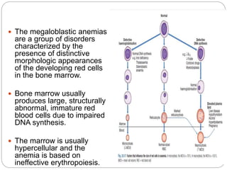
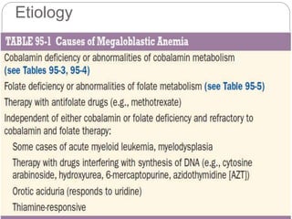
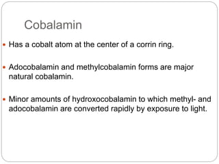
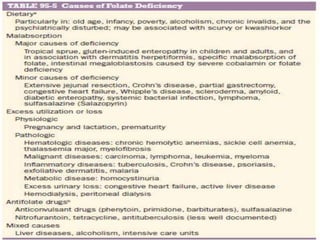
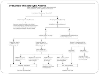
Dr. Bikal Lamichhane
Internal Medicine Resident
Approach to Macrocytic Anemia
 The megaloblastic anemias
are a group of disorders
characterized by the
presence of distinctive
morphologic appearances
of the developing red cells
in the bone marrow.
 Bone marrow usually
produces large, structurally
abnormal, immature red
blood cells due to impaired
DNA synthesis.
 The marrow is usually
hypercellular and the
anemia is based on
ineffective erythropoiesis.
Etiology
Cobalamin
 Has a cobalt atom at the center of a corrin ring.
 Adocobalamin and methylcobalamin forms are major
natural cobalamin.
 Minor amounts of hydroxocobalamin to which methyl- and
adocobalamin are converted rapidly by exposure to light.
Absorption of Cobalamin
Two mechanism,
1) Passive, occurring equally through buccal, duodenal, and
ileal mucosa; it is rapid but extremely inefficient, with <1%
of an oral dose being absorbed by this process.
2) Active; it occurs through the ileum and is efficient for
small (a few micrograms) oral doses of cobalamin, and it is
mediated by gastric intrinsic factor (IF).
Folate
 Folic (pteroylglutamic) acid is a yellow, crystalline, water-
soluble substance.
 The highest concentrations are found in liver, yeast,
spinach, other greens, and nuts (>100 μg/100 g).
 Folate is easily destroyed by heating, particularly in large
volumes of water.
 Total body folate in the adult is ~10 mg, with the liver
containing the largest store.
 Daily adult requirements are ~100 μg, and so stores are
sufficient for only 3–4 months in normal adults.
 Folates are absorbed rapidly from the upper small
intestine.
Clinical Features
 Many symptomless patients are detected through the
finding of a raised mean corpuscular volume (MCV) on a
routine blood count.
 Anorexia is usually marked, and there may be weight loss,
diarrhea, or constipation.
 Glossitis, angular cheilosis, a mild fever in more severely
anemic patients, jaundice (unconjugated), and reversible
melanin skin hyperpigmentation also may occur.
Neurologic Manifestations
 Vitamin B12 is needed for the myelination of the central
nervous system.
 Its deficiency may cause a bilateral peripheral neuropathy
or degeneration (demyelination) of the cervical and
thoracic posterior and lateral (pyramidal) tracts of the
spinal cord and, less frequently, of the cranial nerves and
of the white matter of the brain.
 Optic atrophy and cerebral symptoms including dementia,
depression, psychotic symptoms, and cognitive impairment
may be prominent.
 MRI may show the “spongy” degeneration of the cord.
 Loss of proprioception and vibration sensation with
positive Romberg and Lhermitte signs.
 Gait may be ataxic with spasticity (hyperreflexia).
 Autonomic nervous dysfunction can result in postural
hypotension, impotence, and incontinence
 Cobalamin deficiency in infancy:poor brain
development,impaired intellectual development,feeding
difficulties, lethargy, coma,convulsions and myoclonus.

 Cardiovascular Disease:
severe homocystinuria (blood levels ≥100 μmol/L)
due to deficiency of one of three enzymes,
methionine synthase, MTHFR, or cystathionine
synthase ischemic heart disease, cerebrovascular
disease, or pulmonary embolus or deep vein
thrombosis in young.
HEMATOLOGIC FINDINGS
 PERIPHERAL BLOOD :
 Oval macrocytes, with anisocytosis and
poikilocytosis
 The MCV is usually >100 fL.
 Neutrophils are hypersegmented (more than five
nuclear lobes).
 Leukopenia due to a reduction in granulocytes and
lymphocytes.
 Platelet count may be moderately reduced.
 BONE MARROW :
 Hypercellular with an accumulation of primitive cells
.Cells are larger than normoblasts, and an increased
number of cells with eccentric lobulated nuclei or
nuclear fragments.
 Giant and abnormally shaped metamyelocytes and
enlarged hyperpolyploid megakaryocytes are
characteristic.
CHROMOSOMES
 Proliferating cells in the body show : random breaks,
reduced contraction, spreading of the centromere,
and exaggeration of secondary chromosomal
constrictions and overprominent satellites.
INEFFECTIVE HEMATOPOIESIS :
 unconjugated bilirubin in plasma ,raised urine
urobilinogen, reduced haptoglobins and positive
urine hemosiderin,raised serum lactate
dehydrogenase,weakly positive direct antiglobulin
test due to complement.
DIAGNOSIS OF COBALAMIN AND
FOLATE DEFICIENCIES
COBALAMIN DEFICIENCY :
 Serum Cobalamin normal levels :160–200 ng/L
to 1000 ng/L.
 In patients with megaloblastic anemia due to
cobalamin deficiency, the level is usually <100
ng/L.
 Borderline :100 to 200 ng/L.
 Serum cobalamin level is sufficiently robust, cost-
effective, and most convenient to rule out
cobalamin deficiency .
 But where clinical indications of pernicious
anemia( PA) are strong, a normal serum vitamin
B12 does not rule out the diagnosis.
 Folate deficiency, transcobalamin 1 deficiency, oral
contraceptives, and multiple myeloma have all been
associated with low serum B12 levels that do not
indicate B12 deficiency.
 High serum B12 levels : serum transcobalamin 1
levels raised presence of liver, renal, or
myeloproliferative diseases or to cancer of the
breast, colon, or liver.
Serum Methylmalonate(MMA) and Homocysteine:
 elevated.
 Serum homocysteine: raised
but may be raised in chronic renal disease,
alcoholism, smoking, pyridoxine deficiency,
hypothyroidism,therapy with steroids, cyclosporine,
and other drugs
Tests for the Cause of Cobalamin Deficiency :
 Only strict vegetarians, or people living on a totally
inadequate diet will be B12 deficient because of
inadequate intake.
 Tests to diagnose PA : serum gastrin,( raised) serum
pepsinogen I (low) in PA (90–92%) and gastric
endoscopy.
 Tests for IF and parietal cell antibodies
 Tests for individual intestinal diseases.
FOLATE DEFICIENCY :
 Serum Folate: Normal range 2 μg/L to 15 μg/L.
 The serum folate level is low in all folate-deficient
patients
 Serum folate rises in severe cobalamin deficiency
because of the block in conversion of MTHF to THF
inside cells,also in intestinal stagnant loop syndrome
 Red Cell Folate :Test of body folate stores.
Normal (160–640 μg/L) of packed red cells.
 Serum homocysteine elevated.
Tests for the Cause of Folate Deficiency :
 Diet history
 Tests for transglutaminase antibodies for celiac disease
/duodenal biopsy as needed.
HEMATOLOGICAL FINDINGS
PERIPHERAL BLOOD
 Oval macrocytes, usually with considerable anisocytosis
and poikilocytosis, are the main feature.
 The MCV is usually >100 Fl unless a cause of microcytosis
(e.g., iron deficiency or thalassemia trait) is present.
 Some of the neutrophils are hypersegmented (more than
five nuclear lobes).
 There may be leukopenia due to a reduction in
granulocytes and lymphocytes, but this is usually >1.5 ×
109/L.
 platelet count may be moderately reduced, rarely to <40 ×
109/L.
 The severity of all these changes parallels the degree of
anemia.
OTHER CAUSES OF MACROCYTIC
ANEMIA
 Vitamin B6 deficiency: assessed by a therapeutic
trial of pyridoxine
 Acute viral infections, drug reactions, or chemical
toxicity( transient).
 AML :20% blasts in the marrow
 Genetic diseases:genomic testing
References
 Harrison’s principles of Internal Medicine , 20th
edition.
 Uptodate 2020.

Approach to macrocytic anemia

  • 1.
    Dr. Bikal Lamichhane InternalMedicine Resident Approach to Macrocytic Anemia
  • 2.
     The megaloblasticanemias are a group of disorders characterized by the presence of distinctive morphologic appearances of the developing red cells in the bone marrow.  Bone marrow usually produces large, structurally abnormal, immature red blood cells due to impaired DNA synthesis.  The marrow is usually hypercellular and the anemia is based on ineffective erythropoiesis.
  • 3.
  • 4.
    Cobalamin  Has acobalt atom at the center of a corrin ring.  Adocobalamin and methylcobalamin forms are major natural cobalamin.  Minor amounts of hydroxocobalamin to which methyl- and adocobalamin are converted rapidly by exposure to light.
  • 5.
    Absorption of Cobalamin Twomechanism, 1) Passive, occurring equally through buccal, duodenal, and ileal mucosa; it is rapid but extremely inefficient, with <1% of an oral dose being absorbed by this process. 2) Active; it occurs through the ileum and is efficient for small (a few micrograms) oral doses of cobalamin, and it is mediated by gastric intrinsic factor (IF).
  • 9.
    Folate  Folic (pteroylglutamic)acid is a yellow, crystalline, water- soluble substance.  The highest concentrations are found in liver, yeast, spinach, other greens, and nuts (>100 μg/100 g).  Folate is easily destroyed by heating, particularly in large volumes of water.  Total body folate in the adult is ~10 mg, with the liver containing the largest store.
  • 10.
     Daily adultrequirements are ~100 μg, and so stores are sufficient for only 3–4 months in normal adults.  Folates are absorbed rapidly from the upper small intestine.
  • 12.
    Clinical Features  Manysymptomless patients are detected through the finding of a raised mean corpuscular volume (MCV) on a routine blood count.  Anorexia is usually marked, and there may be weight loss, diarrhea, or constipation.  Glossitis, angular cheilosis, a mild fever in more severely anemic patients, jaundice (unconjugated), and reversible melanin skin hyperpigmentation also may occur.
  • 13.
    Neurologic Manifestations  VitaminB12 is needed for the myelination of the central nervous system.  Its deficiency may cause a bilateral peripheral neuropathy or degeneration (demyelination) of the cervical and thoracic posterior and lateral (pyramidal) tracts of the spinal cord and, less frequently, of the cranial nerves and of the white matter of the brain.  Optic atrophy and cerebral symptoms including dementia, depression, psychotic symptoms, and cognitive impairment may be prominent.
  • 14.
     MRI mayshow the “spongy” degeneration of the cord.  Loss of proprioception and vibration sensation with positive Romberg and Lhermitte signs.  Gait may be ataxic with spasticity (hyperreflexia).  Autonomic nervous dysfunction can result in postural hypotension, impotence, and incontinence  Cobalamin deficiency in infancy:poor brain development,impaired intellectual development,feeding difficulties, lethargy, coma,convulsions and myoclonus. 
  • 15.
     Cardiovascular Disease: severehomocystinuria (blood levels ≥100 μmol/L) due to deficiency of one of three enzymes, methionine synthase, MTHFR, or cystathionine synthase ischemic heart disease, cerebrovascular disease, or pulmonary embolus or deep vein thrombosis in young.
  • 16.
    HEMATOLOGIC FINDINGS  PERIPHERALBLOOD :  Oval macrocytes, with anisocytosis and poikilocytosis  The MCV is usually >100 fL.  Neutrophils are hypersegmented (more than five nuclear lobes).  Leukopenia due to a reduction in granulocytes and lymphocytes.  Platelet count may be moderately reduced.
  • 17.
     BONE MARROW:  Hypercellular with an accumulation of primitive cells .Cells are larger than normoblasts, and an increased number of cells with eccentric lobulated nuclei or nuclear fragments.  Giant and abnormally shaped metamyelocytes and enlarged hyperpolyploid megakaryocytes are characteristic.
  • 18.
    CHROMOSOMES  Proliferating cellsin the body show : random breaks, reduced contraction, spreading of the centromere, and exaggeration of secondary chromosomal constrictions and overprominent satellites. INEFFECTIVE HEMATOPOIESIS :  unconjugated bilirubin in plasma ,raised urine urobilinogen, reduced haptoglobins and positive urine hemosiderin,raised serum lactate dehydrogenase,weakly positive direct antiglobulin test due to complement.
  • 19.
    DIAGNOSIS OF COBALAMINAND FOLATE DEFICIENCIES COBALAMIN DEFICIENCY :  Serum Cobalamin normal levels :160–200 ng/L to 1000 ng/L.  In patients with megaloblastic anemia due to cobalamin deficiency, the level is usually <100 ng/L.  Borderline :100 to 200 ng/L.  Serum cobalamin level is sufficiently robust, cost- effective, and most convenient to rule out cobalamin deficiency .  But where clinical indications of pernicious anemia( PA) are strong, a normal serum vitamin B12 does not rule out the diagnosis.
  • 20.
     Folate deficiency,transcobalamin 1 deficiency, oral contraceptives, and multiple myeloma have all been associated with low serum B12 levels that do not indicate B12 deficiency.  High serum B12 levels : serum transcobalamin 1 levels raised presence of liver, renal, or myeloproliferative diseases or to cancer of the breast, colon, or liver. Serum Methylmalonate(MMA) and Homocysteine:  elevated.  Serum homocysteine: raised but may be raised in chronic renal disease, alcoholism, smoking, pyridoxine deficiency, hypothyroidism,therapy with steroids, cyclosporine, and other drugs
  • 21.
    Tests for theCause of Cobalamin Deficiency :  Only strict vegetarians, or people living on a totally inadequate diet will be B12 deficient because of inadequate intake.  Tests to diagnose PA : serum gastrin,( raised) serum pepsinogen I (low) in PA (90–92%) and gastric endoscopy.  Tests for IF and parietal cell antibodies  Tests for individual intestinal diseases.
  • 22.
    FOLATE DEFICIENCY : Serum Folate: Normal range 2 μg/L to 15 μg/L.  The serum folate level is low in all folate-deficient patients  Serum folate rises in severe cobalamin deficiency because of the block in conversion of MTHF to THF inside cells,also in intestinal stagnant loop syndrome  Red Cell Folate :Test of body folate stores. Normal (160–640 μg/L) of packed red cells.  Serum homocysteine elevated. Tests for the Cause of Folate Deficiency :  Diet history  Tests for transglutaminase antibodies for celiac disease /duodenal biopsy as needed.
  • 23.
    HEMATOLOGICAL FINDINGS PERIPHERAL BLOOD Oval macrocytes, usually with considerable anisocytosis and poikilocytosis, are the main feature.  The MCV is usually >100 Fl unless a cause of microcytosis (e.g., iron deficiency or thalassemia trait) is present.  Some of the neutrophils are hypersegmented (more than five nuclear lobes).
  • 24.
     There maybe leukopenia due to a reduction in granulocytes and lymphocytes, but this is usually >1.5 × 10⁹/L.  platelet count may be moderately reduced, rarely to <40 × 109/L.  The severity of all these changes parallels the degree of anemia.
  • 25.
    OTHER CAUSES OFMACROCYTIC ANEMIA  Vitamin B6 deficiency: assessed by a therapeutic trial of pyridoxine  Acute viral infections, drug reactions, or chemical toxicity( transient).  AML :20% blasts in the marrow  Genetic diseases:genomic testing
  • 27.
    References  Harrison’s principlesof Internal Medicine , 20th edition.  Uptodate 2020.