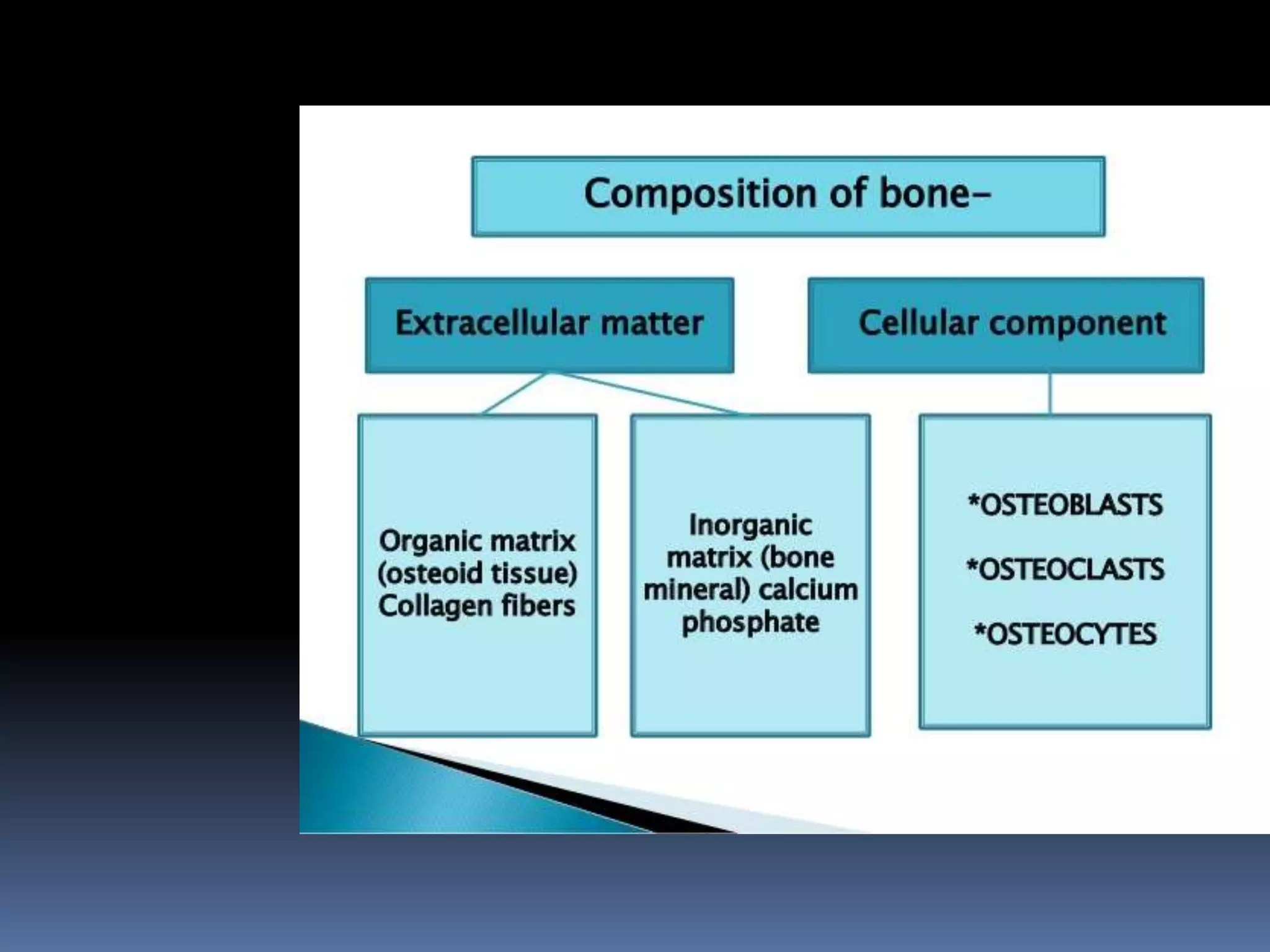
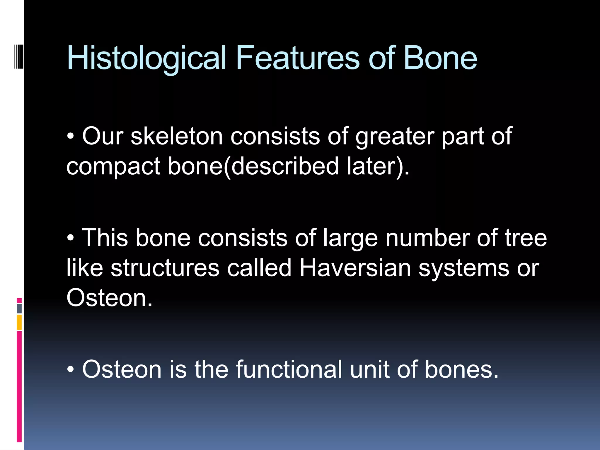
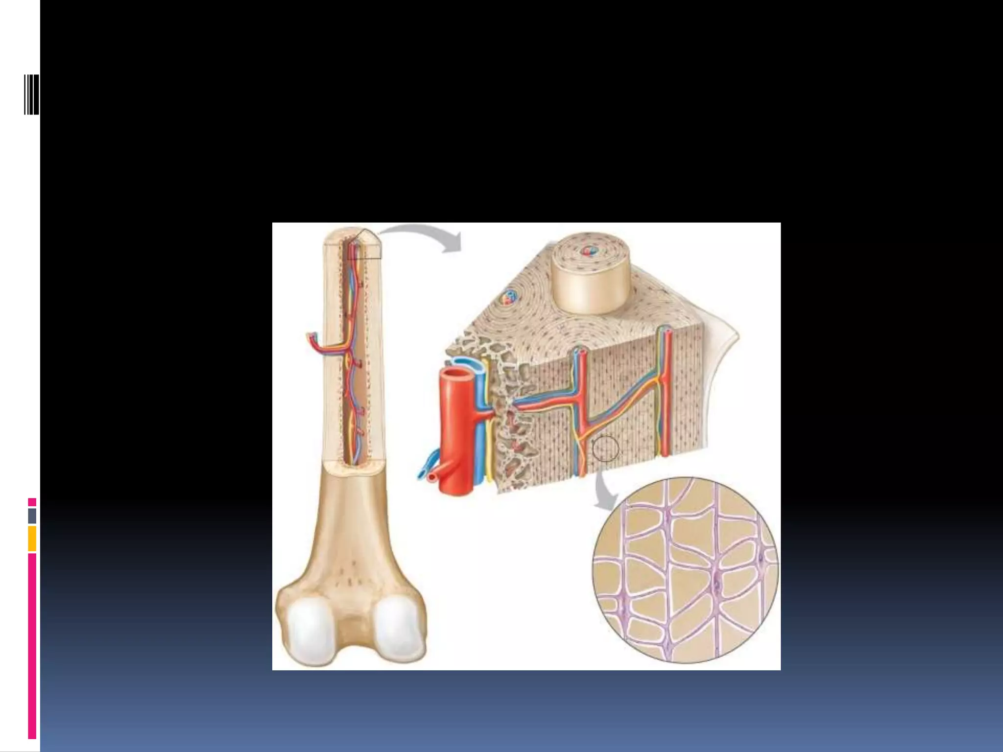
What are bones??
 Bones are rigid bars, that are made up of
connective tissues.
 These are living tissues that are capable of
growth and perform vast number of functions.
 These are the reservoirs of about 99% of
calcium of our body.
 Bones, despite of their hardness, are very much
a living tissue.
 They have greater regenerative power than
any other tissue of the body(except blood).
 It can be subjected to disease, and can be
fractured.
 It can mould itself according to changes is
stress and strain.
 The adult skeleton consists of 206 bones.
 When we are born, we have greater number
of bones, about 275.
 But as time passes, many bones fuse
together, and decrease in count.
 Also at birth, there is greater amount of
cartilage, which is afterwards replaced by
bones.
 Over a period of about 7 years, each bone in
our body is slowly replaced by a new bone.
 This happens by a process called :
Cartilaginous ossification (which will be
described later)
Histological Features of Bone
• Our skeleton consists of greater part of
compact bone(described later).
• This bone consists of large number of tree
like structures called Haversian systems or
Osteon.
• Osteon is the functional unit of bones.
 Lamellae :
 these are plate like structures around the
haversian canal. Edged by Lacunae (small
spaces), where osteocytes reside.
Osteoblasts are bone cells contained in
lamellae of bones( described later)
 Haversian canal :
 Here Nerve and blood supply is present.
Here O2 is supplied, and CO2 is taken
away
 Osteoblasts are bone forming cells. They
secrete bone matrix.
• They are responsible for mineralization the
bone tissue.
• When osteoblasts are trapped in the matrix
which they have secreted, then they become
osteocytes
 Osteoclasts :
• Osteoclasts are bone reabsorbing cells.
• Responsible for demineralization of bone.
• Important role in re- modeling of bone.
• Also recycle the matrix.
 Extracellular matrix
 It contains collagen fibers(organic substance)
and inorganic minerals embedded in it.
 Q) What are collagen fibers?
 Collagen fibers are the most abundant
protein in human body.
 It is a kind of fibrous connective tissue.
 They function as a glue in the body, i.e. it
holds together all the body structures.
 About 60 % of the bones are composed of
inorganic minerals.
 These include:
 1. Calcium
 2. Phosphorus
 3. Magnesium
 4. Potassium
 5. Sodium
 6. Citrate
 Major minerals are calcium and phosphorus.
 Calcium is in the form of calcium salt, mainly
calcium phosphate (CaP04) and partly calcium
carbonate (CaC03).
Significance of Inorganic Minerals
 Inorganic mineral crystals form rod
shaped crystals.
 These mineral crystals are responsible for
hardness, rigidity and great compressive
strength of bone.
 In Strength, bone is comparable to iron
and steel.
 About 25 % of bone is composed of
organic substances, which includes:
1. Collagen fibers -Mainly type I collagen
2. Ground substances Organic substances
 Glycoproteins
 Mucopolysaccharides
Significance of organic substances
 Provide strength and stability to our body.
 Make the bones tough.
 Provide flexibility, which can afford
resistance to tensile forces.
Parts of a Long Bone
20
• Epiphysis
• Distal
• Proximal
• Diaphysis
• Metaphysis
• Compact bone
• Spongy bone
• Articular cartilage
• Periosteum
• Endosteum
• Medullary cavity
• Trabeculae
• Bone marrow
• Red marrow and yellow marrow
Femur
Periosteum
Yellow marrow
Medullary cavity
Space containing
red marrow
Spongy bone
Compact bone
Articular cartilage
Epiphyseal plates
Proximal
epiphysis
Distal
epiphysis
Diaphysis
Endosteum
Copyright © The McGraw-Hill Companies, Inc. Permission required for reproduction or display.
Bone Classification
21
• Bone Classification:
• Long Bones
• Short Bones
• Sesamoid Bones
• Flat Bones
• Irregular Bones
• Wormian Bones
(sutural)
(a) (e)
(b)
(c)
(d)
Copyright © The McGraw-Hill Companies, Inc. Permission required for reproduction or display.
Classification of Bones
Slide 5.4aCopyright © 2003 Pearson Education, Inc. publishing as Benjamin Cummings
 Long bones
Typically longer than wide
Have a shaft with heads at both ends
Contain mostly compact bone
• Examples: Femur, humerus
Classification of Bones
Slide 5.4bCopyright © 2003 Pearson Education, Inc. publishing as Benjamin Cummings
 Short bones
Generally cube-shape
Contain mostly spongy bone
Examples: Carpals, tarsals
Classification of Bones on the
Basis of Shape
Slide 5.4cCopyright © 2003 Pearson Education, Inc. publishing as Benjamin Cummings
Figure 5.1
Classification of Bones
Slide 5.5aCopyright © 2003 Pearson Education, Inc. publishing as Benjamin Cummings
 Flat bones
Thin and flattened
Usually curved
Thin layers of compact bone around a layer
of spongy bone
Examples: Skull, ribs, sternum
Classification of Bones
Slide 5.5bCopyright © 2003 Pearson Education, Inc. publishing as Benjamin Cummings
 Irregular bones
Irregular shape
Do not fit into other bone classification
categories
Example: Vertebrae and hip
Classification of Bones on the
Basis of Shape
Slide 5.5cCopyright © 2003 Pearson Education, Inc. publishing as Benjamin Cummings
Figure 5.1
Bone Development
and Growth
28
• Parts of the skeletal system begin to develop during the first few
weeks of prenatal development
• Bones replace existing connective tissue in one of two ways:
• As intramembranous bones
• As endchondral bones
Intramembranous Bones
29
• Intramembranous Bones
• These bones originate within sheetlike layers of connective tissues
• They are the broad, flat bones
• Skull bones (except mandible)
• Are known as intramembranous bones
Endochondral Bones
30
• Endochondral Bones
• Bones begin as hyaline cartilage
• Form models for future bones
• These are most bones of the skeleton
• Are known as endochondral bones
Endochondral Ossification
31
• Hyaline cartilage model
• Primary ossification center
• Secondary ossification centers
• Epiphyseal plate
• Osteoblasts vs. osteoclasts
(b) (c) (d) (e) (f)(a)
Cartilaginous
model
Calcified
cartilage
Articular
cartilage
Developing
periosteum
Compact bone
developing
Primary
ossification
center
Medullary
cavity
Medullary
cavity
Medullary
cavity
Secondary
ossification
center
Secondary
ossification
center
Blood
vessel
Epiphyseal
plate
Remnant of
epiphyseal
plate
Remnants of
epiphyseal
plates
Epiphyseal
plates
Compact
bone
Spongy
bone
Articular
cartilage
Spongy
bone
Copyright © The McGraw-Hill Companies, Inc. Permission required for reproduction or display.
Growth at the Epiphyseal Plate
32
• First layer of cells
• Closest to the end of
epiphysis
• Resting cells
• Anchors epiphyseal plate
to epiphysis
• Zone of resting cartilage
• Second layer of cells
• Many rows of young
cells
• Undergoing mitosis
• zone of proliferating
cartilage
1
2
3
4
(a) (b)
Bone tissue
of epiphysis
Zone of
resting
cartilage
Zone of
proliferating
cartilage
Zone of
hypertrophic
cartilage
Zone of
calcified
cartilage
Ossified
bone of
diaphysis
Copyright © The McGraw-Hill Companies, Inc. Permission required for reproduction or display.
b: © The McGraw-Hill Companies, Inc./Al Telser, photographer
Growth at the Epiphyseal Plate
33
• Third layer of cells
• Older cells
• Left behind when new
cells appear
• Cells enlarging and
becoming calcified
• zone of hypertrophic
cartilage
• Fourth layer of cells
• Thin
• Dead cells
• Calcified extracellular
matrix
• zone of calcified
cartilage
1
2
3
4
(a) (b)
Bone tissue
of epiphysis
Zone of
resting
cartilage
Zone of
proliferating
cartilage
Zone of
hypertrophic
cartilage
Zone of
calcified
cartilage
Ossified
bone of
diaphysis
Copyright © The McGraw-Hill Companies, Inc. Permission required for reproduction or display.
b: © The McGraw-Hill Companies, Inc./Al Telser, photographer
Homeostasis of Bone Tissue
34
• Bone Resorption – action of osteoclasts and parathyroid hormone aka
parathormone aka PTH
• Bone Deposition – action of osteoblasts and calcitonin
• Occurs by direction of the thyroid and parathyroid glands
Copyright © The McGraw-Hill Companies, Inc. Permission required for reproduction or display.
© Biophoto Associates/Photo Researchers, Inc.
Developing
medullary
cavity
Osteoclast
Factors Affecting Bone
Development, Growth and Repair
35
• Deficiency of Vitamin A – retards bone development
• Deficiency of Vitamin C – results in fragile bones
• Deficiency of Vitamin D – rickets, osteomalacia
• Insufficient Growth Hormone – dwarfism
• Excessive Growth Hormone – gigantism, acromegaly
• Insufficient Thyroid Hormone – delays bone growth
• Sex Hormones – promote bone formation; stimulate ossification of epiphyseal
plates
• Physical Stress – stimulates bone growth
Bone Function
36
• Bones shape, support, and protect body structures
Support, Protection,
and Movement
37
• Support, Movement & Protection
• Gives shape to head, etc.
• Supports body’s weight
• Protects lungs, etc.
• Bones and muscles interact
• When limbs or body parts move
Blood Cell Formation
38
• Blood Cell Formation
• Also known as hematopoiesis
• Occurs in the red bone marrow

Introduction of bones, types, structure

  • 2.
    What are bones?? Bones are rigid bars, that are made up of connective tissues.  These are living tissues that are capable of growth and perform vast number of functions.  These are the reservoirs of about 99% of calcium of our body.
  • 3.
     Bones, despiteof their hardness, are very much a living tissue.  They have greater regenerative power than any other tissue of the body(except blood).  It can be subjected to disease, and can be fractured.  It can mould itself according to changes is stress and strain.
  • 4.
     The adultskeleton consists of 206 bones.  When we are born, we have greater number of bones, about 275.  But as time passes, many bones fuse together, and decrease in count.
  • 5.
     Also atbirth, there is greater amount of cartilage, which is afterwards replaced by bones.  Over a period of about 7 years, each bone in our body is slowly replaced by a new bone.  This happens by a process called : Cartilaginous ossification (which will be described later)
  • 7.
    Histological Features ofBone • Our skeleton consists of greater part of compact bone(described later). • This bone consists of large number of tree like structures called Haversian systems or Osteon. • Osteon is the functional unit of bones.
  • 9.
     Lamellae : these are plate like structures around the haversian canal. Edged by Lacunae (small spaces), where osteocytes reside. Osteoblasts are bone cells contained in lamellae of bones( described later)  Haversian canal :  Here Nerve and blood supply is present. Here O2 is supplied, and CO2 is taken away
  • 12.
     Osteoblasts arebone forming cells. They secrete bone matrix. • They are responsible for mineralization the bone tissue. • When osteoblasts are trapped in the matrix which they have secreted, then they become osteocytes
  • 13.
     Osteoclasts : •Osteoclasts are bone reabsorbing cells. • Responsible for demineralization of bone. • Important role in re- modeling of bone. • Also recycle the matrix.
  • 14.
     Extracellular matrix It contains collagen fibers(organic substance) and inorganic minerals embedded in it.  Q) What are collagen fibers?  Collagen fibers are the most abundant protein in human body.  It is a kind of fibrous connective tissue.  They function as a glue in the body, i.e. it holds together all the body structures.
  • 15.
     About 60% of the bones are composed of inorganic minerals.  These include:  1. Calcium  2. Phosphorus  3. Magnesium  4. Potassium  5. Sodium  6. Citrate  Major minerals are calcium and phosphorus.  Calcium is in the form of calcium salt, mainly calcium phosphate (CaP04) and partly calcium carbonate (CaC03).
  • 16.
    Significance of InorganicMinerals  Inorganic mineral crystals form rod shaped crystals.  These mineral crystals are responsible for hardness, rigidity and great compressive strength of bone.  In Strength, bone is comparable to iron and steel.
  • 17.
     About 25% of bone is composed of organic substances, which includes: 1. Collagen fibers -Mainly type I collagen 2. Ground substances Organic substances  Glycoproteins  Mucopolysaccharides
  • 18.
    Significance of organicsubstances  Provide strength and stability to our body.  Make the bones tough.  Provide flexibility, which can afford resistance to tensile forces.
  • 20.
    Parts of aLong Bone 20 • Epiphysis • Distal • Proximal • Diaphysis • Metaphysis • Compact bone • Spongy bone • Articular cartilage • Periosteum • Endosteum • Medullary cavity • Trabeculae • Bone marrow • Red marrow and yellow marrow Femur Periosteum Yellow marrow Medullary cavity Space containing red marrow Spongy bone Compact bone Articular cartilage Epiphyseal plates Proximal epiphysis Distal epiphysis Diaphysis Endosteum Copyright © The McGraw-Hill Companies, Inc. Permission required for reproduction or display.
  • 21.
    Bone Classification 21 • BoneClassification: • Long Bones • Short Bones • Sesamoid Bones • Flat Bones • Irregular Bones • Wormian Bones (sutural) (a) (e) (b) (c) (d) Copyright © The McGraw-Hill Companies, Inc. Permission required for reproduction or display.
  • 22.
    Classification of Bones Slide5.4aCopyright © 2003 Pearson Education, Inc. publishing as Benjamin Cummings  Long bones Typically longer than wide Have a shaft with heads at both ends Contain mostly compact bone • Examples: Femur, humerus
  • 23.
    Classification of Bones Slide5.4bCopyright © 2003 Pearson Education, Inc. publishing as Benjamin Cummings  Short bones Generally cube-shape Contain mostly spongy bone Examples: Carpals, tarsals
  • 24.
    Classification of Boneson the Basis of Shape Slide 5.4cCopyright © 2003 Pearson Education, Inc. publishing as Benjamin Cummings Figure 5.1
  • 25.
    Classification of Bones Slide5.5aCopyright © 2003 Pearson Education, Inc. publishing as Benjamin Cummings  Flat bones Thin and flattened Usually curved Thin layers of compact bone around a layer of spongy bone Examples: Skull, ribs, sternum
  • 26.
    Classification of Bones Slide5.5bCopyright © 2003 Pearson Education, Inc. publishing as Benjamin Cummings  Irregular bones Irregular shape Do not fit into other bone classification categories Example: Vertebrae and hip
  • 27.
    Classification of Boneson the Basis of Shape Slide 5.5cCopyright © 2003 Pearson Education, Inc. publishing as Benjamin Cummings Figure 5.1
  • 28.
    Bone Development and Growth 28 •Parts of the skeletal system begin to develop during the first few weeks of prenatal development • Bones replace existing connective tissue in one of two ways: • As intramembranous bones • As endchondral bones
  • 29.
    Intramembranous Bones 29 • IntramembranousBones • These bones originate within sheetlike layers of connective tissues • They are the broad, flat bones • Skull bones (except mandible) • Are known as intramembranous bones
  • 30.
    Endochondral Bones 30 • EndochondralBones • Bones begin as hyaline cartilage • Form models for future bones • These are most bones of the skeleton • Are known as endochondral bones
  • 31.
    Endochondral Ossification 31 • Hyalinecartilage model • Primary ossification center • Secondary ossification centers • Epiphyseal plate • Osteoblasts vs. osteoclasts (b) (c) (d) (e) (f)(a) Cartilaginous model Calcified cartilage Articular cartilage Developing periosteum Compact bone developing Primary ossification center Medullary cavity Medullary cavity Medullary cavity Secondary ossification center Secondary ossification center Blood vessel Epiphyseal plate Remnant of epiphyseal plate Remnants of epiphyseal plates Epiphyseal plates Compact bone Spongy bone Articular cartilage Spongy bone Copyright © The McGraw-Hill Companies, Inc. Permission required for reproduction or display.
  • 32.
    Growth at theEpiphyseal Plate 32 • First layer of cells • Closest to the end of epiphysis • Resting cells • Anchors epiphyseal plate to epiphysis • Zone of resting cartilage • Second layer of cells • Many rows of young cells • Undergoing mitosis • zone of proliferating cartilage 1 2 3 4 (a) (b) Bone tissue of epiphysis Zone of resting cartilage Zone of proliferating cartilage Zone of hypertrophic cartilage Zone of calcified cartilage Ossified bone of diaphysis Copyright © The McGraw-Hill Companies, Inc. Permission required for reproduction or display. b: © The McGraw-Hill Companies, Inc./Al Telser, photographer
  • 33.
    Growth at theEpiphyseal Plate 33 • Third layer of cells • Older cells • Left behind when new cells appear • Cells enlarging and becoming calcified • zone of hypertrophic cartilage • Fourth layer of cells • Thin • Dead cells • Calcified extracellular matrix • zone of calcified cartilage 1 2 3 4 (a) (b) Bone tissue of epiphysis Zone of resting cartilage Zone of proliferating cartilage Zone of hypertrophic cartilage Zone of calcified cartilage Ossified bone of diaphysis Copyright © The McGraw-Hill Companies, Inc. Permission required for reproduction or display. b: © The McGraw-Hill Companies, Inc./Al Telser, photographer
  • 34.
    Homeostasis of BoneTissue 34 • Bone Resorption – action of osteoclasts and parathyroid hormone aka parathormone aka PTH • Bone Deposition – action of osteoblasts and calcitonin • Occurs by direction of the thyroid and parathyroid glands Copyright © The McGraw-Hill Companies, Inc. Permission required for reproduction or display. © Biophoto Associates/Photo Researchers, Inc. Developing medullary cavity Osteoclast
  • 35.
    Factors Affecting Bone Development,Growth and Repair 35 • Deficiency of Vitamin A – retards bone development • Deficiency of Vitamin C – results in fragile bones • Deficiency of Vitamin D – rickets, osteomalacia • Insufficient Growth Hormone – dwarfism • Excessive Growth Hormone – gigantism, acromegaly • Insufficient Thyroid Hormone – delays bone growth • Sex Hormones – promote bone formation; stimulate ossification of epiphyseal plates • Physical Stress – stimulates bone growth
  • 36.
    Bone Function 36 • Bonesshape, support, and protect body structures
  • 37.
    Support, Protection, and Movement 37 •Support, Movement & Protection • Gives shape to head, etc. • Supports body’s weight • Protects lungs, etc. • Bones and muscles interact • When limbs or body parts move
  • 38.
    Blood Cell Formation 38 •Blood Cell Formation • Also known as hematopoiesis • Occurs in the red bone marrow