The skeletal system is composed of bones and associated tissues that perform several essential functions:
1. Support - Bones provide structural support for the body and protection for internal organs.
2. Movement - Skeletal muscles use bones as levers to enable movement of the body.
3. Mineral storage - Bones store minerals like calcium and phosphorus.
There are over 200 bones in the human body that are classified as long, short, flat, or irregular. Bones are living tissues composed of cells like osteoblasts, osteocytes, and osteoclasts embedded in an organic bone matrix and inorganic minerals. Compact bone forms the dense outer layer while spongy bone composes the inner layer. Long bones have
CONTENTS
FORMATION OF BONE
CLASSIFICATION OF BONES
STRUCTURE OF BONE
BLOOD SUPPLY
COMPOSITION OF BONE
FRACTURE HEALING
CARTILAGE
TYPES OF CARTILAGE
BONE (syn – Os; Osteon)
Osseous tissue, a specialised form of dense connective
tissue consisting of bone cells (osteocytes)
Embedded in a matrix of calcified intercelluar
substance
Bone matrix contains collagen fibres and the minerals
calcium phosphate and calcium carbonate
The Indian Dental Academy is the Leader in continuing dental education , training dentists in all aspects of dentistry and
offering a wide range of dental certified courses in different formats.for more details please visit
www.indiandentalacademy.com
A detail account of Bones, their histological features, classification, composition, Formation, blood and nerve supply, functions, plus some interesting facts about bones.
BONE – AN INTRODUCTION
A bone is a rigid organ that constitutes part of the vertebrate skeleton.
There are around 270 to 300+ bones in Infants which gets reduced to 206 bones in adults.
Bones are dynamic structures that are undergoing constant change and remodelling in
response to the ever-changing environment.
Bones support and protect the various organs of the body, produce red and white blood cells,
store minerals, provide structure and support for the body, and enable mobility.
It has a honeycomb-like matrix internally, which helps to give the bone rigidity.
The largest bone in the body is the femur or thigh-bone, and the smallest is the stapes in
the middle ear.
Bone tissue is the major structural and supportive connective tissue of the body. Osseous tissue forms the rigid part of the bones that make up the skeletal system.
Osteology, derived from the from Greek ὀστέον (ostéon) 'bones', and λόγος (logos) 'study', is the scientific study of bones, practised by osteologists. A subdiscipline of anatomy, anthropology, and paleontology, osteology is the detailed study of the structure of bones, skeletal elements, teeth, microbone morphology, function, disease, pathology, the process of ossification (from cartilaginous molds), and the resistance and hardness of bones (biophysics).[1]
Osteologists frequently work in the public and private sector as consultants for museums, scientists for research laboratories, scientists for medical investigations and/or for companies producing osteological reproductions in an academic context.
Osteology and osteologists should not be confused with osteopathy and its practitioners, osteopaths.
1. Unit - 7- Skeleton anatomy by Thiru muruganthiru murugan
The Skeletal System
By Thiru murugan. M
Unit – 7: Anatomy - The Musculoskeletal system:
The Skeletal system
Anatomical positions
Bones: types, structure, growth and ossification
Axial and appendicular skeleton
Joints: classification, major joints and structure
Application and implications in nursing
The Muscular system:
Types and structure of muscles
Muscle groups: muscles of the head, neck, thorax, abdomen, pelvis, upper limb and lower limbs
Principal muscles: deltoid, biceps, triceps, respiratory, abdominal, pelvic floor muscles, gluteal muscles and vastus lateralis
Major muscles involved in nursing procedures
Skeletal system:
The human skeletal system consists of all of the bones, cartilage, tendons, and ligaments in the body
It Provide framework of the body.
Altogether, the skeleton makes up about 20% of a person's body weight. An adult's skeleton contains 206 bones.
It providing support and protection for the internal organs
The skeletal system also provides attachment points for muscles to allow movements at the joints.
Components of skeletal system:
Cartilage: This smooth and flexible substance covers the tips of your bones where they meet. It enables bones to move without friction (rubbing against each other).
Functions of Cartilage:
Model for bone growth in embryo & fetus
Provides a smooth cushion between adjacent bones
Provides firm flexible support (nose, ears, ribs & trachea)
Excellent shock absorber
Ligaments: Bands of strong connective tissue called ligaments hold bones together.
Functions of Ligaments:
Attach bones to bones
Provide stability
Tendons: Tendons are bands of tissue that connect the ends of a muscle to your bone.
Functions of Tendons:
Attach muscles to bones
Anchors muscle to bone for movement
Joints: A joint is where two or more bones in the body come together.
Anatomical position:
Anatomical position, or standard anatomical position, refers to the positioning of the body when it is standing upright and facing forward with each arm hanging on either side of the body, and the palms facing forward. The legs are parallel, with feet flat on the floor and facing forward.
Bones – types, structure, growth and ossification:
Bones:
Bone are specialized forms of strong connective tissue that forms the skeleton of the body.
It is composed of calcium phosphate and calcium carbonate.
It also serves as a storage area for calcium, playing a large role in calcium balance in the blood
The smallest bone in the human body is called the stirrup or stapes bone, located deep inside the ear & The longest bone in the human is called the femur.
Classification or types of bones:
Bones are divided into 5 types.
Long Bone
Short Bone
Flat Bone
Irregular Bone
Sesamoid Bone
1. Long Bone:
A long bone is one that is cylindrical in shape, being longer than it is wide.
Shape of a bone, not its size.
Long bones are found in: Arms (humerus, ulna, radius) & fingers (metacarpals, phalanges) and also Legs (femur, tibia, fibula),
Bones and its structure in detail with two different form of bone formationbhartisharma175
It consist of detail content about different types of bone cells, two different type of bone formation and structure of long bone. easy to understand for students. language is simple.
Ozempic: Preoperative Management of Patients on GLP-1 Receptor Agonists Saeid Safari
Preoperative Management of Patients on GLP-1 Receptor Agonists like Ozempic and Semiglutide
ASA GUIDELINE
NYSORA Guideline
2 Case Reports of Gastric Ultrasound
Ethanol (CH3CH2OH), or beverage alcohol, is a two-carbon alcohol
that is rapidly distributed in the body and brain. Ethanol alters many
neurochemical systems and has rewarding and addictive properties. It
is the oldest recreational drug and likely contributes to more morbidity,
mortality, and public health costs than all illicit drugs combined. The
5th edition of the Diagnostic and Statistical Manual of Mental Disorders
(DSM-5) integrates alcohol abuse and alcohol dependence into a single
disorder called alcohol use disorder (AUD), with mild, moderate,
and severe subclassifications (American Psychiatric Association, 2013).
In the DSM-5, all types of substance abuse and dependence have been
combined into a single substance use disorder (SUD) on a continuum
from mild to severe. A diagnosis of AUD requires that at least two of
the 11 DSM-5 behaviors be present within a 12-month period (mild
AUD: 2–3 criteria; moderate AUD: 4–5 criteria; severe AUD: 6–11 criteria).
The four main behavioral effects of AUD are impaired control over
drinking, negative social consequences, risky use, and altered physiological
effects (tolerance, withdrawal). This chapter presents an overview
of the prevalence and harmful consequences of AUD in the U.S.,
the systemic nature of the disease, neurocircuitry and stages of AUD,
comorbidities, fetal alcohol spectrum disorders, genetic risk factors, and
pharmacotherapies for AUD.
Title: Sense of Taste
Presenter: Dr. Faiza, Assistant Professor of Physiology
Qualifications:
MBBS (Best Graduate, AIMC Lahore)
FCPS Physiology
ICMT, CHPE, DHPE (STMU)
MPH (GC University, Faisalabad)
MBA (Virtual University of Pakistan)
Learning Objectives:
Describe the structure and function of taste buds.
Describe the relationship between the taste threshold and taste index of common substances.
Explain the chemical basis and signal transduction of taste perception for each type of primary taste sensation.
Recognize different abnormalities of taste perception and their causes.
Key Topics:
Significance of Taste Sensation:
Differentiation between pleasant and harmful food
Influence on behavior
Selection of food based on metabolic needs
Receptors of Taste:
Taste buds on the tongue
Influence of sense of smell, texture of food, and pain stimulation (e.g., by pepper)
Primary and Secondary Taste Sensations:
Primary taste sensations: Sweet, Sour, Salty, Bitter, Umami
Chemical basis and signal transduction mechanisms for each taste
Taste Threshold and Index:
Taste threshold values for Sweet (sucrose), Salty (NaCl), Sour (HCl), and Bitter (Quinine)
Taste index relationship: Inversely proportional to taste threshold
Taste Blindness:
Inability to taste certain substances, particularly thiourea compounds
Example: Phenylthiocarbamide
Structure and Function of Taste Buds:
Composition: Epithelial cells, Sustentacular/Supporting cells, Taste cells, Basal cells
Features: Taste pores, Taste hairs/microvilli, and Taste nerve fibers
Location of Taste Buds:
Found in papillae of the tongue (Fungiform, Circumvallate, Foliate)
Also present on the palate, tonsillar pillars, epiglottis, and proximal esophagus
Mechanism of Taste Stimulation:
Interaction of taste substances with receptors on microvilli
Signal transduction pathways for Umami, Sweet, Bitter, Sour, and Salty tastes
Taste Sensitivity and Adaptation:
Decrease in sensitivity with age
Rapid adaptation of taste sensation
Role of Saliva in Taste:
Dissolution of tastants to reach receptors
Washing away the stimulus
Taste Preferences and Aversions:
Mechanisms behind taste preference and aversion
Influence of receptors and neural pathways
Impact of Sensory Nerve Damage:
Degeneration of taste buds if the sensory nerve fiber is cut
Abnormalities of Taste Detection:
Conditions: Ageusia, Hypogeusia, Dysgeusia (parageusia)
Causes: Nerve damage, neurological disorders, infections, poor oral hygiene, adverse drug effects, deficiencies, aging, tobacco use, altered neurotransmitter levels
Neurotransmitters and Taste Threshold:
Effects of serotonin (5-HT) and norepinephrine (NE) on taste sensitivity
Supertasters:
25% of the population with heightened sensitivity to taste, especially bitterness
Increased number of fungiform papillae
Report Back from SGO 2024: What’s the Latest in Cervical Cancer?bkling
Are you curious about what’s new in cervical cancer research or unsure what the findings mean? Join Dr. Emily Ko, a gynecologic oncologist at Penn Medicine, to learn about the latest updates from the Society of Gynecologic Oncology (SGO) 2024 Annual Meeting on Women’s Cancer. Dr. Ko will discuss what the research presented at the conference means for you and answer your questions about the new developments.
New Directions in Targeted Therapeutic Approaches for Older Adults With Mantl...i3 Health
i3 Health is pleased to make the speaker slides from this activity available for use as a non-accredited self-study or teaching resource.
This slide deck presented by Dr. Kami Maddocks, Professor-Clinical in the Division of Hematology and
Associate Division Director for Ambulatory Operations
The Ohio State University Comprehensive Cancer Center, will provide insight into new directions in targeted therapeutic approaches for older adults with mantle cell lymphoma.
STATEMENT OF NEED
Mantle cell lymphoma (MCL) is a rare, aggressive B-cell non-Hodgkin lymphoma (NHL) accounting for 5% to 7% of all lymphomas. Its prognosis ranges from indolent disease that does not require treatment for years to very aggressive disease, which is associated with poor survival (Silkenstedt et al, 2021). Typically, MCL is diagnosed at advanced stage and in older patients who cannot tolerate intensive therapy (NCCN, 2022). Although recent advances have slightly increased remission rates, recurrence and relapse remain very common, leading to a median overall survival between 3 and 6 years (LLS, 2021). Though there are several effective options, progress is still needed towards establishing an accepted frontline approach for MCL (Castellino et al, 2022). Treatment selection and management of MCL are complicated by the heterogeneity of prognosis, advanced age and comorbidities of patients, and lack of an established standard approach for treatment, making it vital that clinicians be familiar with the latest research and advances in this area. In this activity chaired by Michael Wang, MD, Professor in the Department of Lymphoma & Myeloma at MD Anderson Cancer Center, expert faculty will discuss prognostic factors informing treatment, the promising results of recent trials in new therapeutic approaches, and the implications of treatment resistance in therapeutic selection for MCL.
Target Audience
Hematology/oncology fellows, attending faculty, and other health care professionals involved in the treatment of patients with mantle cell lymphoma (MCL).
Learning Objectives
1.) Identify clinical and biological prognostic factors that can guide treatment decision making for older adults with MCL
2.) Evaluate emerging data on targeted therapeutic approaches for treatment-naive and relapsed/refractory MCL and their applicability to older adults
3.) Assess mechanisms of resistance to targeted therapies for MCL and their implications for treatment selection
Tom Selleck Health: A Comprehensive Look at the Iconic Actor’s Wellness Journeygreendigital
Tom Selleck, an enduring figure in Hollywood. has captivated audiences for decades with his rugged charm, iconic moustache. and memorable roles in television and film. From his breakout role as Thomas Magnum in Magnum P.I. to his current portrayal of Frank Reagan in Blue Bloods. Selleck's career has spanned over 50 years. But beyond his professional achievements. fans have often been curious about Tom Selleck Health. especially as he has aged in the public eye.
Follow us on: Pinterest
Introduction
Many have been interested in Tom Selleck health. not only because of his enduring presence on screen but also because of the challenges. and lifestyle choices he has faced and made over the years. This article delves into the various aspects of Tom Selleck health. exploring his fitness regimen, diet, mental health. and the challenges he has encountered as he ages. We'll look at how he maintains his well-being. the health issues he has faced, and his approach to ageing .
Early Life and Career
Childhood and Athletic Beginnings
Tom Selleck was born on January 29, 1945, in Detroit, Michigan, and grew up in Sherman Oaks, California. From an early age, he was involved in sports, particularly basketball. which played a significant role in his physical development. His athletic pursuits continued into college. where he attended the University of Southern California (USC) on a basketball scholarship. This early involvement in sports laid a strong foundation for his physical health and disciplined lifestyle.
Transition to Acting
Selleck's transition from an athlete to an actor came with its physical demands. His first significant role in "Magnum P.I." required him to perform various stunts and maintain a fit appearance. This role, which he played from 1980 to 1988. necessitated a rigorous fitness routine to meet the show's demands. setting the stage for his long-term commitment to health and wellness.
Fitness Regimen
Workout Routine
Tom Selleck health and fitness regimen has evolved. adapting to his changing roles and age. During his "Magnum, P.I." days. Selleck's workouts were intense and focused on building and maintaining muscle mass. His routine included weightlifting, cardiovascular exercises. and specific training for the stunts he performed on the show.
Selleck adjusted his fitness routine as he aged to suit his body's needs. Today, his workouts focus on maintaining flexibility, strength, and cardiovascular health. He incorporates low-impact exercises such as swimming, walking, and light weightlifting. This balanced approach helps him stay fit without putting undue strain on his joints and muscles.
Importance of Flexibility and Mobility
In recent years, Selleck has emphasized the importance of flexibility and mobility in his fitness regimen. Understanding the natural decline in muscle mass and joint flexibility with age. he includes stretching and yoga in his routine. These practices help prevent injuries, improve posture, and maintain mobilit
Flu Vaccine Alert in Bangalore Karnatakaaddon Scans
As flu season approaches, health officials in Bangalore, Karnataka, are urging residents to get their flu vaccinations. The seasonal flu, while common, can lead to severe health complications, particularly for vulnerable populations such as young children, the elderly, and those with underlying health conditions.
Dr. Vidisha Kumari, a leading epidemiologist in Bangalore, emphasizes the importance of getting vaccinated. "The flu vaccine is our best defense against the influenza virus. It not only protects individuals but also helps prevent the spread of the virus in our communities," he says.
This year, the flu season is expected to coincide with a potential increase in other respiratory illnesses. The Karnataka Health Department has launched an awareness campaign highlighting the significance of flu vaccinations. They have set up multiple vaccination centers across Bangalore, making it convenient for residents to receive their shots.
To encourage widespread vaccination, the government is also collaborating with local schools, workplaces, and community centers to facilitate vaccination drives. Special attention is being given to ensuring that the vaccine is accessible to all, including marginalized communities who may have limited access to healthcare.
Residents are reminded that the flu vaccine is safe and effective. Common side effects are mild and may include soreness at the injection site, mild fever, or muscle aches. These side effects are generally short-lived and far less severe than the flu itself.
Healthcare providers are also stressing the importance of continuing COVID-19 precautions. Wearing masks, practicing good hand hygiene, and maintaining social distancing are still crucial, especially in crowded places.
Protect yourself and your loved ones by getting vaccinated. Together, we can help keep Bangalore healthy and safe this flu season. For more information on vaccination centers and schedules, residents can visit the Karnataka Health Department’s official website or follow their social media pages.
Stay informed, stay safe, and get your flu shot today!
TEST BANK for Operations Management, 14th Edition by William J. Stevenson, Ve...kevinkariuki227
TEST BANK for Operations Management, 14th Edition by William J. Stevenson, Verified Chapters 1 - 19, Complete Newest Version.pdf
TEST BANK for Operations Management, 14th Edition by William J. Stevenson, Verified Chapters 1 - 19, Complete Newest Version.pdf
micro teaching on communication m.sc nursing.pdfAnurag Sharma
Microteaching is a unique model of practice teaching. It is a viable instrument for the. desired change in the teaching behavior or the behavior potential which, in specified types of real. classroom situations, tends to facilitate the achievement of specified types of objectives.
Couples presenting to the infertility clinic- Do they really have infertility...Sujoy Dasgupta
Dr Sujoy Dasgupta presented the study on "Couples presenting to the infertility clinic- Do they really have infertility? – The unexplored stories of non-consummation" in the 13th Congress of the Asia Pacific Initiative on Reproduction (ASPIRE 2024) at Manila on 24 May, 2024.
The prostate is an exocrine gland of the male mammalian reproductive system
It is a walnut-sized gland that forms part of the male reproductive system and is located in front of the rectum and just below the urinary bladder
Function is to store and secrete a clear, slightly alkaline fluid that constitutes 10-30% of the volume of the seminal fluid that along with the spermatozoa, constitutes semen
A healthy human prostate measures (4cm-vertical, by 3cm-horizontal, 2cm ant-post ).
It surrounds the urethra just below the urinary bladder. It has anterior, median, posterior and two lateral lobes
It’s work is regulated by androgens which are responsible for male sex characteristics
Generalised disease of the prostate due to hormonal derangement which leads to non malignant enlargement of the gland (increase in the number of epithelial cells and stromal tissue)to cause compression of the urethra leading to symptoms (LUTS
These lecture slides, by Dr Sidra Arshad, offer a quick overview of physiological basis of a normal electrocardiogram.
Learning objectives:
1. Define an electrocardiogram (ECG) and electrocardiography
2. Describe how dipoles generated by the heart produce the waveforms of the ECG
3. Describe the components of a normal electrocardiogram of a typical bipolar leads (limb II)
4. Differentiate between intervals and segments
5. Enlist some common indications for obtaining an ECG
Study Resources:
1. Chapter 11, Guyton and Hall Textbook of Medical Physiology, 14th edition
2. Chapter 9, Human Physiology - From Cells to Systems, Lauralee Sherwood, 9th edition
3. Chapter 29, Ganong’s Review of Medical Physiology, 26th edition
4. Electrocardiogram, StatPearls - https://www.ncbi.nlm.nih.gov/books/NBK549803/
5. ECG in Medical Practice by ABM Abdullah, 4th edition
6. ECG Basics, http://www.nataliescasebook.com/tag/e-c-g-basics
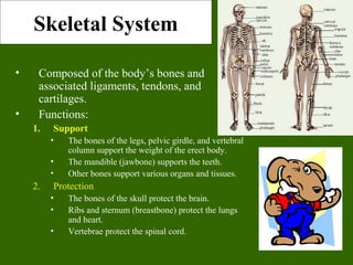
1. Skeletal System
• Composed of the body’s bones and
associated ligaments, tendons, and
cartilages.
• Functions:
1. Support
• The bones of the legs, pelvic girdle, and vertebral
column support the weight of the erect body.
• The mandible (jawbone) supports the teeth.
• Other bones support various organs and tissues.
2. Protection
• The bones of the skull protect the brain.
• Ribs and sternum (breastbone) protect the lungs
and heart.
• Vertebrae protect the spinal cord.
2. Skeletal System
• Functions:
1. Movement
• Skeletal muscles use the bones as levers to
move the body.
2. Reservoir for minerals and adipose tissue
• 99% of the body’s calcium is stored in bone.
• 85% of the body’s phosphorous is stored in
bone.
• Adipose tissue is found in the marrow of
certain bones.
– What is really being stored in this case? (hint –
it starts with an E)
3. Hematopoiesis
• A.k.a. blood cell formation.
• All blood cells are made in the marrow of
certain bones.
3. Bone Classification• There are 206 named bones in
the human body.
• Each belongs to one of 2 large
groups:
– Axial skeleton
• Forms long axis of the body.
• Includes the bones of the skull,
vertebral column, and rib cage.
• These bones are involved in
protection, support, and
carrying other body parts.
– Appendicular skeleton
• Bones of upper & lower limbs
and the girdles (shoulder bones
and hip bones) that attach them
to the axial skeleton.
• Involved in locomotion and
manipulation of the
environment.
4. Bone Classification
• 4 types of bones:
1. Long Bones
• Much longer than they are wide.
• All bones of the limbs except for
the patella (kneecap),
and the bones of the wrist and ankle.
• Consists of a shaft plus 2
expanded ends.
• Your finger bones are long bones
even though they’re
very short – how can this be?
1. Short Bones
• Roughly cube shaped.
• Bones of the wrist and the ankle.
Femur
Carpal Bones
5. Bone Classification
• Types of bones:
1. Flat Bones
• Thin, flattened, and usually
a bit curved.
• Scapulae, sternum,
(shoulder blades), ribs and
most bones of the skull.
2. Irregular Bones
• Have weird shapes that fit
none of the 3 previous
classes.
• Vertebrae, hip bones, 2
skull bones ( sphenoid
and the ethmoid bones).
Sternum
Sphenoid
Bone
6. Bone Structure
• Bones are organs. Thus, they’re composed of
multiple tissue types. Bones are composed of:
– Bone tissue (a.k.a. osseous tissue).
– Fibrous connective tissue.
– Cartilage.
– Vascular tissue.
– Lymphatic tissue.
– Adipose tissue.
– Nervous tissue.
7. • All bones consist of a
dense, solid outer
layer known as
compact bone and an
inner layer of spongy
bone – a honeycomb
of flat, needle-like
projections called
trabeculae.
• Bone is an extremely
dynamic tissue!!!!
Above: Note the relationship btwn the
compact and spongy bone.
Below: Close up of spongy bone.
8. Note the gross differences between the spongy bone and the
compact bone in the above photo.
Do you see the trabeculae?
10. Bone Structure
• Bone tissue is a type of
connective tissue, so it must
consist of cells plus a
significant amount of
extracellular matrix.
• Bone cells:
1. Osteoblasts
• Bone-building cells.
• Synthesize and secrete
collagen fibers and other
organic components of
bone matrix.
• Initiate the process of
calcification.
• Found in both the
periosteum and the
endosteum
The blue arrows indicate the
osteoblasts. The yellow arrows indicate
the bone matrix they’ve just secreted.
11. Bone Structure
2.Osteocytes
• Mature bone cells.
• Osteoblasts that have
become trapped by the
secretion of matrix.
• No longer secrete
matrix.
• Responsible for
maintaining the bone
tissue.
Yellow arrows indicate
osteocytes – notice
how they are
surrounded by the
pinkish bone matrix.
Blue arrow shows an
osteoblast in the
process of becoming an
osteocyte.
On the right, notice how the osteocyte
is “trapped” within the pink matrix
12. 3. Osteoclasts
– Huge cells derived from the fusion of as many as 50 monocytes (a type of
white blood cell).
– Cells that digest bone matrix – this process is called bone resorption and is
part of normal bone growth, development, maintenance, and repair.
– Concentrated in the endosteum.
– On the side of the cell that faces the bone surface, the PM is deeply folded
into a ruffled border. Here, the osteoclast secretes digestive enzymes (how
might this occur?) to digest the bone matrix. It also pumps out hydrogen
ions (how might this occur?) to create an acid environment that eats away at
the matrix. What advantage might a ruffled border confer?
– Why do we want a cell that eats away at bone? (Hint: bone is a very
dynamic tissue.)
13. •Here, we see a cartoon showing all 3 cell types. Osteoblasts and osteoclasts are indicated.
•Note the size of the osteoclast (compare it to the osteoblast), and note the ruffled border.
•Why is there a depression underneath the osteoclast?
•What is the name of the third cell type shown here?
•What do you think the tan material represents?
14. Bone Structure
• Bone Matrix:
– Consists of organic and inorganic
components.
– 1/3 organic and 2/3 inorganic by
weight.
• Organic component consists of several
materials that are secreted by the
osteoblasts:
– Collagen fibers and other organic materials
» These (particularly the collagen) provide
the bone with resilience and the ability
to resist stretching and twisting.
15. • Inorganic component
of bone matrix
– Consists mainly of 2
salts: calcium
phosphate and calcium
hydroxide. These 2
salts interact to form a
compound called
hydroxyapatite.
– Bone also contains
smaller amounts of
magnesium, fluoride,
and sodium.
– These minerals give
bone its characteristic
hardness and the
ability to resist
compression.
Three-dimensional array of
collagen molecules. The rod-
shaped molecules lie in a
staggered arrangement which
acts as a template for bone
mineralization. Bone mineral is
laid down in the gaps.
Note collagen fibers in longitudinal & cross section
and how they occupy space btwn the black bone cells.
16. This bone:
a. Has been demineralized
b. Has had its organic component removed
17. Long Bone Structure
• Shaft plus 2 expanded ends.
• Shaft is known as the diaphysis.
– Consists of a thick collar of compact
bone surrounding a central marrow
cavity
• In adults, the marrow cavity contains
fat - yellow bone marrow.
• Expanded ends are epiphyses
– Thin layer of compact bone covering
an interior of spongy bone.
– Joint surface of each epiphysis is
covered w/ a type of hyaline cartilage
known as articular cartilage. It
cushions the bone ends and reduces
friction during movement.
18. Long Bone
Structure
• The external surface of the entire
bone except for the joint surfaces of
the epiphyses is covered by a
double-layered membrane known as
the periosteum.
– Outer fibrous layer is dense irregular
connective tissue.
– Inner cellular layer contains
osteoprogenitor cells and osteoblasts.
– Periosteum is richly supplied with
nerve fibers, lymphatic vessels and
blood vessels.
• These enter the bone of the shaft via a
nutrient foramen.
– Periosteum is connected to the bone
matrix via strong strands of collagen.
19. Long Bone
Structure
• Internal bone surfaces are covered with a delicate
connective tissue membrane known as the
endosteum.
– Covers the trabeculae of spongy bone in the marrow
cavities and lines the canals that pass through compact
bone.
– Contains both osteoblasts and osteoclasts.
20. Structure of Short, Irregular, and
Flat Bones
• Thin plates of periosteum-covered
compact bone on the outside and
endosteum-covered spongy bone
within.
• Have no diaphysis or epiphysis
because they are not cylindrical.
• Contain bone marrow between
their trabeculae, but no marrow
cavity.
• In flat bones, the internal spongy
bone layer is known as the diploë,
and the whole arrangement
resembles a stiffened sandwich.
21. Bone Marrow
• Bone marrow is a general term for the
soft tissue occupying the medullary
cavity of a long bone, the spaces amid
the trabeculae of spongy bone, and the
larger haversian canals.
• There are 2 main types: red & yellow.
• Red bone marrow = blood cell
forming tissue = hematopoietic tissue
• Red bone marrow looks like blood but
with a thicker consistency.
• It consists of a delicate mesh of reticular
tissue saturated with immature red blood
cells and scattered adipocytes.
Notice the red marrow
and the compact bone
22. Distribution of
Marrow
• In a child, the medullary
cavity of nearly every bone is
filled with red bone marrow.
• In young to middle-aged
adults, the shafts of the long
bones are filled with fatty
yellow bone marrow.
– Yellow marrow no longer
produces blood, although in
the event of severe or chronic
anemia, it can transform back
into red marrow
• In adults, red marrow is
limited to the axial skeleton,
pectoral girdle, pelvic girdle,
and proximal heads of the
humerus and the femur.
Note the compact bone on the
bottom and marrow on the bottom.
23. Microscopic
Structure of
Compact Bone
• Consists of multiple
cylindrical structural
units known as
osteons or haversian
systems.
• Imagine these osteons
as weight-bearing
pillars that are
arranged parallel to
one another along the
long axis of a
compact bone.
The diagram below represents a long
bone shaft in cross-section. Each
yellow circle represents an osteon. The
blue represents additional matrix filling
in the space btwn osteons. The white in
the middle is the marrow cavity.
24. Osteons
• Each osteon consists of a single
central canal, known as a
haversian canal, surrounded by
concentric layers of calcified
bone matrix.
– Haversian canals allow the passage
of blood vessels, lymphatic vessels,
and nerve fibers.
– Each of the concentric matrix
“tubes” that surrounds a haversian
canal is known as a lamella.
– All the collagen fibers in a particular
lamella run in a single direction,
while collagen fibers in adjacent
lamellae will run in the opposite
direction. This allows bone to better
withstand twisting forces.
25. Running perpendicular to the haversian canals are Volkmann’s canals.
They connect the blood and nerve supply in the periosteum to those
in the haversian canals and the medullary cavity.
26. Osteons
• Lying in between intact
osteons are incomplete
lamellae called
interstitial lamellae.
These fill the gaps
between osteons or are
remnants of bone
remodeling.
• There are also circumferential lamellae that extend around the
circumference of the shaft. There are inner circumferential
lamellae surrounding the endosteum and outer circumferential
lamellae just inside the periosteum.
27. • Spider-shaped
osteocytes occupy small
cavities known as
lacunae at the junctions
of the lamellae. Hairlike
canals called canaliculi
connect the lacunae to
each other and to the
central canal.
• Canaliculi allow the
osteocytes to exchange
nutrients, wastes, and
chemical signals to each
other via intercellular
connections known as
gap junctions.
28.
29. Here, we have a close up and a far
away view of compact bone. You
should be able to identify haversian
canals, concentric lamellae,
interstitial lamellae, lacunae, and
canaliculi.
30. Microscopic
Structure of Spongy
Bone
• Appears poorly organized
compared to compact bone.
• Lacks osteons.
• Trabeculae align along
positions of stress and
exhibit extensive cross-
bracing.
• Trabeculae are a few cell
layers thick and contain
irregularly arranged
lamellae and osteocytes
interconnected by
canaliculi.
• No haversian or Volkmann’s
canals are necessary. Why?
31. Bone Development
• Osteogenesis (a.k.a.
ossification) is the
process of bone tissue
formation.
• In embryos this leads to
the formation of the
bony skeleton.
• In children and young
adults, ossification
occurs as part of bone
growth.
• In adults, it occurs as
part of bone remodeling
and bone repair.
32. Formation of the Bony Skeleton
• Before week 8, the human
embryonic skeleton is made of
fibrous membranes and hyaline
cartilage.
• After week 8, bone tissue
begins to replace the fibrous
membranes and hyaline
cartilage.
– The development of bone from a
fibrous membrane is called
intramembranous ossification.
Why?
– The replacement of hyaline
cartilage with bone is known as
endochondral ossification. Why?
33. Intramembranous Ossification
• Some bones of the skull (frontal, parietal, temporal, and occipital
bones), the facial bones, the clavicles, the pelvis, the scapulae, and
part of the mandible are formed by intramembranous ossification
• Prior to ossification, these structures exist as fibrous membranes
made of embryonic connective tissue known as mesenchyme.
34. • Mesenchymal cells first
cluster together and start
to secrete the organic
components of bone
matrix which then
becomes mineralized
through the crystallization
of calcium salts. As
calcification occurs, the
mesenchymal cells
differentiate into
osteoblasts.
• The location in the tissue
where ossification begins
is known as an
ossification center.
• Some osteoblasts are
trapped w/i bony pockets.
These cells differentiate
into osteocytes.
35. • The developing bone grows outward from the ossification center
in small struts called spicules.
• Mesenchymal cell divisions provide additional osteoblasts.
• The osteoblasts require a reliable source of oxygen and
nutrients. Blood vessels trapped among the spicules meet these
demands and additional vessels branch into the area. These
vessels will eventually become entrapped within the growing
bone.
36. • Initially, the intramembranous bone consists only of
spongy bone. Subsequent remodeling around trapped
blood vessels can produce osteons typical of compact
bone.
• As the rate of growth slows, the connective tissue around
the bone becomes organized into the fibrous layer of the
periosteum. Osteoblasts close to the bone surface become
the inner cellular layer of the periosteum.
37. Endochondral Ossification
• Begins with the formation of a hyaline cartilage model which
will later be replaced by bone.
• Most bones in the body develop via this model.
• More complicated than intramembranous because the hyaline
cartilage must be broken down as ossification proceeds.
• We’ll follow limb bone development as an example.
38. Endochondral Ossification – Step 1
• Chondrocytes near the center
of the shaft of the hyaline
cartilage model increase
greatly in size. As these cells
enlarge, their lacunae expand,
and the matrix is reduced to a
series of thin struts. These
struts soon begin to calcify.
• The enlarged chondrocytes
are now deprived of nutrients
(diffusion cannot occur
through calcified cartilage)
and they soon die and
disintegrate.
39. Endochondral Ossification – Step 2
• Blood vessels grow into the perichondrium surrounding the shaft
of the cartilage. The cells of the inner layer of the
perichondrium in this region then differentiate into osteoblasts.
• The perichondrium is now a periosteum and the inner osteogenic
layer soon produces a thin layer of bone around the shaft of the
cartilage. This bony collar provides support.
40. Endochondral Ossification – Step 3
• Blood supply to the periosteum, and
capillaries and fibroblasts migrate into
the heart of the cartilage, invading the
spaces left by the disintegrating
chondrocytes.
• The calcified cartilaginous matrix
breaks down; the fibroblasts
differentiate into osteoblasts that replace
it with spongy bone.
• Bone development begins at this
primary center of ossification and
spreads toward both ends of the
cartilaginous model.
• While the diameter is small, the entire
diaphysis is filled with spongy bone.
Notice the primary
ossification centers in the
thigh and forearm bones
of the above fetus.
41. Endochondral Ossification – Step 4
• The primary ossification center enlarges
proximally and distally, while osteoclasts break
down the newly formed spongy bone and open up
a medullary cavity in the center of the shaft.
• As the osteoblasts move towards the epiphyses,
the epiphyseal cartilage is growing as well. Thus,
even though the shaft is getting longer, the
epiphyses have yet to be transformed into bone.
42. Endochondral Ossification – Step 5
• Around birth, most long bones
have a bony diaphysis surrounding
remnants of spongy bone, a
widening medullary cavity, and 2
cartilaginous epiphyses.
• At this time, capillaries and
osteoblasts will migrate into the
epiphyses and create secondary
ossification centers. The
epiphysis will be transformed into
spongy bone. However, a small
cartilaginous plate, known as the
epiphyseal plate, will remain at
the juncture between the epiphysis
and the diaphysis.
Articular
cartilage
Epiphyseal plate
43.
44. Growth in Bone
Length
• Epiphyseal cartilage
(close to the epiphysis)
of the epiphyseal plate
divides to create more
cartilage, while the
diaphyseal cartilage
(close to the diaphysis)
of the epiphyseal plate is
transformed into bone.
This increases the length
of the shaft.
45. •As a result osteoblasts begin
producing bone faster than the
rate of epiphyseal cartilage
expansion. Thus the bone grows
while the epiphyseal plate gets
narrower and narrower and
ultimately disappears. A remnant
(epiphyseal line) is visible on X-
rays (do you see them in the
adjacent femur, tibia, and fibula?)
At puberty, growth in bone length
is increased dramatically by the
combined activities of growth
hormone, thyroid hormone, and
the sex hormones.
46. Growth in Bone Thickness
• Osteoblasts beneath the periosteum secrete bone
matrix on the external surface of the bone. This
obviously makes the bone thicker.
• At the same time, osteoclasts on the endosteum
break down bone and thus widen the medullary
cavity.
• This results in an increase in shaft diameter even
though the actual amount of bone in the shaft is
relatively unchanged.
47. Fractures
• Despite its mineral strength,
bone may crack or even break
if subjected to extreme loads,
sudden impacts, or stresses
from unusual directions.
– The damage produced constitutes
a fracture.
• The proper healing of a
fracture depends on whether or
not, the blood supply and
cellular components of the
periosteum and endosteum
survive.
48. Fracture
Repair
• Step 1:
A. Immediately after
the fracture,
extensive
bleeding occurs.
Over a period of
several hours, a
large blood clot,
or fracture
hematoma,
develops.
B. Bone cells at the
site become
deprived of
nutrients and die.
The site becomes
swollen, painful,
and inflamed.
• Step 2:
A. Granulation tissue is formed as the hematoma is
infiltrated by capillaries and macrophages, which begin
to clean up the debris.
B. Some fibroblasts produce collagen fibers that span the
break , while others differentiate into chondroblasts and
begin secreting cartilage matrix.
C. Osteoblasts begin forming spongy bone.
D. This entire structure is known as a fibrocartilaginous callus
and it splints the broken bone.
49. • Step 3:
A. Bone trabeculae
increase in number
and convert the
fibrocartilaginous
callus into a bony
callus of spongy
bone. Typically
takes about 6-8
weeks for this to
occur.
Fracture
Repair
• Step 4:
A. During the next several months, the bony callus is continually
remodeled.
B. Osteoclasts work to remove the temporary supportive structures
while osteoblasts rebuild the compact bone and reconstruct the
bone so it returns to its original shape/structure.
50. Fracture Types
• Fractures are often classified according to the position of the
bone ends after the break:
Open (compound) bone ends penetrate the skin.
Closed (simple) bone ends don’t penetrate the skin.
Comminuted bone fragments into 3 or more pieces.
Common in the elderly (brittle
bones).
Greenstick bone breaks incompletely. One side bent,
one side broken. Common in
children whose bone contains more
collagen and are less mineralized.
Spiral ragged break caused by excessive twisting
forces. Sports injury/Injury of abuse.
51.
52. What kind of fracture is this?
It’s kind of tough to tell, but
this is a _ _ _ _ _ _ fracture.
53. Bone Remodeling
• Bone is a
dynamic tissue.
– What does that
mean?
• Wolff’s law
holds that bone
will grow or
remodel in
response to the
forces or
demands placed
on it. Examine
this with the
bone on the left.
54. Check out the mechanism of
remodeling on the right!
Why might you suspect
someone whose been a
powerlifter for 15 years to
have heavy, massive bones,
especially at the point of
muscle insertion?
Astronauts tend to experience
bone atrophy after they’re in
space for an extended period
of time. Why?
55. Nutritional Effects on Bone
• Normal bone growth/maintenance
cannot occur w/o sufficient dietary
intake of calcium and phosphate
salts.
• Calcium and phosphate are not
absorbed in the intestine unless the
hormone calcitriol is present.
Calcitriol synthesis is dependent on
the availability of the steroid
cholecalciferol (a.k.a. Vitamin D)
which may be synthesized in the skin
or obtained from the diet.
• Vitamins C, A, K, and B12 are all
necessary for bone growth as well.
56. Hormonal Effects
on Bone
• Growth hormone, produced
by the pituitary gland, and
thyroxine, produced by the
thyroid gland, stimulate bone
growth.
– GH stimulates protein synthesis
and cell growth throughout the
body.
– Thyroxine stimulates cell
metabolism and increases the
rate of osteoblast activity.
– In proper balance, these
hormones maintain normal
activity of the epiphyseal plate
(what would you consider
normal activity?) until roughly
the time of puberty.
57. Hormonal Effects on Bone
• At puberty, the rising levels of sex hormones (estrogens in
females and androgens in males) cause osteoblasts to
produce bone faster than the epiphyseal cartilage can
divide. This causes the characteristic growth spurt as well
as the ultimate closure of the epiphyseal plate.
• Estrogens cause faster closure of the epiphyseal growth
plate than do androgens.
• Estrogen also acts to stimulate osteoblast activity.
58. Hormonal Effects on Bone
• Other hormones that affect bone growth include
insulin and the glucocorticoids.
– Insulin stimulates bone formation
– Glucocorticoids inhibit osteoclast activity.
• Parathyroid hormone and calcitonin are 2
hormones that antagonistically maintain blood
[Ca2+
] at homeostatic levels.
– Since the skeleton is the body’s major calcium
reservoir, the activity of these 2 hormones affects bone
resorption and deposition.
59. Calcitonin
• Released by the C cells of the thyroid gland in response to high
blood [Ca2+
].
• Calcitonin acts to “tone down” blood calcium levels.
• Calcitonin causes decreased osteoclast activity which results in
decreased break down of bone matrix and decreased calcium
being released into the blood.
• Calcitonin also stimulates osteoblast activity which means
calcium will be taken from the blood and deposited as bone
matrix.
Notice the thyroid
follicles on the
right. The arrow
indicates a C cell
61. Parathyroid Hormone
• PTH increases calcitriol synthesis which increases Ca2+
absorption in the small intestine.
• PTH decreases urinary Ca2+
excretion and increases urinary
phosphate excretion.
• Released by the cells of the
parathyroid gland in response to low
blood [Ca2+
].Causes blood [Ca2+
] to
increase.
• PTH will bind to osteoblasts and this
will cause 2 things to occur:
• The osteoblasts will decrease their activity
and they will release a chemical known as
osteoclast-stimulating factor.
• Osteoclast-stimulating factor will increase
osteoclast activity.
62. Increased PTH release
by parathyroid gland
Binds to osteoblast
causing decreased
osteoblast activity and
release of osteoclast-
stimulating factor
OSF causes increased
osteoclast activity
Decreased bone
deposition and increased
bone resorption
Increased calcitriol
synthesis
Increased intestinal
Ca2+
absorption
Decreased Ca2+
excretion
Increased Blood [Ca2+
]
Decreased Blood [Ca2+
]
63. Clinical Conditions
• Osteomalacia
– Literally “soft bones.”
– Includes many disorders in which
osteoid is produced but
inadequately mineralized.
• Causes can include insufficient
dietary calcium
• Insufficient vitamin D fortification
or insufficient exposure to sun
light.
• Rickets
– Children's form of osteomalacia
– More detrimental due to the fact
that their bones are still growing.
– Signs include bowed legs, and
deformities of the pelvis, ribs, and
skull.
What about the above x-ray is
indicative of rickets?
64. Clinical Conditions
• Osteomyelitis
– Osteo=bone +
myelo=marrow +
itis=inflammation.
– Inflammation of bone and
bone marrow caused by
pus-forming bacteria that
enter the body via a
wound (e.g., compound
fracture) or migrate from a
nearby infection.
– Fatal before the advent of
antibiotics.
65. Clinical Conditions
• Osteoporosis
– Group of diseases in which
bone resorption occurs at a
faster rate than bone deposition.
– Bone mass drops and bones
become increasingly porous.
– Compression fractures of the
vertebrae and fractures of the
femur are common.
– Often seen in postmenopausal
women because they
experience a rapid decline in
estrogen secretion; estrogen
stimulates osteoblast and
inhibits osteoclast activity.
• Based on the above, what
preventative measures might
you suggest?
66. Clinical Conditions
• Gigantism
– Childhood hypersecretion
of growth hormone by the
pituitary gland causes
excessive growth.
• Acromegaly
– Adulthood hypersecretion
of GH causes overgrowth
of bony areas still
responsive to GH such as
the bones of the face, feet,
and hands.
• Pituitary dwarfism
– GH deficiency in children
resulting in extremely short
long bones and maximum
stature of 4 feet.