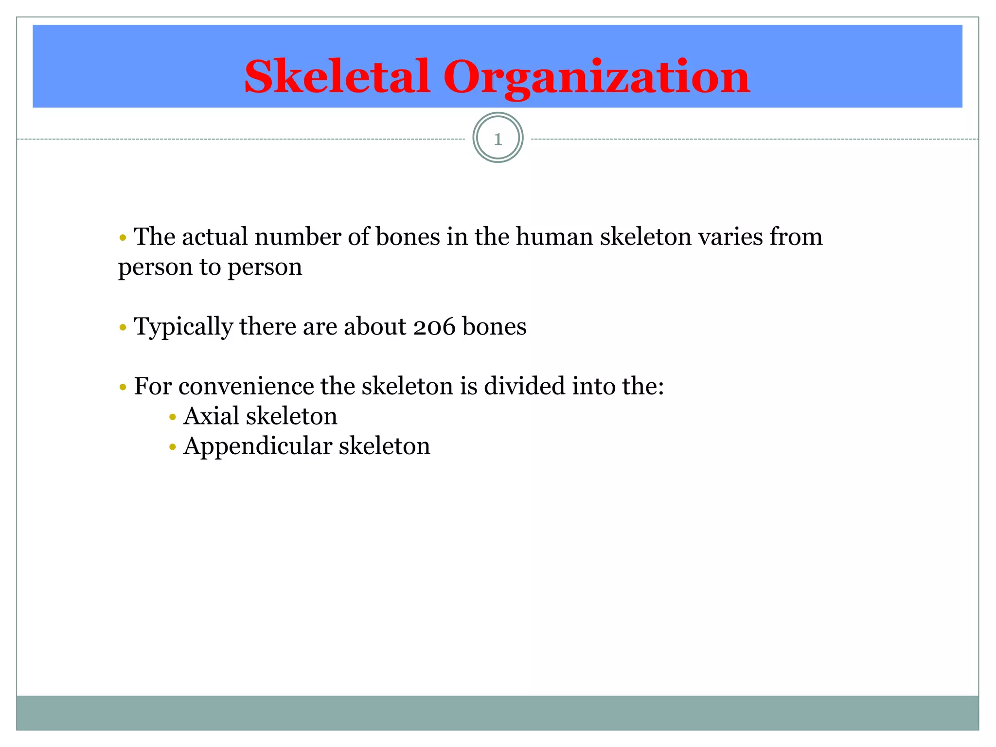
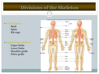
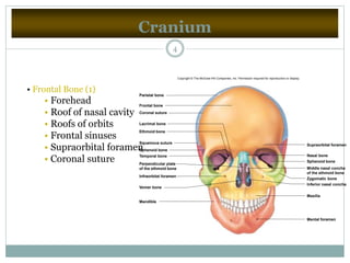
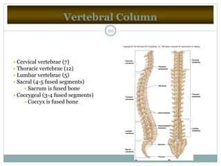
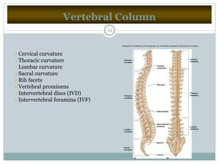
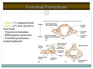
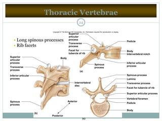
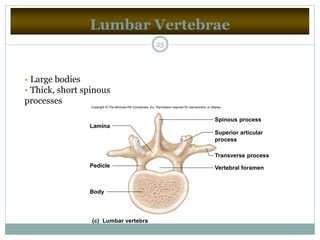
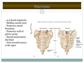
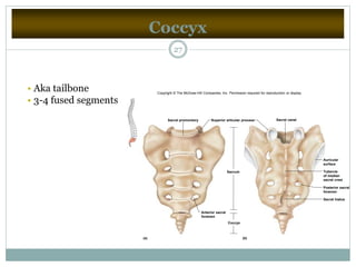
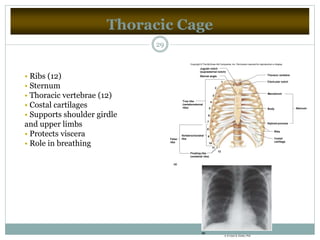
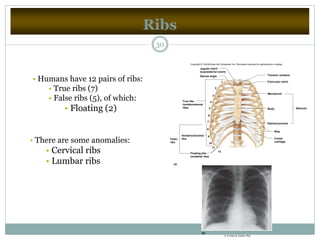
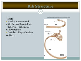
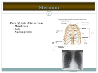
Skeletal Organization
1
• The actual number of bones in the human skeleton varies from
person to person
• Typically there are about 206 bones
• For convenience the skeleton is divided into the:
• Axial skeleton
• Appendicular skeleton
Divisions of the Skeleton
2
• Axial Skeleton
• Skull
• Spine
• Rib cage
• Appendicular Skeleton
• Upper limbs
• Lower limbs
• Shoulder girdle
• Pelvic girdle
Hyoid
Cranium
Face
Clavicle
Scapula
Sternum
Ribs
Humerus
Ulna
Hip
bone
Radius
Femur
Patella
Tibia
Fibula
Tarsals
Metatarsals
Phalanges
Phalanges
Skull
Vertebral
column
Vertebral
column
Sacrum
Coccyx
Carpals
Metacarpals
(a) (b)
Copyright © The McGraw-Hill Companies, Inc. Permission required for reproduction or display.
Skull
3
• Is composed of the cranium (brain case) and the facial bones
Cranium
4
• Frontal Bone (1)
• Forehead
• Roof of nasal cavity
• Roofs of orbits
• Frontal sinuses
• Supraorbital foramen
• Coronal suture
Parietal bone
Frontal bone
Coronal suture
Lacrimal bone
Ethmoid bone
Squamous suture
Sphenoid bone
Temporal bone
Perpendicular plate
of the ethmoid bone
Infraorbital foramen
Vomer bone
Mandible
Supraorbital foramen
Nasal bone
Sphenoid bone
Middle nasal concha
of the ethmoid bone
Inferior nasal concha
Maxilla
Mental foramen
Zygomatic bone
Copyright © The McGraw-Hill Companies, Inc. Permission required for reproduction or display.
Cranium
5
• Parietal Bones (2)
• Side walls of cranium
• Roof of cranium
• Sagittal suture
Coronal suture
Frontal bone
Sphenoid bone
Ethmoid bone
Lacrimal bone
Nasal bone
Zygomatic bone
Maxilla
Mental foramen
Mandible
Coronoid process
Styloid process
Mandibular condyle
Mastoid process
External acoustic meatus Temporal process
of zygomatic bone
Zygomatic process
of temporal bone
Occipital bone
Temporal bone
Parietal bone
Lambdoid suture
Squamous suture
Copyright © The McGraw-Hill Companies, Inc. Permission required for reproduction or display.
Cranium
6
• Occipital Bone (1)
• Back of skull
• Base of cranium
• Foramen magnum
• Occipital condyles
• Lambdoidal suture
Zygomatic bone
Palatine process of maxilla
Palatine bone
Occipital bone
Foramen magnum
Lambdoid suture
Zygomatic arch
Mandibular fossa
Styloid process
Occipital condyle
External acoustic meatus
Sphenoid bone
Vomer bone
Temporal bone
Mastoid foramen
Frontal bone
Median palatine suture
Incisive foramen
Greater palatine foramen
Stylomastoid foramen
Foramen spinosum
Carotid canal
Jugular foramen
Foramen ovale
Foramen lacerum
Condylar canal
Copyright © The McGraw-Hill Companies, Inc. Permission required for reproduction or display.
Cranium
7
• Temporal Bones (2)
• Side walls of cranium
• Floor of cranium
• Floors and sides of orbits
• Squamous suture
• External acoustic meatus
• Mandibular fossa
• Mastoid process
• Styloid process
• Zygomatic process
Coronal suture
Frontal bone
Sphenoid bone
Ethmoid bone
Lacrimal bone
Nasal bone
Zygomatic bone
Maxilla
Mental foramen
Mandible
Coronoid process
Styloid process
Mandibular condyle
Mastoid process
External acoustic meatus
Temporal process
of zygomatic bone
Zygomatic process
of temporal bone
Occipital bone
Temporal bone
Parietal bone
Lambdoid suture
Squamous suture
Copyright © The McGraw-Hill Companies, Inc. Permission required for reproduction or display.
Cranium
8
• Sphenoid Bone (1)
• Base of cranium
• Sides of skull
• Floors and sides
of orbits
• Sella turcica
• Sphenoid sinusesTransverse section
Lesser wing
Optic canal
Greater
wing
Sella turcica Foramen ovale
Foramen spinosum
Foramen rotundum
Lesser wing
Greater wing
Superior
orbital fissure
Foramen
rotundum
Lateral pterygoid plate
Medial pterygoid plate
(b)
(a)
Cranium
9
• Ethmoid Bone (1)
• Roof and walls of nasal cavity
• Floor of cranium
• Wall of orbits
• Cribiform plates
• Perpendicular plate
• Superior and middle
nasal conchae
• Ethmoid sinuses
• Crista galli
Transverse section
Crista galli
Orbital
surface
Ethmoidal
sinuses
Cribriform
plate
Crista galli
Perpendicular
plate
Superior
nasal concha
Middle nasal
concha
Perpendicular
plate
(a) (b)
Facial Skeleton
10
• Maxillary Bones (2)
• Upper jaw
• Anterior roof of mouth
• Floors of orbits
• Sides of nasal cavity
• Floors of nasal cavity
• Alveolar processes
• Maxillary sinuses
• Palatine process
Coronal suture
Frontal bone
Sphenoid bone
Ethmoid bone
Lacrimal bone
Nasal bone
Zygomatic bone
Maxilla
Mental foramen
Mandible
Coronoid process
Styloid process
Mandibular condyle
Mastoid process
External acoustic meatus
Temporal process
of zygomatic bone
Zygomatic process
of temporal bone
Occipital bone
Temporal bone
Parietal bone
Lambdoid suture
Squamous suture
Copyright © The McGraw-Hill Companies, Inc. Permission required for reproduction or display.
Facial Skeleton
11
Frontal sinus
Ethmoidal sinuses
Sphenoidal sinus
Maxillary sinus
Copyright © The McGraw-Hill Companies, Inc. Permission required for reproduction or display.
Facial Skeleton
12
• Palatine Bones (2)
• ‘L’ shaped bones located
behind the maxillae
• Posterior section of hard
palate
• Floor of nasal cavity
• Lateral walls of
nasal cavity
Coronal section
Horizontal portion
Perpendicular
portion
Copyright © The McGraw-Hill Companies, Inc. Permission required for reproduction or display.
Facial Skeleton
13
• Zygomatic Bones (2)
• Prominences of cheeks
• Lateral walls of orbits
• Floors of orbits
• Temporal process
Coronal suture
Frontal bone
Sphenoid bone
Ethmoid bone
Lacrimal bone
Nasal bone
Zygomatic bone
Maxilla
Mental foramen
Mandible
Coronoid process
Styloid process
Mandibular condyle
Mastoid process
External acoustic meatus
Temporal process
of zygomatic bone
Zygomatic process
of temporal bone
Occipital bone
Temporal bone
Parietal bone
Lambdoid suture
Squamous suture
Copyright © The McGraw-Hill Companies, Inc. Permission required for reproduction or display.
Facial Skeleton
14
• Lacrimal Bones (2)
• Medial walls of orbits
• Groove from orbit to
nasal cavity
• Nasal Bones (2)
• Bridge of nose
Coronal suture
Frontal bone
Sphenoid bone
Ethmoid bone
Lacrimal bone
Nasal bone
Zygomatic bone
Maxilla
Mental foramen
Mandible
Coronoid process
Styloid process
Mandibular condyle
Mastoid process
External acoustic meatus
Temporal process
of zygomatic bone
Zygomatic process
of temporal bone
Occipital bone
Temporal bone
Parietal bone
Lambdoid suture
Squamous suture
Copyright © The McGraw-Hill Companies, Inc. Permission required for reproduction or display.
Facial Skeleton
15
• Vomer Bone (1)
• Inferior portion of nasal
septum
Copyright © The McGraw-Hill Companies, Inc. Permission required for reproduction or display.
Coronal suture
Frontal bone
Nasal bone
Palatine bone
Vomer bone
Alveolar processes
Inferior nasal concha
Maxilla
Palatine process
of maxilla
Styloid process
Sphenoidal sinus
Mandible
Perpendicular plate
(nasal septum)
Frontal sinus
Crista galli
Cribriform plate
Foramen magnum
Jugular foramen
Internal acoustic meatus
Sella turcica
Occipital bone
Parietal bone
Temporal bone
Lambdoid suture
Squamous suture
Sphenoid bone
Hypoglossal canal
Mastoid process
Ethmoid
bone
Facial Skeleton
16
• Inferior Nasal Conchae (2)
• Extend from lateral
walls of nasal cavity
Copyright © The McGraw-Hill Companies, Inc. Permission required for reproduction or display.
Coronal suture
Frontal bone
Nasal bone
Palatine bone
Vomer bone
Alveolar processes
Inferior nasal concha
Maxilla
Palatine process
of maxilla
Styloid process
Sphenoidal sinus
Mandible
Perpendicular plate
(nasal septum)
Frontal sinus
Crista galli
Cribriform plate
Foramen magnum
Jugular foramen
Internal acoustic meatus
Sella turcica
Occipital bone
Parietal bone
Temporal bone
Lambdoid suture
Squamous suture
Sphenoid bone
Hypoglossal canal
Mastoid process
Ethmoid
bone
Facial Skeleton
17
• Mandible Bone (1)
• Lower jaw
• Body
• Ramus
• Mandibular condyle
• Coronoid process
• Alveolar process
• Mandibular foramen
• Mental foramen
Coronoid
process
Mandibular
foramen Mandibular condyle
Ramus
Alveolar
border
Mental foramen
Body
Body Alveolar
arch
Mandibular
foramen
Coronoid process
(a)
(b)
Copyright © The McGraw-Hill Companies, Inc. Permission required for reproduction or display.
Infantile Skull
18
• Fontanels – fibrous membranes
(b)
Anterior fontanel
Posterior fontanel
Frontal bone
Frontal suture
(metopic suture)
Sagittal suture
Copyright © The McGraw-Hill Companies, Inc. Permission required for reproduction or display.
Vertebral Column
19
• The vertebral column, or spinal column, consists of many
vertebrae separated by cartilaginous intervertebral discs.
Vertebral Column
20
• Cervical vertebrae (7)
• Thoracic vertebrae (12)
• Lumbar vertebrae (5)
• Sacral (4-5 fused segments)
• Sacrum is fused bone
• Coccygeal (3-4 fused segments)
• Coccyx is fused bone
(b)(a)
Cervical
curvature
Thoracic
curvature
Lumbar
curvature
Lumbar
vertebrae
Thoracic
vertebrae
Cervical
vertebrae
Sacral
curvature
Vertebra
prominens
Rib facet
Intervertebral
Intervertebral
foramina
Sacrum
Coccyx
Copyright © The McGraw-Hill Companies, Inc. Permission required for reproduction or display.
Vertebral Column
21
• Cervical curvature
• Thoracic curvature
• Lumbar curvature
• Sacral curvature
• Rib facets
• Vertebral prominens
• Intervertebral discs (IVD)
• Intervertebral foramina (IVF)
(b)(a)
Cervical
curvature
Thoracic
curvature
Lumbar
curvature
Lumbar
vertebrae
Thoracic
vertebrae
Cervical
vertebrae
Sacral
curvature
Vertebra
prominens
Rib facet
Intervertebral
Intervertebral
foramina
Sacrum
Coccyx
Copyright © The McGraw-Hill Companies, Inc. Permission required for reproduction or display.
Typical Vertebrae
22
• Includes the following parts:
• Vertebral body
• Pedicles
• Lamina
• Spinous process
• Transverse processes
• Vertebral foramen
• Facets
Cervical Vertebrae
23
• Atlas – 1st; supports head
• Axis – 2nd; dens pivots to
turn head
• Transverse foramina
• Bifid spinous processes
• Vertebral prominens –
useful landmark
(b) (c)
(a)
Anterior
Posterior
Atlas
Axis
Body
Dens (odontoid
process)
Spinous processDens
Inferior articular
process
Facet that articulates
with dens (odontoid process)
of axis
Facet that articulates
with occipital condyle
Spinous
process
Anterior articular
facet for atlas
Transverse
foramen
Transverse
process
Superior
articular facet
Vertebral
foramen
Transverse
process
Transverse
foramen
Copyright © The McGraw-Hill Companies, Inc. Permission required for reproduction or display.
Thoracic Vertebrae
24
BodySuperior
articular
process
Spinous
process
Transverse
process
Inferior articular
process
Intervertebral
disc
Anterior
Posterior
Body
Pedicle
Vertebral foramen
Superior articular process
Facet for tubercle of rib
Transverse process
Lamina
Spinous process
Inferior articular
process
Intervertebral notch
Body
Pedicle
Superior
articular
process
Transverse
process
Facet for
tubercle of rib
Spinous
process
(a)
(c)
(b)
• Long spinous processes
• Rib facets
Copyright © The McGraw-Hill Companies, Inc. Permission required for reproduction or display.
Lumbar Vertebrae
25
• Large bodies
• Thick, short spinous
processes
(c) Lumbar vertebra
Lamina
Pedicle
Body
Vertebral foramen
Spinous process
Superior articular
process
Transverse process
Copyright © The McGraw-Hill Companies, Inc. Permission required for reproduction or display.
Sacrum
26
• 4-5 fused segments
• Median sacral crest
• Posterior sacral
foramina
• Posterior wall of
pelvic cavity
• Sacral promontory
aka base
• Area toward coccyx
is the apex
Sacral canal
Tubercle
of median
sacral crest
Auricular
surface
Posterior sacral
foramen
Sacral hiatus
Coccyx
Sacrum
Superior articular processSacral promontory
Anterior sacral
foramen
(a) (b)
Copyright © The McGraw-Hill Companies, Inc. Permission required for reproduction or display.
Coccyx
27
• Aka tailbone
• 3-4 fused segments
Sacral canal
Tubercle
of median
sacral crest
Auricular
surface
Posterior sacral
foramen
Sacral hiatus
Coccyx
Sacrum
Superior articular processSacral promontory
Anterior sacral
foramen
(a) (b)
Copyright © The McGraw-Hill Companies, Inc. Permission required for reproduction or display.
Thoracic Cage
28
• The thoracic cage includes the ribs, the thoracic vertebrae, the
sternum, and the costal cartilages that attach the ribs to the sternum.
Thoracic Cage
29
• Ribs (12)
• Sternum
• Thoracic vertebrae (12)
• Costal cartilages
• Supports shoulder girdle
and upper limbs
• Protects viscera
• Role in breathing
1
2
3
4
5
6
7
8
9
10
11
12
True ribs
(vertebrosternal
ribs)
Vertebrochondral
ribsFalse
ribs
(a)
Floating ribs
(vertebral ribs)
SternumBody
Manubrium
Ribs
Costal
cartilage
Xiphoid process
Thoracic vertebra
Clavicular notch
Sternal angle
Jugular notch
(suprasternal notch)
(b)
b: © Victor B. Eichler, PhD
Copyright © The McGraw-Hill Companies, Inc. Permission required for reproduction or display.
Ribs
30
• Humans have 12 pairs of ribs:
• True ribs (7)
• False ribs (5), of which:
• Floating (2)
• There are some anomalies:
• Cervical ribs
• Lumbar ribs
1
2
3
4
5
6
7
8
9
10
11
12
True ribs
(vertebrosternal
ribs)
Vertebrochondral
ribsFalse
ribs
(a)
Floating ribs
(vertebral ribs)
SternumBody
Manubrium
Ribs
Costal
cartilage
Xiphoid process
Thoracic vertebra
Clavicular notch
Sternal angle
Jugular notch
(suprasternal notch)
(b)
b: © Victor B. Eichler, PhD
Copyright © The McGraw-Hill Companies, Inc. Permission required for reproduction or display.
Rib Structure
31
• Shaft
• Head – posterior end;
articulates with vertebrae
• Tubercle – articulates
with vertebrae
• Costal cartilage – hyaline
cartilage
Anterior end
(sternal end)
Shaft
Facet
Head
Neck
Tubercle
Facet
Spinous process
Costal groove
ShaftAnterior end
Head
Tubercle
Neck
(a)
(b)
Copyright © The McGraw-Hill Companies, Inc. Permission required for reproduction or display.
Sternum
32
• Three (3) parts of the sternum:
• Manubrium
• Body
• Xiphoid process
1
2
3
4
5
6
7
8
9
10
11
12
True ribs
(vertebrosternal
ribs)
Vertebrochondral
ribsFalse
ribs
(a)
Floating ribs
(vertebral ribs)
SternumBody
Manubrium
Ribs
Costal
cartilage
Xiphoid process
Thoracic vertebra
Clavicular notch
Sternal angle
Jugular notch
(suprasternal notch)
(b)
b: © Victor B. Eichler, PhD
Copyright © The McGraw-Hill Companies, Inc. Permission required for reproduction or display.

Skeletal organization

  • 1.
    Skeletal Organization 1 • Theactual number of bones in the human skeleton varies from person to person • Typically there are about 206 bones • For convenience the skeleton is divided into the: • Axial skeleton • Appendicular skeleton
  • 2.
    Divisions of theSkeleton 2 • Axial Skeleton • Skull • Spine • Rib cage • Appendicular Skeleton • Upper limbs • Lower limbs • Shoulder girdle • Pelvic girdle Hyoid Cranium Face Clavicle Scapula Sternum Ribs Humerus Ulna Hip bone Radius Femur Patella Tibia Fibula Tarsals Metatarsals Phalanges Phalanges Skull Vertebral column Vertebral column Sacrum Coccyx Carpals Metacarpals (a) (b) Copyright © The McGraw-Hill Companies, Inc. Permission required for reproduction or display.
  • 3.
    Skull 3 • Is composedof the cranium (brain case) and the facial bones
  • 4.
    Cranium 4 • Frontal Bone(1) • Forehead • Roof of nasal cavity • Roofs of orbits • Frontal sinuses • Supraorbital foramen • Coronal suture Parietal bone Frontal bone Coronal suture Lacrimal bone Ethmoid bone Squamous suture Sphenoid bone Temporal bone Perpendicular plate of the ethmoid bone Infraorbital foramen Vomer bone Mandible Supraorbital foramen Nasal bone Sphenoid bone Middle nasal concha of the ethmoid bone Inferior nasal concha Maxilla Mental foramen Zygomatic bone Copyright © The McGraw-Hill Companies, Inc. Permission required for reproduction or display.
  • 5.
    Cranium 5 • Parietal Bones(2) • Side walls of cranium • Roof of cranium • Sagittal suture Coronal suture Frontal bone Sphenoid bone Ethmoid bone Lacrimal bone Nasal bone Zygomatic bone Maxilla Mental foramen Mandible Coronoid process Styloid process Mandibular condyle Mastoid process External acoustic meatus Temporal process of zygomatic bone Zygomatic process of temporal bone Occipital bone Temporal bone Parietal bone Lambdoid suture Squamous suture Copyright © The McGraw-Hill Companies, Inc. Permission required for reproduction or display.
  • 6.
    Cranium 6 • Occipital Bone(1) • Back of skull • Base of cranium • Foramen magnum • Occipital condyles • Lambdoidal suture Zygomatic bone Palatine process of maxilla Palatine bone Occipital bone Foramen magnum Lambdoid suture Zygomatic arch Mandibular fossa Styloid process Occipital condyle External acoustic meatus Sphenoid bone Vomer bone Temporal bone Mastoid foramen Frontal bone Median palatine suture Incisive foramen Greater palatine foramen Stylomastoid foramen Foramen spinosum Carotid canal Jugular foramen Foramen ovale Foramen lacerum Condylar canal Copyright © The McGraw-Hill Companies, Inc. Permission required for reproduction or display.
  • 7.
    Cranium 7 • Temporal Bones(2) • Side walls of cranium • Floor of cranium • Floors and sides of orbits • Squamous suture • External acoustic meatus • Mandibular fossa • Mastoid process • Styloid process • Zygomatic process Coronal suture Frontal bone Sphenoid bone Ethmoid bone Lacrimal bone Nasal bone Zygomatic bone Maxilla Mental foramen Mandible Coronoid process Styloid process Mandibular condyle Mastoid process External acoustic meatus Temporal process of zygomatic bone Zygomatic process of temporal bone Occipital bone Temporal bone Parietal bone Lambdoid suture Squamous suture Copyright © The McGraw-Hill Companies, Inc. Permission required for reproduction or display.
  • 8.
    Cranium 8 • Sphenoid Bone(1) • Base of cranium • Sides of skull • Floors and sides of orbits • Sella turcica • Sphenoid sinusesTransverse section Lesser wing Optic canal Greater wing Sella turcica Foramen ovale Foramen spinosum Foramen rotundum Lesser wing Greater wing Superior orbital fissure Foramen rotundum Lateral pterygoid plate Medial pterygoid plate (b) (a)
  • 9.
    Cranium 9 • Ethmoid Bone(1) • Roof and walls of nasal cavity • Floor of cranium • Wall of orbits • Cribiform plates • Perpendicular plate • Superior and middle nasal conchae • Ethmoid sinuses • Crista galli Transverse section Crista galli Orbital surface Ethmoidal sinuses Cribriform plate Crista galli Perpendicular plate Superior nasal concha Middle nasal concha Perpendicular plate (a) (b)
  • 10.
    Facial Skeleton 10 • MaxillaryBones (2) • Upper jaw • Anterior roof of mouth • Floors of orbits • Sides of nasal cavity • Floors of nasal cavity • Alveolar processes • Maxillary sinuses • Palatine process Coronal suture Frontal bone Sphenoid bone Ethmoid bone Lacrimal bone Nasal bone Zygomatic bone Maxilla Mental foramen Mandible Coronoid process Styloid process Mandibular condyle Mastoid process External acoustic meatus Temporal process of zygomatic bone Zygomatic process of temporal bone Occipital bone Temporal bone Parietal bone Lambdoid suture Squamous suture Copyright © The McGraw-Hill Companies, Inc. Permission required for reproduction or display.
  • 11.
    Facial Skeleton 11 Frontal sinus Ethmoidalsinuses Sphenoidal sinus Maxillary sinus Copyright © The McGraw-Hill Companies, Inc. Permission required for reproduction or display.
  • 12.
    Facial Skeleton 12 • PalatineBones (2) • ‘L’ shaped bones located behind the maxillae • Posterior section of hard palate • Floor of nasal cavity • Lateral walls of nasal cavity Coronal section Horizontal portion Perpendicular portion Copyright © The McGraw-Hill Companies, Inc. Permission required for reproduction or display.
  • 13.
    Facial Skeleton 13 • ZygomaticBones (2) • Prominences of cheeks • Lateral walls of orbits • Floors of orbits • Temporal process Coronal suture Frontal bone Sphenoid bone Ethmoid bone Lacrimal bone Nasal bone Zygomatic bone Maxilla Mental foramen Mandible Coronoid process Styloid process Mandibular condyle Mastoid process External acoustic meatus Temporal process of zygomatic bone Zygomatic process of temporal bone Occipital bone Temporal bone Parietal bone Lambdoid suture Squamous suture Copyright © The McGraw-Hill Companies, Inc. Permission required for reproduction or display.
  • 14.
    Facial Skeleton 14 • LacrimalBones (2) • Medial walls of orbits • Groove from orbit to nasal cavity • Nasal Bones (2) • Bridge of nose Coronal suture Frontal bone Sphenoid bone Ethmoid bone Lacrimal bone Nasal bone Zygomatic bone Maxilla Mental foramen Mandible Coronoid process Styloid process Mandibular condyle Mastoid process External acoustic meatus Temporal process of zygomatic bone Zygomatic process of temporal bone Occipital bone Temporal bone Parietal bone Lambdoid suture Squamous suture Copyright © The McGraw-Hill Companies, Inc. Permission required for reproduction or display.
  • 15.
    Facial Skeleton 15 • VomerBone (1) • Inferior portion of nasal septum Copyright © The McGraw-Hill Companies, Inc. Permission required for reproduction or display. Coronal suture Frontal bone Nasal bone Palatine bone Vomer bone Alveolar processes Inferior nasal concha Maxilla Palatine process of maxilla Styloid process Sphenoidal sinus Mandible Perpendicular plate (nasal septum) Frontal sinus Crista galli Cribriform plate Foramen magnum Jugular foramen Internal acoustic meatus Sella turcica Occipital bone Parietal bone Temporal bone Lambdoid suture Squamous suture Sphenoid bone Hypoglossal canal Mastoid process Ethmoid bone
  • 16.
    Facial Skeleton 16 • InferiorNasal Conchae (2) • Extend from lateral walls of nasal cavity Copyright © The McGraw-Hill Companies, Inc. Permission required for reproduction or display. Coronal suture Frontal bone Nasal bone Palatine bone Vomer bone Alveolar processes Inferior nasal concha Maxilla Palatine process of maxilla Styloid process Sphenoidal sinus Mandible Perpendicular plate (nasal septum) Frontal sinus Crista galli Cribriform plate Foramen magnum Jugular foramen Internal acoustic meatus Sella turcica Occipital bone Parietal bone Temporal bone Lambdoid suture Squamous suture Sphenoid bone Hypoglossal canal Mastoid process Ethmoid bone
  • 17.
    Facial Skeleton 17 • MandibleBone (1) • Lower jaw • Body • Ramus • Mandibular condyle • Coronoid process • Alveolar process • Mandibular foramen • Mental foramen Coronoid process Mandibular foramen Mandibular condyle Ramus Alveolar border Mental foramen Body Body Alveolar arch Mandibular foramen Coronoid process (a) (b) Copyright © The McGraw-Hill Companies, Inc. Permission required for reproduction or display.
  • 18.
    Infantile Skull 18 • Fontanels– fibrous membranes (b) Anterior fontanel Posterior fontanel Frontal bone Frontal suture (metopic suture) Sagittal suture Copyright © The McGraw-Hill Companies, Inc. Permission required for reproduction or display.
  • 19.
    Vertebral Column 19 • Thevertebral column, or spinal column, consists of many vertebrae separated by cartilaginous intervertebral discs.
  • 20.
    Vertebral Column 20 • Cervicalvertebrae (7) • Thoracic vertebrae (12) • Lumbar vertebrae (5) • Sacral (4-5 fused segments) • Sacrum is fused bone • Coccygeal (3-4 fused segments) • Coccyx is fused bone (b)(a) Cervical curvature Thoracic curvature Lumbar curvature Lumbar vertebrae Thoracic vertebrae Cervical vertebrae Sacral curvature Vertebra prominens Rib facet Intervertebral Intervertebral foramina Sacrum Coccyx Copyright © The McGraw-Hill Companies, Inc. Permission required for reproduction or display.
  • 21.
    Vertebral Column 21 • Cervicalcurvature • Thoracic curvature • Lumbar curvature • Sacral curvature • Rib facets • Vertebral prominens • Intervertebral discs (IVD) • Intervertebral foramina (IVF) (b)(a) Cervical curvature Thoracic curvature Lumbar curvature Lumbar vertebrae Thoracic vertebrae Cervical vertebrae Sacral curvature Vertebra prominens Rib facet Intervertebral Intervertebral foramina Sacrum Coccyx Copyright © The McGraw-Hill Companies, Inc. Permission required for reproduction or display.
  • 22.
    Typical Vertebrae 22 • Includesthe following parts: • Vertebral body • Pedicles • Lamina • Spinous process • Transverse processes • Vertebral foramen • Facets
  • 23.
    Cervical Vertebrae 23 • Atlas– 1st; supports head • Axis – 2nd; dens pivots to turn head • Transverse foramina • Bifid spinous processes • Vertebral prominens – useful landmark (b) (c) (a) Anterior Posterior Atlas Axis Body Dens (odontoid process) Spinous processDens Inferior articular process Facet that articulates with dens (odontoid process) of axis Facet that articulates with occipital condyle Spinous process Anterior articular facet for atlas Transverse foramen Transverse process Superior articular facet Vertebral foramen Transverse process Transverse foramen Copyright © The McGraw-Hill Companies, Inc. Permission required for reproduction or display.
  • 24.
    Thoracic Vertebrae 24 BodySuperior articular process Spinous process Transverse process Inferior articular process Intervertebral disc Anterior Posterior Body Pedicle Vertebralforamen Superior articular process Facet for tubercle of rib Transverse process Lamina Spinous process Inferior articular process Intervertebral notch Body Pedicle Superior articular process Transverse process Facet for tubercle of rib Spinous process (a) (c) (b) • Long spinous processes • Rib facets Copyright © The McGraw-Hill Companies, Inc. Permission required for reproduction or display.
  • 25.
    Lumbar Vertebrae 25 • Largebodies • Thick, short spinous processes (c) Lumbar vertebra Lamina Pedicle Body Vertebral foramen Spinous process Superior articular process Transverse process Copyright © The McGraw-Hill Companies, Inc. Permission required for reproduction or display.
  • 26.
    Sacrum 26 • 4-5 fusedsegments • Median sacral crest • Posterior sacral foramina • Posterior wall of pelvic cavity • Sacral promontory aka base • Area toward coccyx is the apex Sacral canal Tubercle of median sacral crest Auricular surface Posterior sacral foramen Sacral hiatus Coccyx Sacrum Superior articular processSacral promontory Anterior sacral foramen (a) (b) Copyright © The McGraw-Hill Companies, Inc. Permission required for reproduction or display.
  • 27.
    Coccyx 27 • Aka tailbone •3-4 fused segments Sacral canal Tubercle of median sacral crest Auricular surface Posterior sacral foramen Sacral hiatus Coccyx Sacrum Superior articular processSacral promontory Anterior sacral foramen (a) (b) Copyright © The McGraw-Hill Companies, Inc. Permission required for reproduction or display.
  • 28.
    Thoracic Cage 28 • Thethoracic cage includes the ribs, the thoracic vertebrae, the sternum, and the costal cartilages that attach the ribs to the sternum.
  • 29.
    Thoracic Cage 29 • Ribs(12) • Sternum • Thoracic vertebrae (12) • Costal cartilages • Supports shoulder girdle and upper limbs • Protects viscera • Role in breathing 1 2 3 4 5 6 7 8 9 10 11 12 True ribs (vertebrosternal ribs) Vertebrochondral ribsFalse ribs (a) Floating ribs (vertebral ribs) SternumBody Manubrium Ribs Costal cartilage Xiphoid process Thoracic vertebra Clavicular notch Sternal angle Jugular notch (suprasternal notch) (b) b: © Victor B. Eichler, PhD Copyright © The McGraw-Hill Companies, Inc. Permission required for reproduction or display.
  • 30.
    Ribs 30 • Humans have12 pairs of ribs: • True ribs (7) • False ribs (5), of which: • Floating (2) • There are some anomalies: • Cervical ribs • Lumbar ribs 1 2 3 4 5 6 7 8 9 10 11 12 True ribs (vertebrosternal ribs) Vertebrochondral ribsFalse ribs (a) Floating ribs (vertebral ribs) SternumBody Manubrium Ribs Costal cartilage Xiphoid process Thoracic vertebra Clavicular notch Sternal angle Jugular notch (suprasternal notch) (b) b: © Victor B. Eichler, PhD Copyright © The McGraw-Hill Companies, Inc. Permission required for reproduction or display.
  • 31.
    Rib Structure 31 • Shaft •Head – posterior end; articulates with vertebrae • Tubercle – articulates with vertebrae • Costal cartilage – hyaline cartilage Anterior end (sternal end) Shaft Facet Head Neck Tubercle Facet Spinous process Costal groove ShaftAnterior end Head Tubercle Neck (a) (b) Copyright © The McGraw-Hill Companies, Inc. Permission required for reproduction or display.
  • 32.
    Sternum 32 • Three (3)parts of the sternum: • Manubrium • Body • Xiphoid process 1 2 3 4 5 6 7 8 9 10 11 12 True ribs (vertebrosternal ribs) Vertebrochondral ribsFalse ribs (a) Floating ribs (vertebral ribs) SternumBody Manubrium Ribs Costal cartilage Xiphoid process Thoracic vertebra Clavicular notch Sternal angle Jugular notch (suprasternal notch) (b) b: © Victor B. Eichler, PhD Copyright © The McGraw-Hill Companies, Inc. Permission required for reproduction or display.