This document provides an overview of the skeletal system chapter from an anatomy and physiology textbook. It covers the following key points in 3 sentences:
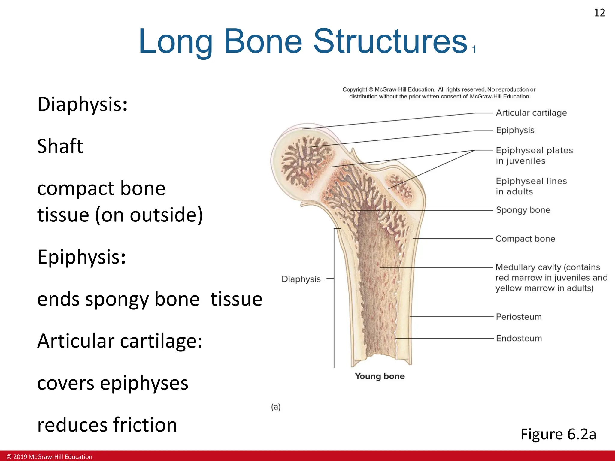
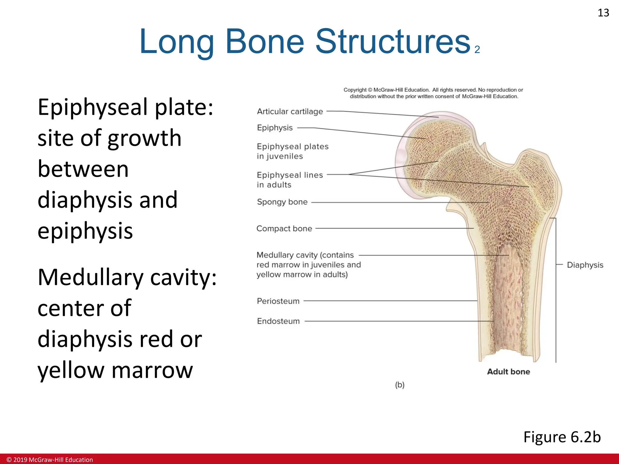
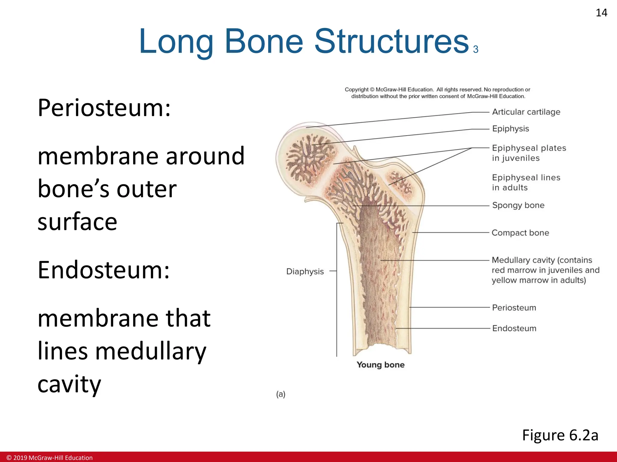
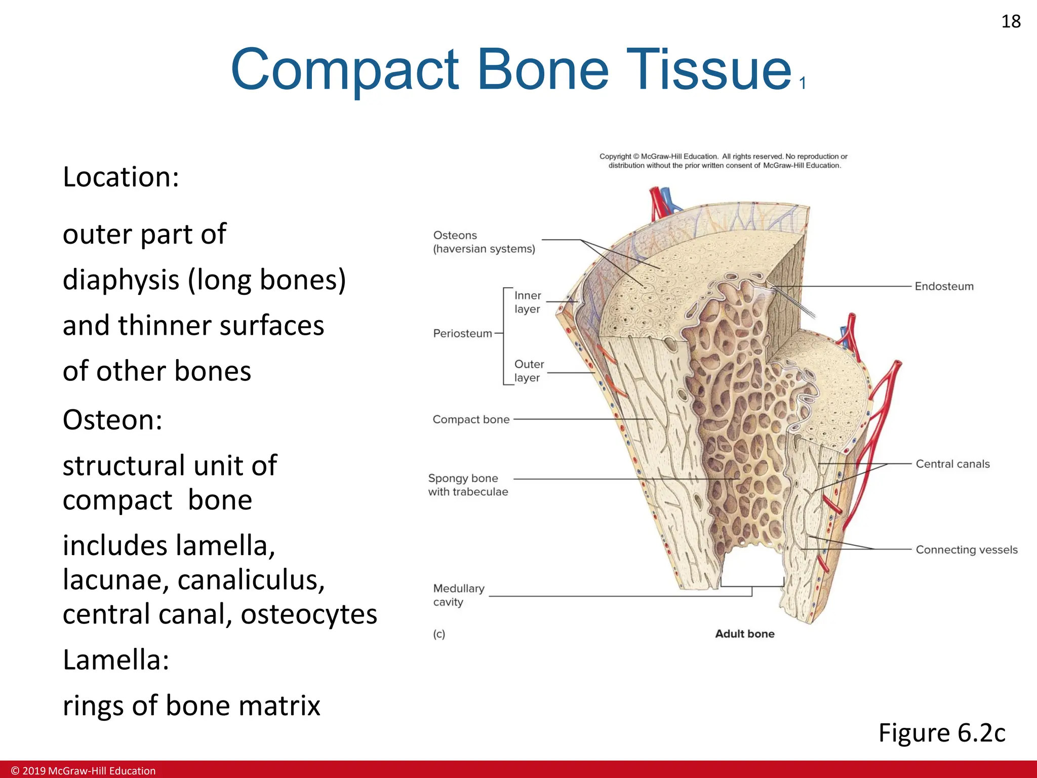
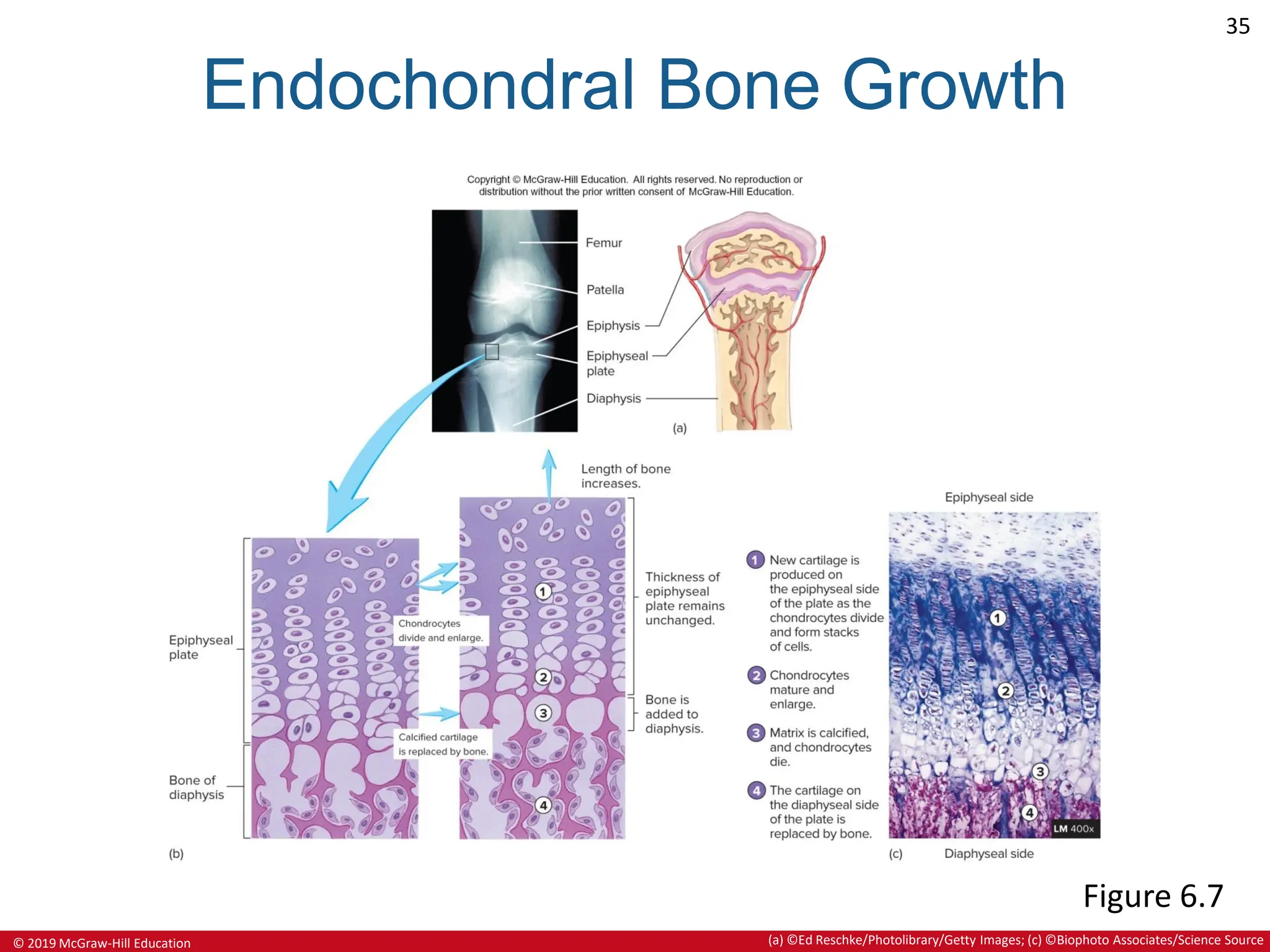
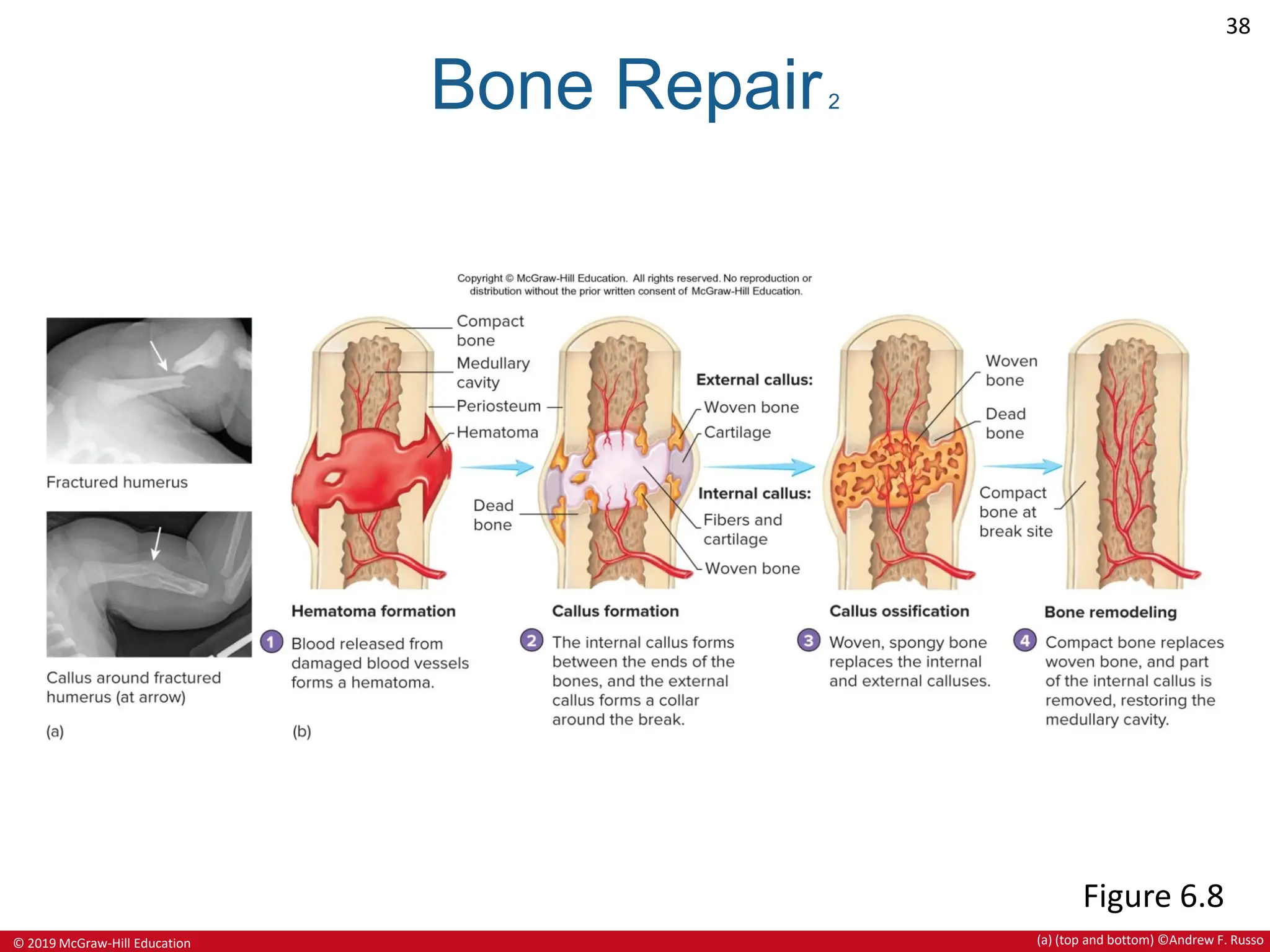
It defines the components of the skeletal system as bones, cartilages, tendons, and ligaments. It describes the functions of the skeletal system as support, protection, movement, storage, and blood cell production. It provides details on the structure of bones, including their extracellular matrix composition, bone cell types, and the processes of bone formation, growth, remodeling and repair.