Bones, Joints, and the Architecture of Movement: Exploring the Skeletal System

Title: Bones, Joints, and the Architecture of Movement: Exploring the Skeletal System Introduction: Welcome to our engaging SlideShare presentation on the Skeletal System & Joints, where we embark on a fascinating exploration of the framework that supports our bodies and facilitates movement. Join us as we delve into the intricate world of bones, joints, and the dynamic interplay that shapes our mobility and stability. The skeletal system serves as the foundation of our anatomy, providing structural support, protection for vital organs, and a framework for locomotion. Comprised of bones, cartilage, ligaments, and tendons, this complex system forms the structural scaffold upon which our bodies are built. In this presentation, we'll take a closer look at the anatomy of bones, from their composition and classification to their roles in mineral storage and blood cell production. Through detailed illustrations and interactive diagrams, we'll explore the dynamic nature of bone remodeling and the factors that influence bone health and density. But bones alone do not account for the versatility of human movement. Joints, the articulations where two or more bones meet, play a crucial role in facilitating motion and absorbing impact. From synovial joints like the knee and shoulder to fibrous and cartilaginous joints, we'll unravel the mechanics of joint structure and function. Together, the skeletal system and joints form a complex network that enables us to walk, run, jump, and perform a myriad of daily activities. Whether you're a student studying anatomy, a fitness enthusiast seeking to optimize performance, or simply curious about the mechanics of the human body, our presentation offers valuable insights into the wonders of the skeletal system and joints. Join us as we embark on a journey through the bony landmarks and articulations that define our physical form and discover the intricate architecture of movement hidden beneath our skin.

Recommended
More Related Content
Similar to Bones, Joints, and the Architecture of Movement: Exploring the Skeletal System
Similar to Bones, Joints, and the Architecture of Movement: Exploring the Skeletal System (20)
Recently uploaded
Recently uploaded (20)
Bones, Joints, and the Architecture of Movement: Exploring the Skeletal System
- 2. INTRODUCTION The Skeleton is the name given to the collection of bones that holds the rest of our body up. * Human skeleton initially cartilages and fibrous membranes. * Hyaline cartilage is the most abundant cartilage. * By age 25 the skeleton is completely hardened. * 206 bones make up the adult skeleton (20% of body mass). - 80 bones of the axial skeleton - 126 bones of the appendicular skeleton * Parts of the skeletal system include: - Bones (skeleton) - Joints - Cartilages - Ligaments * Divided into two divisions: - Axial skeleton (skull, ribs and vertebra) - Appendicular skeleton (pelvis, extremities- Upper or Lower)
- 4. Bones Highly vascular mineralized connective tissue. Consists of cells and dense intercellular organic matrix impregnated with inorganic salts Organic material- collagen fibers. Provides tensile strength. Inorganic – calcium phosphate, traces of other salts. Provides hardness & rigidity Bones Functions PROTECTION : It protects our vital organs such as brain, heart, lungs etc... SUPPORT & SHAPE : It gives us our shape that we have. Without our skeleton, we would just be a blob of blood and tissue on the floor. MOVEMENT : It allows us to move. Because our muscles are attached to our bones, when our muscles move, they move the bones, and we move. HEMOPOIESIS (IN RED BONE MARROW) : It makes blood cells so the rest of the body can use them. MINERAL HOMEOSTASIS (STORAGE AND RELEASE) : Calcium, and other minerals. TRIGLYCERIDE STORAGE (IN YELLOW BONE MARROW) : It is a warehouse for fat cells.
- 5. Composition of bone SOLID WATER (10%) Organic Inorganic Substance (25%) Substance (65%) Bone Cells Protein Ca2+ (99% of total body Ca2+ is present in bones), Po 4 2- , Na+ , Cl- , Co3 - , K+ etc… Osteoprogenitor cells (Bone-forming stem cells) Osteoblast (Bone-forming cells) Osteocytes (Mature bone cells) Osteoclast (Bone-destroying / remodeling cells) Glycoprotein Collagen fiber type-I
- 7. Classification of bone According to shape : Long Bones - Metacarples, Metatarsals, Phelangies, Humerus, Femur, Ulna, Radius, Tibia, Fibula Short Bones - Carpals, Tarsals Flat Bones - Ribs, Scapula, Skull, Sternum Irregular Bones - Vertebrae, Some facial bones Sesamoid Bone – Patella Long bones Typically longer than wide Have a shaft with heads at both ends Contain mostly compact bone
- 8. (e) Sesamoid Bone (e.g., patella)
- 9. Examples: Femur, humerus Short bones Generally cube-shape Contain mostly spongy bone Examples: Carpals, Tarsals etc... Flat bones Thin and flattened Usually curved Thin layers of compact bone around a layer of spongy bone Examples: Skull, ribs, sternum etc…
- 10. Irregular bones Irregular shape Do not fit into other bone classification categories Example: Vertebrae and hip Sesamoid bone Sort bones within tendones Example: Patella
- 11. According to structure : Macroscopically - Compact Bone, Cancellous (Spongy Bone) Microscopically- Woven, lamellar etc…. • Compact bone Outer layer of bone, very hard and dense. Organized in structural unit called Haversian systems. Matrix is composed of Ca salts (Ca carbonate and Ca phosphate) Osteocytes – living bone cells that live in matrix.
- 12. • Porous (Spongy) bone Located in the ends of long bones. Many spaces that are filled with red bone marrow which produces bone cells. Trabeculae – needle-like threads of spongy bone that surround the spaces. Add strength to this portion of the bone. According to development :
- 13. Femur Periosteum Yellow marrow Medullary cavity Space containing red marrow Spongy bone Compact bone Articular cartilage Epiphyseal plates Proximal epiphysis Distal epiphysis Diaphysis Endosteum Gross Anatomy of a Long Bone Parts of a Long Bone • Epiphysis • Distal • Proximal • Diaphysis • Metaphysis • Compact bone • Spongy bone • Articular cartilage • Periosteum • Endosteum • Medullary cavity • Trabeculae • Bone marrow • Red marrow and yellow marrow
- 14. Distal epiphysis Proximal epiphysis Diaphysis Yellow marrow Epiphyseal line Periosteum Compact bone Spongy bone Endosteum Hyaline cartilage Sharpey’s fibers Distal epiphysis
- 15. Diaphysis Shaft Composed of compact bone Epiphysis Ends of the bone Composed mostly of spongy bone
- 16. Periosteum Outside covering of the diaphysis Fibrous connective tissue membrane Sharpey’s fibers Secure periosteum to underlying bone Arteries Supply bone cells with nutrients
- 17. Articular cartilage Covers the external surface of the epiphyses Made of hyaline cartilage Decreases friction at joint surfaces Medullary cavity Cavity of the shaft Contains yellow marrow (mostly fat) in adults Contains red marrow (for blood cell formation) in infants
- 18. Microscopic Structure • Bone cells are called osteocytes ( in a lacuna ) • Osteocytes transport nutrients and wastes by cellular processes in canaliculi. • The extracellular matrix of bone is largely collagen and inorganic salts • Collagen gives bone resilience & strength • Inorganic salts make bone hard Compact Bone • Osteon aka Haversian System (Structural and basic unit of compact bone) • Central canal • Perforating canal aka Volkmann’s canal • Osteocytes • Lamellae • Lacunae • Bone matrix • Canaliculi Nerve Osteon Nerve Nerve Canaliculus Osteocyte Periosteum Endosteum Trabeculae Pores Bone matrix Blood vessels Compact bone Lacuna (space) Blood vessels Perforating canal Central canal containing blood vessels and nerves Central canal
- 20. Osteon (Haversian System) : A unit of bone Central (Haversian) canal : Opening in the center of an osteon Carries blood vessels and nerves Perforating (Volkman’s) canal : Canal perpendicular to the central canal Carries blood vessels and nerves
- 21. Lacunae Cavities containing bone cells (osteocytes) Arranged in concentric rings Lamellae Rings around the central canal Sites of lacunae Canaliculi Tiny canals Radiate from the central canal to lacunae Form a transport system
- 23. (a) (c) Spongy bone Compact bone (b) Spongy bone Compact bone Remnant of epiphyseal plate Spongy Compact bone Spongy Bone • Spongy bone is aka cancellous bone. • Trabeculae is the basic structural unit of spongy bone. • There is no calcification in spongy bone so hematopoiesis occur. • Trabeculae is the tubular shape structure present in spongy bone, it is soft like honey womb. • The outer covering of spongy bone is covered by compact bone for its protection.
- 25. Bone Development and Growth As an infant, most of your skeleton is cartilage. Process of bone formation called ossification & Calcification From mesenchyme Cartilage is a strong flexible tissue. Over time the cartilage is replaced by solid bone, usually complete by the time you stop growing. Not all cartilage is replaced in adults. Many joints contain cartilage, protecting the ends of bones (ears and the end of the nose is also cartilage). Epiphyseal plates allow for growth of long bone during childhood. New cartilage is continuously formed. Older cartilage becomes ossified. Bone replaces cartilage.
- 26. Bones are remodeled and lengthened until growth stops Bones change shape somewhat Bones grow in width
- 27. • Parts of the skeletal system begin to develop during the first few weeks of prenatal development • Bones replace existing connective tissue in one of two ways: • As intramembranous bones • As endchondral bones Intramembranous Bones • These bones originate within sheetlike layers of connective tissues • They are the broad, flat bones • Skull bones (except mandible) • Are known as intramembranous bones Endochondral Bones • Bones begin as hyaline cartilage • Form models for future bones • These are most bones of the skeleton • Are known as endochondral bones
- 28. 28 Endochondral Ossification Hyaline cartilage model • Primary ossification center • Secondary ossification centers • Epiphyseal plate • Osteoblasts vs. osteoclasts (b) (c) (d) (e) (f) (a) Cartilaginous model Calcified cartilage Articular cartilage Developing periosteum Compact bone developing Primary ossification center Medullary cavity Medullary cavity Medullary cavity Secondary ossification center Secondary ossification center Blood vessel Epiphyseal plate Remnant of epiphyseal plate Remnants of epiphyseal plates Epiphyseal plates Compact bone Spongy bone Articular cartilage Spongy bone
- 29. Types of Bone Cells Osteogenic cells: Respond to traumas, such as fractures, by giving rise to bone-forming cells and bone-destroying cells Osteoblasts (Bone-forming cells) Bone forming cells synthesize and secrete unmineralized ground substance and are found in areas of high metabolism within the bone Bone lining cells - made from osteoblasts along the surface of most bones in an adult. Bone-lining cells are thought to regulate the movement of calcium and phosphate into and out of the bone Osteocytes (Mature bone cells) Mature bone cells made from osteoblasts that have made bone tissue around themselves. They maintain healthy bone tissue by secreting enzymes and controlling the bone mineral content; they also control the calcium release from the bone tissue to the blood.
- 30. Osteoclasts (Bone-destroying cells) Break down bone matrix for remodeling and release of calcium Bone absorbing cell – large cells that break down bone tissue – important to growth, healing, remodeling Bone remodeling is a process by both osteoblasts and osteoclasts Homeostasis of Bone Tissue • Bone Resorption – action of osteoclasts and parathyroid hormone aka parathormone aka PTH • Bone Deposition – action of osteoblasts and calcitonin • Occurs by direction of the thyroid and parathyroid glands • Calcitonin decreases blood ca++ level by increasing osteoblastic activity. • PTH increases blood ca++ level by increasing osteoclastic activity.
- 31. Factors Affecting Bone Development, Growth and Repair • Deficiency of Vitamin A – retards bone development • Deficiency of Vitamin C – results in fragile bones • Deficiency of Vitamin D – Rickets, Osteomalacia • Insufficient Growth Hormone – Dwarfism • Excessive Growth Hormone – Gigantism, Acromegaly • Insufficient Thyroid Hormone – delays bone growth • Sex Hormones – promote bone formation; stimulate ossification of epiphyseal plates • Physical Stress – stimulates bone growth
- 32. Cartilage Specialized connective tissue having rigidity with elasticity Consists of chondrocytes embedded in gel like matrix Provide rigidity & support Smooth gliding surface for articulation Enables development and growth of long bones Structure Cells- chondrogenic, chondroblasts, chondrocytes Fibers- type I collagen, type II collagen, elastic Ground substance- proteoglycans in the form of keratan sulphate & chondroitin sulphate, glycoprotein, water, no mineral component Features Avascular No lymphatics No nerves Surrounded by perichondrium Growth by appos. & inter. When it calcifies, chondrocytes die Articular cartilage & fibrocartilage have no perichondrium
- 33. Hyaline Cartilages: Fine collagen fiber matrix- most abundant type- found in articular (movable joint) cartilages, costal cartilages (connect ribs to sternum), respiratory cartilages (in larynx & upper respiratory passageways) & nasal cartilages Elastic Cartilages: Similar to hyaline cartilage, more elastic fibers (very flexible) – found in external ear & epiglottis (larynx covering) Fibrocartilage: Rows of chondrocytes with thick collagen fibers; highly compressible with great tensile strength- found in menisci of knee, intervertebral discs & pubic symphysis
- 35. Bone Fractures & Repair of fracture A break in the continuity of a bone Types of bone fractures Closed (simple) fracture – break that does not penetrate the skin Open (compound) fracture – broken bone penetrates through the skin Bone fractures are treated by reduction and immobilization (Realignment of the bone) Fracture reduction : Closed reduction ,no surgery is needed Open reduction ,surgery is needed
- 36. Repair of fracture Healing time for simple fracture is 6-8 weeks (longer in elderly people) It occurs in FOUR major events 1- Hematoma formation (blood-filled swelling is formed) 2- Fibrocartilage callus formation (Break is splinted by fibrocartilage to form a callus) 3- Bony callus formation (Fibrocartilage callus is replaced by a bony callus) 4- Bone remodeling (Bony callus is remodeled to form a permanent patch)
- 37. Stages in the Healing of a Bone Fracture
- 38. Common Types of Fractures
- 39. Skeletal Organization Typically there are about 206 bones Divisions of the Skeletal System Bones of the skeleton are grouped into two principal divisions: Axial skeleton Consists of the bones that lie around the longitudinal axis of the human body Skull bones, Auditory ossicles (ear bones), Hyoid bone, Ribs, Sternum (breastbone), and Bones of the vertebral column Appendicular skeleton Consists of the bones of the upper and lower limbs (extremities), plus the bones forming the girdles that connect the limbs to the axial skeleton.
- 42. Axial Skeleton
- 43. A p p e n d i c u l a r S ke l e t o n
- 44. Bone Surface Markings Bones have characteristic surface markings Structural features adapted for specific functions There are two major types of surface markings: 1) Depressions and openings : Allow the passage of blood vessels and nerves or form joints 2) Processes Projections or outgrowths that form joints or serve as attachment points for ligaments and tendons
- 46. Axial Skeleton
- 47. Skull (Cranium) Consists of 22 bones Bones of the skull are grouped into two categories: Cranial bones (8) Paired bones : - Parietal bones (2) -Temporal bones (2) Unpaired bones : - Frontal bone (1) - Occipital bone (1) - Sphenoid bone (1) - Ethmoid bone (1) Facial bones (14) Paired bones : - Nasal bones (2) - Maxilla (2) - Zygomatic (2) - Palatine (2) - Lacrimal (2) - Inferior nasal concha (2) Unpaired bones : - Mandible (1) - vomer (1)
- 56. The cranial and facial bones protect and support special sense organs and the brain. Besides forming the large cranial cavity, the skull also forms several smaller cavities : • Nasal cavity • Orbits (eye sockets) • Para - nasal sinuses • Small cavities which house organs involved in hearing and equilibrium Immovable joints called sutures fuse most of the skull bones together. The skull provides large areas of attachment for muscles that move various parts of the head & produce facial expressions. The facial bones form the framework of the face and provide support for the entrances to the digestive and respiratory systems.
- 57. Cranial Bones Frontal Bone (1) : • Forehead • Roof of nasal cavity • Roofs of orbits • Frontal sinuses • Supraorbital foramen • Coronal suture • Supraorbital ridge/ margin
- 58. Parietal Bones (2) : • Side walls of cranium • Roof of cranium • Sagittal suture Temporal Bones (2) : • Side walls of cranium • Floor of cranium • Floors and sides of orbits • Squamous suture • External acoustic meatus • Mandibular fossa • Mastoid process • Styloid process • Zygomatic process
- 59. Occipital Bone (1) : • Forms the posterior part and most of the base of the cranium • Back of skull • Base of cranium • Foramen magnum • Occipital condyles • Lambdoidal suture
- 61. Sphenoid Bone (1) : • Lies at the middle part of the base of the skull • Base of cranium • Sides of skull Transverse section Lesser wing Optic canal Greater wing Sella turcica Foramen ovale Foramen spinosum Foramen rotundum Lesser wing Greater wing Superior orbital fissure Foramen rotundum Lateral pterygoid plate Medial pterygoid plate (b) (a) • Floors and sides of orbits • Sella turcica • Sphenoid sinuses
- 64. Ethmoid Bone (1) • Located on the midline in the anterior part of the cranial floor medial to the orbits • Roof and walls of nasal cavity • Floor of cranium • Wall of orbits Transverse section Crista galli Orbital surface Ethmoidal sinuses Cribriform plate Crista galli Perpendicular plate Superior nasal concha Middle nasal concha Perpendicular plate (a) (b) • Cribiform plates • Perpendicular plate • Superior and middle nasal concha • Ethmoid sinuses • Crista galli
- 66. Facial Bones (14) : Zygomatic Bones (2) : • Commonly called cheekbones • Prominences of cheeks • Lateral walls of orbits • Floors of orbits • Temporal process Nasal Bones (2) : • Form the bridge of the nose
- 67. Maxilla (2) : • Form the upper jawbone • Form most of the hard palate • Separates the nasal cavity from the oral cavity • Anterior roof of mouth • Floors of orbits • Sides of nasal cavity • Floors of nasal cavity • Alveolar processes • Maxillary sinuses • Palatine process
- 69. Palatine Bones (2) : • Form the posterior portion of the hard palate • ‘L’ shaped bones located behind the maxillae • Posterior section of hard palate • Floor of nasal cavity • Lateral walls of nasal cavity Coronal section Perpendicular portion Horizontal portion
- 70. Lacrimal Bones (2) : • Medial walls of orbits • Groove from orbit to nasal cavity Vomer Bone (1) : • Inferior portion of nasal septum
- 71. Inferior Nasal Conchae (2) : • Form a part of the inferior lateral wall of the nasal cavity.
- 72. Mandible (1) : • Lower jawbone • The largest, strongest facial bone • The only movable skull bone • Lower jaw • Body • Ramus • Mandibular condyle • Coronoid process • Alveolar process • Mandibular foramen • Mental foramen
- 74. Sutures An immovable joint that holds most skull bones together Coronal Suture : Between frontal and parietal bone Saggital Suture : Between both parietal bones Lambdoid Suture : Between occipital and parietal bones Squamous Suture : Between temporal and parietal bones Unique Features of the Skull Sutures, Paranasal sinuses, Fontanels
- 75. Paranasal Sinuses Cavities within cranial and facial bones near the nasal cavity. Secretions produced by the mucous membranes which line the sinuses, drain into the nasal cavity. Serve as resonating chambers that intensify and prolong sounds Frontal sinuses Sphenoid sinuses Ethmoid sinuses Maxillary sinuses
- 77. Frontal Sinus Ethmoid Sinus Sphenoid Sinus Maxillary Sinus Functions of paranasal sinuses : Lighten the skull Give resonance and amplification to voice
- 78. Fontanels The fetal skull is large compared to the infants total body length. Fontanel's are fibrous Membranes connecting the Cranial bones. Allow the brain to grow Convert to bone within 24 months after birth. (a) Anterior fontanel (Bregma) (b) Posterior fontanel (Lambdoid)
- 80. Hyoid Bone (1) The only bone that does not articulate with another bone. Supports the tongue, providing attachment sites for some tongue muscles and for muscles of the neck and pharynx. The hyoid bone also helps to keep the larynx (voice box) open at all times
- 81. Ear Ossicles The middle ear contains the three smallest bones in the body, and these are called the ossicles. Malleus (hammer) Incus (anvil) Stapes (stirrup) The malleus Malleus (hammer) is attached to the eardrum. Largest ear ossicles Parts : Manubrium Neck Head Lateral process
- 83. The incus Incus (anvil) is bound by ligaments to the malleus and to the stapes. Parts : Short process Long process Lenticular process Incudostapedial joint The stapes Stapes (stirrup) is bound to and strikes against the oval window, like a bass drum pedal. Smallest bone of the body Parts :* Head * Neck * Footplate *Anterior crus *Posterior cru
- 85. Also called the spine, backbone, or spinal column Functions to: Protect the spinal cord Support the head Serve as a point of attachment for the ribs, pelvic girdle, and muscles The vertebral column is curved to varying degrees in different locations. Curves increase the column strength. Help maintain balance in the upright position, Absorb shocks during walking, and help protect the vertebrae from fracture. Vertebral Column
- 86. Primary curvatures Secondary curvatures
- 87. Primary Curvatures The vertebral curvatures provide a flexible support (shock-absorbing resilience) for the body. The thoracic and sacral (pelvic) curvatures are concave anteriorly and are referred to as kyphoses (singular: kyposis). They appear during the fetal period of embryonic development, hence they are also termed primary or developmental curvatures. Secondary Curvatures The cervical and lumbar curvatures are concave posteriorly and convex anteriorly, being referred to as lordoses (singular: lordosis). They appear later (although before birth) and are accentuated in infancy by support of the head and by the adoption of an upright or erect human posture. As a result, they are termed secondary or
- 88. Various conditions may exaggerate the normal curves of the vertebral column *Scoliosis * Kyphosis * Lordosis Composed of a series of bones called vertebrae (Adult=26) 7 cervical (neck region) 12 thoracic (posterior to the thoracic cavity) 5 lumbar (lower back) 1 sacrum (five fused sacral vertebrae) 1 coccyx (four fused coccygeal vertebrae)
- 90. Includes the following parts: • Vertebral body • Pedicles • Lamina • Spinous process • Transverse processes • Vertebral foramen • Facets Typical Vertebrae
- 91. Vertebral Column (Regions) Cervical Region (Neck Region) Cervical vertebrae (C1–C7) The atlas (C1) is the first cervical vertebra The axis (C2) is the second cervical vertebra Atlas – 1st; supports head Axis – 2nd; dens pivots to turn head Transverse foramina Bifid spinous processes Vertebral prominence – useful landmark
- 93. Thoracic Region Thoracic vertebrae (T1–T12) Articulate with the ribs Long spinous processes Rib facets
- 94. Lumbar Region Lumbar vertebrae (L1–L5) Provide for the attachment of the large back muscles Large bodies Thick, short spinous processes
- 95. Sacrum The sacrum is a triangular bone formed by the union of five sacral vertebrae (S1–S5) Serves as a strong foundation for the pelvic girdle Median sacral crest Posterior sacral foramina Posterior wall of pelvic cavity Sacral promontory aka base Area toward coccyx is the apex Coccyx The coccyx, like the sacrum, is triangular in shape It is formed by the fusion of usually four coccygeal vertebrae Aka tailbone 3-4 fused segments
- 98. Intervertebral Discs Found between the bodies of adjacent vertebrae
- 101. Functions to: Form strong joints Permit various movements of the vertebral column Absorb vertical shock
- 102. Thoracic Cage Forms the framework of the chest Components of the bony thorax • Thoracic vertebrae – posteriorly • Ribs – laterally • Sternum and costal cartilage – anteriorly Functions to: Enclose and protect the organs in the thoracic and abdominal cavities Provide support for the bones of the upper limbs Play a role in breathing Provides attachment sites for muscles Role in breathing
- 104. Sternum (Breastbone) Located in the center & anterior of the thoracic wall Three (3) parts of the sternum : Manubrium : * Superior part * Articulates with medial end of clavicles Body : * Bulk of sternum * Sides are articulate for costal cartilage of ribs 2–7 Xiphoid process : * Inferior end of sternum * Ossifies around age 40
- 106. Ribs Humans have 12 pairs of ribs: • True ribs (7) • False ribs (5), of which: • Floating (2) Rib Structure • Shaft • Head – posterior end; articulates with vertebrae • Tubercle – articulates with vertebrae • Costal cartilage – hyaline cartilage Anterior end (sternal end) Shaft Facet Head Neck Tubercle Facet Spinous process Costal groove Shaft Anterior end Head Tubercle Neck (a) (b)
- 109. APPENDICULAR SKELETON It includes bones of the upper and lower limbs Girdles attach the limbs to the axial skeleton The primary function is movement • Limbs (Upper & Lower) • Pectoral girdle • Pelvic girdle Skeleton of the Upper Limb Each upper limb has 32 bones Two separate regions 1. The pectoral (shoulder) girdle (2 bones) 2. The free part (30 bones)
- 110. Humerus Ulna Radius 8 Carpals 14 Phalanges 5 Metacarpals
- 111. Upper Limb The pectoral girdle consists of two bones, the scapula and the clavicle The free part has 30 bones 1 Humerus (arm) 1 Ulna (forearm) 1 Radius (forearm) 8 Carpals (wrist) 5 Metacarpal and 14 Phalanges (hand)
- 112. The Pectoral (Shoulder) Girdle Composed of two bones Clavicle – collarbone Scapula – shoulder blade These bones allow the upper limb to have exceptionally free movement. Clavicle (Collarbone / Beauty bone) The clavicle is “S” shaped. Only horizontal bone of the body. The medial end articulates with the manubrium of the sternum forming the sternoclavicular joint. The lateral end articulates with the acromion process (process of scapula bone) forming the acromioclavicular joint.
- 114. The clavicle is convex in shape anteriorly near the sternal junction. The clavicle is concave anteriorly on its lateral edge near the acromion.
- 115. Clinical Connection - Fractured Clavicle A fall on an outstretched arm (F.O.O.S.H.) injury can lead to a fractured clavicle. The clavicle is weakest at the junction of the two curves. Forces are generated through the upper limb to the trunk during a fall. Therefore, most breaks occur approximately in the middle of the clavicle.
- 116. Scapula (Shoulder blade) Also called the shoulder blade. Triangular in shape. Most notable features include the spine, acromion, coracoid process and the glenoid cavity. Spine - A large process on the posterior of the scapula that ends laterally as the acromion. Acromion - The flattened lateral portion of the spine of the scapula. Coracoid process - A protruding projection on the anterior surface just inferior to the lateral aspect of the clavicle. Glenoid cavity - Shallow concavity that articulates with the head of the humerus. The medial (vertebral) border -Closest to the vertebral spine Lateral border - Closest to the arm.
- 117. Superior border - Superior edge Inferior angle - Where medial and lateral borders meet inferiorly Superior angle - Uppermost aspect of scapula where medial border meets superior border Subscapular fossa - Anterior concavity where the subscapularis muscle attaches Supraspinous fossa - Posterior concavity superior to the scapular spine, attachment site for supraspinatus muscle Infraspinous fossa - Posterior concavity inferior to the scapular spine, site of infraspinatus muscle
- 120. Humerus Longest and largest bone of the free part of the upper limb. The proximal ball-shaped end articulates with the glenoid cavity of the scapula. The distal end articulates at the elbow with the radius and ulna. The head of the humerus has two unequal-sized projections. The greater tubercle lies more laterally. The lesser tubercle lies more anteriorly. Between the tubercles lies the intertubercular groove or sulcus (bicipital groove) where the long head of the biceps brachii tendon is located.
- 121. Just distal to the head is the anatomical neck. The surgical neck is where the tubular shaft begins and is a common area of fracture. About mid-shaft on the lateral aspect is a roughened area, the deltoid tuberosity where the deltoid tendon attaches. Capitulum - A round knob- like process on the lateral distal humerus. Trochlea - Medial to the capitulum, is a spool- shaped projection on the distal humerus
- 122. Coronoid fossa - Anterior depression that receives the coronoid process of the ulna during forearm flexion. Olecranon fossa - Posterior depression that receives the olecranon of the ulna during forearm extension. The medial and lateral epicondyles are bony projections to which the forearm muscles attach.
- 123. Also called the antebrachium Consists of 2 long bones: Ulna (Medial) Radius (Lateral) Skeleton of the Forearm – Ulna The longer of the two forearm bones Located medial to the radius. Olecranon - The large, prominent proximal end, the “tip of your elbow”. Coronoid process - The anterior “lip” of the proximal ulna. Trochlear notch - The deep fossa that receives the trochlea of the humerus during elbow flexion. Styloid process - The thin cylindrical projection on the posterior side of the ulna’s head
- 124. Head – The head of ulna at distal end. Ulnar tuberosity – A tuberosity at proximal end inferior to radial notch of ulna. Radial notch - Notch lateral to coronoid process for radial bone at proximal end of ulna
- 126. Radius Lies lateral to the ulna (thumb side of the forearm). The head (disc-shaped) and neck are at the proximal end. The head articulates with the capitulum of the humerus and the radial notch of the ulna. Radial tuberosity - Medial and inferior to neck, attachment site for biceps brachii muscle. Styloid process - Large distal projection on lateral side of radius. Ulnar notch - Notch medial to distal end of ulna for ulnar head.
- 127. Ulna and Radius The shaft of these bones are connected by an interosseus membrane There is a proximal radioulnar joint and a distal radioulnar joint Proximally, the head of the radius articulates with the radial notch of the ulna Distally, the head of the ulna articulates with the ulnar notch of the radius
- 128. Skeleton of the Hand The hand Carpals – Wrist Metacarpals – Palm Phalanges – Fingers CARPALS (WRIST BONES) The carpus (wrist) consists of 8 small bones (carpals). Also called wrist bones. Two rows of carpal bones : Proximal row - scaphoid, lunate, triquetrum, pisiform Distal row - trapezium, trapezoid, capitate, hamate Scaphoid - Most commonly fractured. Carpal tunnel - Space between carpal bones and flexor retinaculum.
- 129. Metacarpals and Phalanges Five metacarpals - numbered I-V, lateral to medial 14 phalanges - two in the thumb (pollex) and three in each of the other fingers Each phalanx has a base, shaft, and head Joints - Carpometacarpal, Metacarpophalangeal, Interphalangeal .
- 131. Skeleton of the Lower Limb Two separate regions: 1. A single pelvic girdle (2 bones) 2. The free part (30 bones) Pelvic (Hip) Girdle Each coxal (hip) bone consists of three bones that fuse together: ilium, pubis, and ischium. The two coxal bones are joined anteriorly by the pubic symphysis (fibrocartilage). Joined posteriorly by the sacrum forming the sacroiliac joints. Protects several organs: Reproductive organs Urinary bladder Part of the large intestine
- 133. The Ilium Largest of the three hip bones. Ilium is the superior part of the hip bone. Consists of a superior ala and inferior body which forms the acetabulum (the socket for the head of the femur). Superior border - Iliac crest. Hip pointer - Anterior superior & inferior iliac spine. Posterior superior & inferior iliac spine. Greater sciatic notch - Allows passage of sciatic nerve Ischium Inferior and posterior part of the hip bone Most prominent feature is the ischial tuberosity, it is the part that meets the chair when you are sitting Lesser sciatic notch
- 135. Pubis Inferior and anterior part of the hip bone Superior and inferior rami and body Obturator foramen Symphysis pubis Pubic arch Pubic tubercle
- 136. Divisions of the Pelvis False and True Pelves : Pelvic brim - A line from the sacral promontory to the upper part of the pubic symphysis. False pelvis - lies above this line. Contains no pelvic organs except urinary bladder (when full) and uterus during pregnancy. True pelvis – The bony pelvis inferior to the pelvic brim, has an inlet, an outlet and a cavity. Pelvic axis - Path of baby during birth.
- 139. Male and Female Pelves Males – • Bone are larger and heavier • Pelvic inlet is smaller and heart shaped • Pubic arch is less the 90° Female – • Wider and shallower • Pubic arch is greater than 90° • More space in the true pelvis
- 140. Comparing Male and Female Pelves
- 142. Lower Limb Bones of the Lower Limbs : Femur (Thigh) Patella (Kneecap) Tibia and fibula (Leg) Tarsals (Ankle) Metatarsals (Foot) Phalanges (Toes & Fingers)
- 143. Skeleton of the Thigh – Femur Longest, heaviest, and strongest bone in the body. Proximally, the head articulates with the acetabulum of the hip bone forming the hip (coxal) joint. Neck - distal to head, common site of fracture. Distally, the medial and lateral condyles articulate with the condyles of the tibia forming the knee joint. Also articulates with patella. Greater and lesser trochanters are projections where large muscles attach. Gluteal tuberosity and linea aspera - attachment sites for the large hip muscles. Intercondylar fossa - depression between the condyles. Medial and lateral epicondyles - muscle site attachments for the knee muscles.
- 144. Fovea capitis – Small depression at the center on the head of the femur (Ligament attach to fovea capitis to acetabulum).
- 145. Patella Largest sesamoid bone in the body. Forms the patellofemoral joint. Superior surface is the base. Inferior, narrower surface is the apex. Thick articular cartilage lines the posterior surface. Increases the leverage of the quadriceps femoris muscle. Patellofemoral stress syndrome - “runner’s knee”
- 146. Tibia (shin bone) The larger, medial weight-bearing bone of the leg. The lateral and medial condyles at the proximal end articulate with the femur. Lateral condyle makes joint with the head of fibula – Proximal tibiofibular joint. It articulates distally with the talus and fibula. Tibial tuberosity - attachment site for the patellar ligament. Medial malleolus - medial surface of distal end (medial surface of ankle joint). Anterior crest – A line / border on the anterior surface of tibia. 3 borders – Anterior, medial & lateral. 3 Surfaces – Lateral, medial & posterior.
- 147. Fibula The smaller, laterally placed bone of the leg. Non-weight bearing. The head forms the proximal tibiofibular joint. Facet for tibia – articular surface for lateral condyle of tibia. Lateral malleolus - distal end, articulates with the tibia and the talus at the ankle. Tibia & Fibula The shaft of these bones are connected by an interosseus membrane. There is a proximal tibiofibular joint and a distal tibiofibular joint. Proximally, the head of the fibula articulates with the tibia at lateral condyle. Distally, the fibula (distal end) articulates with the fibular notch of the tibia (medial surface of distal end).
- 150. Foot • Tarsal Bones (14) • Calcaneus (the heel bone, the largest and strongest), • Talus • Navicular • Cuboid • Lateral (3rd) cuneiform • Intermediate (2nd) cuneiform • Medial (1st) cuneiform • Metatarsal Bones (10) • Phalanges (28) • Proximal • Middle • Distal
- 152. Arches of the Foot Bones of the foot are arranged to form three strong arches • Two longitudinal • One transverse Two arches support the weight of the body Provide spring and leverage to the foot when walking The arches flex when body weight applied Flatfoot - the arches decrease or “fall” Clawfoot - too much arch occurs due to various pathologies
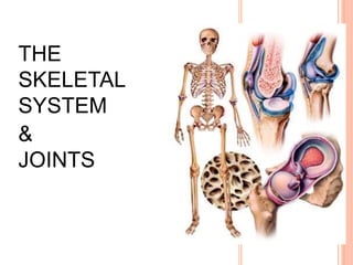
- 155. With the exception of the hyoid bone, every bone in the body is connected to or forms a joint. A joint is a point of contact between: Two or more bones Cartilage and bone Teeth and bone Functions of joints Hold bones together Allow for mobility Joint ( Articulation) The junction between two or more bones or cartilages.
- 156. Ways joints are classified Functionally Structurally Arthrology : study of joints
- 157. Structural Classification of Joints Fibrous Joint Sutures – dense fibrous CT Ex. Suture Syndesmoses – more dense fibrous CT than a suture Ex. Gomphosis Interosseous membranes – a broad sheet of dense fibrous CT Ex. Between radius and ulna Cartilaginous Synchondrosis – hyaline cartilage; no movement Ex. Epiphyseal plate Symphysis – fibrocartilage; some movement Ex. Pubic symphysis Synovial Joint Articular cartilage on ends of long bones and a synovial cavity between articulating bones surrounded by accessory ligaments; freely moveable Ex. Hip, knee, shoulder,elbow
- 158. FUNCTIONAL CLASSIFICATION OF JOINTS Synarthroses Allow no movement Ex. Suture, gomphosis, Syndesmosis Amphiarthroses Allow little movement Ex. Epiphyseal plate (synchondrosis), Pubic symphysis, intervertebral discs (symphysis) Diarthroses Freely moveable Ex. Hip, knee, shoulder, elbow
- 162. What is a Ligament? Group of dense connective tissues connects bone to bone. What is a Tendon? Group of dense connective tissues or ends of muscles which connects muscle to bone.
- 163. Fibrous Joints Lack a synovial cavity. Articulating bones are held together with dense fibrous connective tissue. Permit little or no movement. Types: Sutures Gomphosis Syndesmoses
- 164. EXAMPLES OF FIBROUS JOINTS
- 165. EXAMPLES OF FIBROUS JOINTS
- 166. Lack a synovial cavity. Articulating bones are held together with cartilage connective tissue. Permit little movement. Types: Synchondroses (Primary cartilaginous joint) Symphyses (Secondary cartilaginous joint) CARTILAGINOUS JOINTS Copyright © 2014 John Wiley & Sons, Inc. All rights reserved.
- 167. EXAMPLES OF CARTILAGINOUS JOINTS
- 168. Have a synovial cavity Articulating bones are covered with articular cartilage, held together by ligaments, contain synovial fluid, have a nerve and blood supply, and are surrounded by an articular capsule Permit a large range of movement SYNOVIAL JOINTS
- 169. GENERAL STRUCTURE - Articular cartilage Articular ligaments (Capsule) Synovial membrane Joint (synovial) cavity Synovial fluid Reinforcing ligaments Articular discs Menisci Bursae and Tendon Sheaths
- 170. STRUCTURE OF A SYNOVIAL JOINT
- 171. ARTICULAR CARTILAGE Hyaline cartilage covering the bone surfaces 171
- 172. CAPSULE Fibrous capsule lined by synovial membrane Continuous with periosteum 172
- 173. SYNOVIAL CAVITY Joint cavity is synovial cavity Surrounded by synovial membrane 173
- 174. SYNOVIAL MEMBRANE Lines the cavity of joint except over the articular cartilage. 174
- 175. SYNOVIAL FLUID Viscous slippery fluid rich in albumin & hyaluronic acid & similar to raw egg white 175
- 176. ARTICULAR DISC Circular rim of fibrous cartilage between articular surfaces of two bones 176
- 177. MENISCUS Meniscus is an incomplete rim of white fibrous cartilage between articular cartilages. Shock absorber Enhancement of congruence Protection of edges Weight distribution Facilitation of movement 177
- 178. BURSA Lubricating device consist of a closed fibrous sac. Present wherever tendon rub against bones,ligaments or other tendons 178
- 179. Bursae and tendon sheaths can be found at many synovial joints. Bursae – flattened, fibrous sacs lined with synovial membranes and containing synovial fluid Tendon sheath – elongated (tubelike) bursa that wraps completely around tendons subject to a great deal of friction BURSAE AND TENDON SHEATHS Copyright © 2014 John Wiley & Sons, Inc. All rights reserved.
- 180. Synovial Joints: Range of Motion 1. Nonaxial – Gliding movements only 2. Uniaxial – Movement in one plane 3. Biaxial – Movement in two planes 4. Multiaxial – Movement in or around all three planes
- 181. Types of Movement at Synovial Joints
- 192. Types of Synovial Joints