
Recommended
More Related Content
What's hot
What's hot (20)
Similar to IVP by Dr.Anil.ppt
Similar to IVP by Dr.Anil.ppt (20)
Recently uploaded
Recently uploaded (20)
IVP by Dr.Anil.ppt
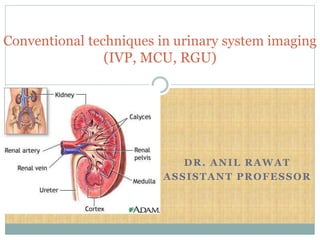
- 1. DR. ANIL RAWAT ASSISTANT PROFESSOR Conventional techniques in urinary system imaging (IVP, MCU, RGU)
- 2. History In 1896, for first time, X-ray demonstration of renal calculi in a patient was done. Techniques of cystography, retrograde pyelography and retrograde urethrography were described within 15 years after that. With introduction of RGP, renal PCS and ureters were made available for X-ray study. In 1929, Moses Swick invented uroselectan (5-iodo-2- pyridone-N-acetic acid ) which led to revolutionary advent of IVP.
- 3. Progressive improvement in the chemistry of contrast media made IVP an excellent method for studying PCS and renal parenchyma. Subsequent development of better and safe contrast media like iohexol, iopamidol etc. brought a current era of lower osmolality radio-opaque contrast media. Currently, the use of USG, CT, MRI and radionuclide scanning has superseded older conventional techniques at many fronts in the form of high sensitivity, specificity and better safety margins.
- 4. EXCRETORY UROGRAPHY Excretory urography refers to visualization of the kidney parenchyma, calyces and pelvis after iv. Injection of contrast. The excretory Urogram is the classic routine investigation of Uroradiology. Technically satisfactory IVU demonstrates clearly and completely both the renal parenchyma & the collecting system including the calyces, renal pelvis, ureters and the urinary bladder and gives an indication of their function.
- 5. INDICATIONS: Hematuria Renal colic Renal trauma Persistent pyuria Prior to percutaneous urological procedures to define renal anatomy Prior to surgery involving risk of significant ureteric injury After surgery e.g. Ureteric surgery Ureteric strictures or fistulas Complex urinary tract infection (including tuberculosis) Work up of live donor in renal transplant To screen for renal anomalies in patient with multiple congenital anomalies.
- 6. CONTRAINDICATIONS: No absolute contraindication Relative contraindications • Previous reaction to contrast media • Asthma • Renal & hepatic failure • Multiple myeloma • Pregnancy • H/o allergy to other drugs • Thyroid disease • Diabetes • Sickle cell disease
- 7. PATIENT PREPARATION Dehydration : Overhydration should be avoided but dehydration is unecessary. It is a potentiating factor for contrast induced nephrotoxicity. Bowel preparation 2 tab charcol or gasex TDS X 2days prior to examination & 25 ml Castor oil or 2 tab Dulcolax at night X 2 days prior Ambulatory for 2 hrs prior to examination to reduce bowel gas A group of children in which we never perform bowel preparation are those with myelomeningocele. • Fasting on the morning of the study
- 8. EXPOSURE FACTORS Kvp 65-75 mA Sufficiently high to allow short exposure times(mA-600-1000 mA or 60 MAS), Exposure time < 0.1 sec (approx 80 msec in adults, < 50 msec in children) Film/Screen combination Medium speed All exposures are to be made in suspended expiration to minimize geometric distortion of renal image because deep inspiration causes descent and ventral rotation of lower poles of the kidneys
- 9. History taking Clinical history of patient Co-morbid illnesses eg. diabetes, hypertension, cardiac disease, thyroid disease and other risk factors Renal function (S. creatinine) LMP in females Drugs Written informed consent before contrast administration
- 10. Technique KUB radiograph, an indispensable part of sequence. Appropriate technique (65–75 kVp, high mA, short exposure time) to maximize inherent soft-tissue contrast and optimize visualization of calcium-containing lesions that are potentially of urinary tract origin. Proper KUB may require additional images for evaluating portions of urinary tract not seen on standard 14* 17-inch image (area from suprarenal region to a level below symphysis pubis). Patient should void immediately prior to examination.
- 11. PROCEDURE Plain film of the abdomen (Scout film) Area to be included from the suprarenal region to the level below the symphysis pubis. The patient should void immediately prior to undergoing this examination. Center the rays at the level of the iliac crest in the midsagittal plane. Gonadal shields should be used routinely throughout the examination.
- 15. Things to look for in plain abdominal radiograph: 1. Calculus/ calcifications: Assessment of probable location of calcifications in abdomen with respect to urinary tract should be made prior to contrast injection, which can obscure a calcification. Oblique radiographs helpful. 2. Skeletal abnormalities: spinal deformity, metabolic bone changes, skeletal metastasis. 3.Intestinal gas pattern: Ileus, obstruction. 4.Abdominal masses: May be seen better on scout view. 5.Foreign bodies: Opaque foreign bodies or barium.
- 17. HOCM-high osmolality Diatrizoate -urografin Iothalamate -conray Iodamide -uromiro Metrizoate -triosil/isopaque
- 18. LOCM –low osmolality Iohexol -omnipaque Iopromide –ultravist Iopamidol –niopam Ioversol -optiray
- 19. Indications for LOCM Those at high risk of anaphylactoid reaction: Previous reactors Asthamatics Atopics allergy to other drugs
- 20. Those unable to tolerate a high osmotic load Poor cardiac reserve Infants and elderly patients Sickle cell disease or trait Pre-existing renal impairment
- 21. Contrast administration Bolus infusion of 350-370 strength water-soluble contrast is done with 18 gauge needle into antecubital vein. adult dose 50 ml elderly and obese patient 75 ml small children 1.5 ml/kg neonate 4ml/kg
- 22. Although modern contrast medium is exceptionally safe, there is a small risk of serious reactions. Most dangerous of these are anaphylactoid -type hypersensitivity reactions. To minimise the risks of these a routine inquiry about previous contrast exposure and allergy is recommended. Injection should be through some form of indwelling cannula or needle that can he taped into place for duration of investigation. Most adverse events are likely to take place within first few minutes after injection. Emergency drugs, oxygen and resuscitation equipment should also be readily available.
- 23. Filming sequence 1 min –nephrogram (often omitted as renal outline are adequately visualised on 5 min radiograph) 5 min- collecting system Abdominal compression at 5 min 10 min - proximal ureters 15 min ‘release’ film- entire urinary tract Full bladder film Post micturation film
- 25. In our department Scout film 5 min film with compression for PCS 15min film with release of compression for ureter 20 min film in prone position (if lower ureter not visualized in 15 min film) full bladder Empty bladder All films are taken in suspended respiration at end- expiration.
- 26. 1 min film (Nephrogram) Nephrograms are produced primarily by filtered contrast material within the nephron, with optimal visualization of the renal parenchyma 1–3 minutes after bolus injection. Features to be seen here are: Size Contour position Axis of kidney
- 27. 5 min film Focused to the renal area (to assess temporal symmetry and progress of opacification).
- 28. Abdominal compression Applied after 5 min film with compression pad placed between ASISs (ureters are compressed against the sacrum as the ureter traverses the sacral ala) Produces partial ureteric obstruction Distension of calyceal system Improved calyceal detail Reliable ureteric opapification on release
- 29. Value of compression. (a) On a radiograph obtained 5 minutes after administration of low- osmolar urographic contrast material, the collecting system is bilaterally underfilled and poorly demonstrated. (b) On a radiograph obtained 5 minutes after compression was applied, distention of the collecting system is significantly improved (arrows).
- 30. Contraindications to compression Urinary tract obstruction Severe abdominal pain e.g.. Ureteric colic Abdominal mass or abdominal aortic aneurysm Recent trauma or abdominal surgery IVC filter Presence of urinary diversion e.g.. nephrostomy Renal transplant Severe hypertension
- 31. 15 min film Ureter -bladder images Full film is taken immediately after release of compression to visualise ureter and filling bladder
- 32. Bladder Films Central ray is angulated by 15 degrees caudally and centered in midline to a point 5 cm above pubic symphysis or 2.5 cm below ASIS. Exposure is made on suspended expiration.
- 33. Post micturation film Residual urine Diverticula Bladder hernia Bladder tumor Obstructive urethral lesion Distal ureteral process e.g. Ureterocele Primary mega ureter
- 34. Additional films Oblique radiographs Prone radiograph Erect radiograph Full length post- micturation radiograph Fluoroscopy
- 35. Patient Aftercare General psychological reassurance. Needle wound site dressed and checked for extravasation. Tell to patient how to receive the report of examination Ensure patient has understood preparation instructions are finished
- 36. Examination Effective dose (mSv) Equivalent no. of chest x-rays Limbs and joints (except hip) < 0.01 <0.5 Chest PA 0.02 1 Skull 0.06 3 Thoracic spine 0.7 35 Lumbar spine 1 50 Hip 0.4 20 Abdomen or Pelvis 0.7 35 IVU 2.4 120 Ba swallow 1.5 75 Ba meal 2.6 130 BMFT 3 150 Ba enema 7.2 360 CT head 2.0 100 CT chest 8.8 400 CT abdomen or pelvis 10 500
- 37. Micturiting Cystourethrogram/MCU/VCU Commonly used examination for UB and urethral evaluation in both children and adult. Preliminary abdominal imaging precedes catheterization. If an abdominal radiograph obtained within past 3– 6 months , a scout image may be unnecessary. Abdominal radiograph may reveal bone abnormalities, calcifications, foreign bodies, or other disease processes.
- 38. Indications for VCU in children Main is UTI, esp. in girls <5 yrs age. In neonate, MC indication is hydronephrosis detected on prenatal USG. To look for VUR (30 – 50% children with UTI have VUR) For congenital lower urinary tract anomalies.
- 39. Indications for VCU in adults Trauma UTI To document reflux nephropathy Evaluation and follow up of in spinal cord injury or voiding difficulties Following renal transplantation to see for bladder capacity, VUR or BOO. In females to look for urethral diverticula.
- 40. For catheterization, a 5-F feeding tube is appropriate in children under 3 months of age and an 8-F feeding tube in all other children. In female child, after cleansing intralabial region with povidone iodine solution, a small drop of solution remains pooled in midline, depressed, slightly oval meatus, which thus becomes easily visible. It helps prevent vaginal catheterization. In boys, external sphincter is MC site of resistance to catheter advancement. Gentle, steady pressure rather than intermittent poking at this level permits advancement into the bladder.
- 41. In older boys, retrograde injection of 2% lidocaine jelly into urethra several minutes before catheterization may be used to diminish sensation. Once catheter has safely reached UB, filling with diluted contrast material can begin. During early filling, minimally filled bladder is imaged in AP projection. A ureterocele or bladder tumor well seen during early filling may become obscured as more contrast material enters bladder.
- 42. Older children may indicate when voiding is imminent. In younger children, as bladder capacity is reached, flow of contrast material may slow, stop, or even reverse in tubing, indicating an abrupt rise in intravesical pressure and complete bladder filling. At this stage steep oblique images of bladder centered on UVJ should be obtained. Bladder Capacity = (Age [Years] + 2) * 30 Volume of contrast instilled should be recorded.
- 43. URINARY BLADDER CAPACITY (in ml) ACCORDING TO AGE : For <2 years- weight(kg)*7 For>2 to 12 years- ( age(years)+2)*30 For adults around 500ml
- 44. Male Urethra can also be divided into: Anterior Urethra- Penile urethra Bulbar urethra Posterior Urethra- Membranous urethra Prostatic urethra
- 45. VUR can be seen on oblique radiographs obtained just before voiding and can be graded after voiding. If reflux is observed during late bladder filling, ipsilateral renal fossa may be imaged in AP projection prior to voiding. Catheter may be removed as voiding is initiated, but voiding around it is recommended as it allows desired cyclic voiding in neonates, repeat filling if needed, and bladder drainage when unable to empty bladder completely.
- 46. Approximately 20% of reflux will be missed if voiding does not occur. Urethral disease is very rare in girls, and one AP image of urethra is usually sufficient. Voiding while supine, especially with legs in close apposition, can produce vaginal reflux. In boys, entire urethra must be imaged in steep oblique position because disease can occur anywhere from the bladder base to the urethral meatus. After voiding, each renal fossa should be imaged.
- 47. Causes of VUR: Anatomical causes – Posterior urethral valves(PUV’s)-most common anatomical cause Prostatomegaly Ureteroceles Ureteral duplication Neurofunctional causes- Neurogenic bladder Dysfunctional voiding Uninhibited bladder contractions-most common urodynamic abnormality associated with reflux
- 48. GRADES OF VUR GRADE 1-reflux limited to ureter GRADE2-reflux upto the renal pelvis GRADE 3-mild dilatation of ureter and pelvicalyceal system GRADE4-tortuous ureter with moderate dilatation blunting of fornices with preserved papillary impressions GRADE5-tortuous ureter with severe dilatation of ureter and pelvicalyceal system loss of fornices and papillary impressions
- 49. GRADE 1-reflux limited to ureter
- 50. GRADE 2-reflux upto the renal pelvis
- 51. GRADE 3-mild dilatation of ureter and pelvicalyceal system
- 52. GRADE 4-tortuous ureter with moderate dilatation blunting of fornices with preserved papillary impressions
- 53. GRADE 5-tortuous ureter with severe dilatation of ureter and pelvicalyceal system loss of fornices and papillary impressions
- 54. Points to be assessed and reported by radiologist at end of examinations: 1. status of spine and pelvis 2. presence of masses or opaque calculi 3. bladder capacity and contour and emptying capability 4. presence and grade of reflux and obstruction of a refluxing segment 5. insertion site of a refluxing ureter 6. appearance of entire urethra
- 55. Retrograde urethrography (RGU) Best initial study for urethral and periurethral imaging in men, indicated in evaluation of urethral injuries, strictures, and fistulas. Not a physiological examination. External meatus prepared in standard sterile fashion for placement of a conventional 16- or 18-F Foley catheter. Catheter, with both irrigating syringe and inflating (saline solution) syringe attached, should be flushed before use. When balloon portion of catheter is seated in fossa navicularis of penile urethra, balloon is inflated with 1.0–1.5 mL of saline solution.
- 56. Lubrication not recommended, may prevent balloon from remaining in place for optimal occlusion. Patient placed in a supine 45° oblique position, penis placed laterally over proximal thigh with moderate traction. 20–30 mL of 60% iodinated contrast material is injected so that anterior urethra is filled. Commonly, spasm of external urethral sphincter will be encountered preventing filling of deep bulbar, membranous, and prostatic urethras. Slow, gentle pressure is usually needed to overcome this resistance.
- 57. Contrast material can seen to jet through bladder neck into bladder. Verumontanum seen as ovoid filling defect in posterior part of prostatic urethra. Distal end of it marks proximal boundary of membranous urethra Identification of bulbomembranous junction (1–1.5 cm distal to the inferior margin of verumontanum) on a RGU is important for assessing patients with urethral disease as well as for planning urologic procedures. With poorly opacified posterior urethra , bulbomembranous junction can be localized by an imaginary line connecting inferior margins of obturator foramina intersects urethra.
- 58. Filming: Under fluoroscopic control, contrast medium is injected and spot films are taken in the following positions- 30-degree right anterior oblique with left leg abducted and knee flexed Supine AP 30-degree left anterior oblique with right leg abducted and knee flexed
- 60. Anterior urethra extends from its origin at end of membranous urethra to urethral meatus, divided into bulbar segment and penile segment. There is usually mild angulation of the urethra where these two segments join at penoscrotal junction. Contraction of constrictor nudae muscle, a deep musculotendinous sling of bulbocavernous muscle, may cause anterior or circumferential indentation of proximal bulbous urethra at RGU (should not be confused with urethral stricture) If the membranous urethra can be identified, it should not be confused with a stricture.
- 61. Narrowing elsewhere in urethra will be clearly defined as separate from membranous urethra and, therefore, representative of a pathologic stricture. If the patient is not positioned sufficiently oblique, the bulbous urethra will appear foreshortened and will therefore not be adequately evaluated. Filling of Cowper ducts should not be misinterpreted as extravasation. If integrity of urethral mucosal lining is disrupted by increased pressure during contrast material injection, intravasation of contrast material with opacification of corpora and draining veins may occur.
- 62. Extravasation: Intramural extravasation can occur in bladder esp. during voiding, but is self limited and requires no treatment. Inadvertent catheterization of vagina or ureter: If vagina is catheterized, tubular shape with reflux into uterus , fallopian tubes or peritoneal cavity may occur. Catheter can also enter a ureter with an ectopic ending, of a double collecting system or one with a very dilated orifice. Radiation effect Autonomic dysreflexia
- 63. Complication of MCU/RGU Infections: Bacteria may be introduced via the catheter and high fever can develop after cystography. Trauma: Dysuria, urinary retention and penetration of bladder wall may develop following catheterization. Reaction to contrast medium: Very rare, when contrast can get absorbed through bladder wall or from the ureteral or pyelocalyceal epithelium or intravasation during RGU. In children with ventriculoperitoneal shunt, nonionic contrast is to be used. Knotting of catheter within bladder
- 64. Urethral Trauma Urethral injuries are divided into anterior and posterior urethral injuries . Causes: Blunt Trauma-due to shearing/straddle injuries posterior urethral injury is caused by a crushing force to the pelvis and is associated with pelvic fractures and bladder injury . anterior urethral injury is usually caused by a straddle injury and is an isolated injury Penetrating trauma-due to stab wounds,gunshot wounds more commonly affects anterior urethra Iatrogenic-Catheterization,Cystoscopy Post surgical(surgery for Benign Prostatic Hyperplasia)
- 65. Retrograde Urethrography is the modality of choice to investigate the anterior part of urethra .It will demonstrate extraluminal contrast which has extravasated from the urethra at the site of injury. VCUG is the most appropriate way to evaluate the posterior part of male urethra and injuries to female urethra.
- 66. Goldman system for Classification of urethral injuries: Type 1- Stretching of prostatic urethra due to disruption of puboprostatic ligaments but the urethra is intact. Urethrographic appearance shows intact but stretched urethra
- 67. Type 2- Posterior urethral injury above urogenital diaphragm while the membranous segment remains intact . Urethrographic appearance shows contrast material extravasation above urogenital diaphragm only.
- 68. Type 3-(most common) Injury to membranous urethra extending into proximal bulbous urethra(with laceration of urogenital diaphragm). Urethrographic appearance shows contrast material extravasation below the urogenital diaphragm,possibly extending into pelvis or peritoneum with intact bladder neck.
- 69. Type 4- Bladder base injury involving bladder neck extending into proximal urethra.Internal Sphincter is injured hence incontinence can occur. Extraperitoneal contrast agent extravasation,bladder neck disruption
- 70. Type 4a- Bladder base injury but not involving bladder neck . Cannot be differentiated from Type 4 radiologically. Periurethral contrast material extravasation ,bladder base disruption.
- 71. Type 5- Anterior urethral injury (isolated). Contrast material extravasation below urogenital diaphragm in and around penile soft tissue.
- 72. MCU VS RGU MCU is performed to demonstrate posterior urethral abnormalities ,in addition it shows bladder pathologies and VUR also. RGU is performed to demonstrate anterior urethral abnormalities. In cases of Trauma, RGU is performed before MCU to avoid more injury while introducing Foley catheter blindly into bladder.
- 73. THANK YOU