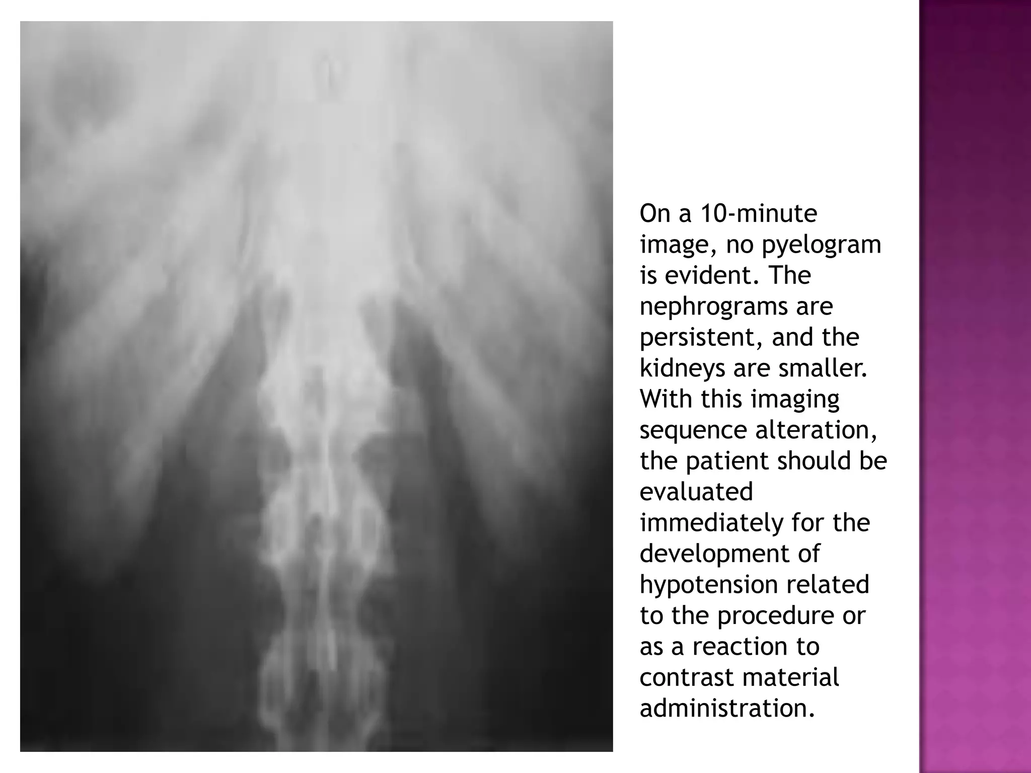
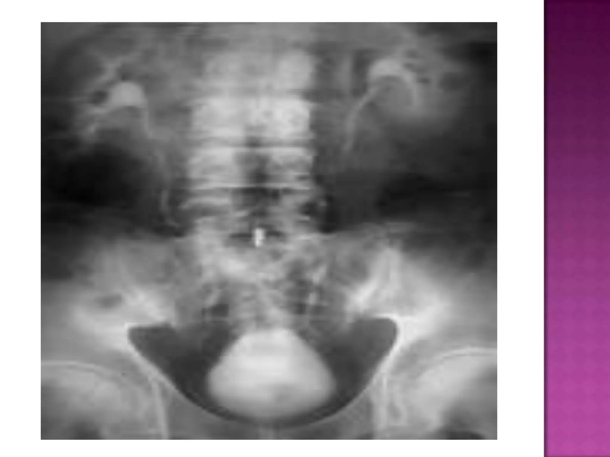
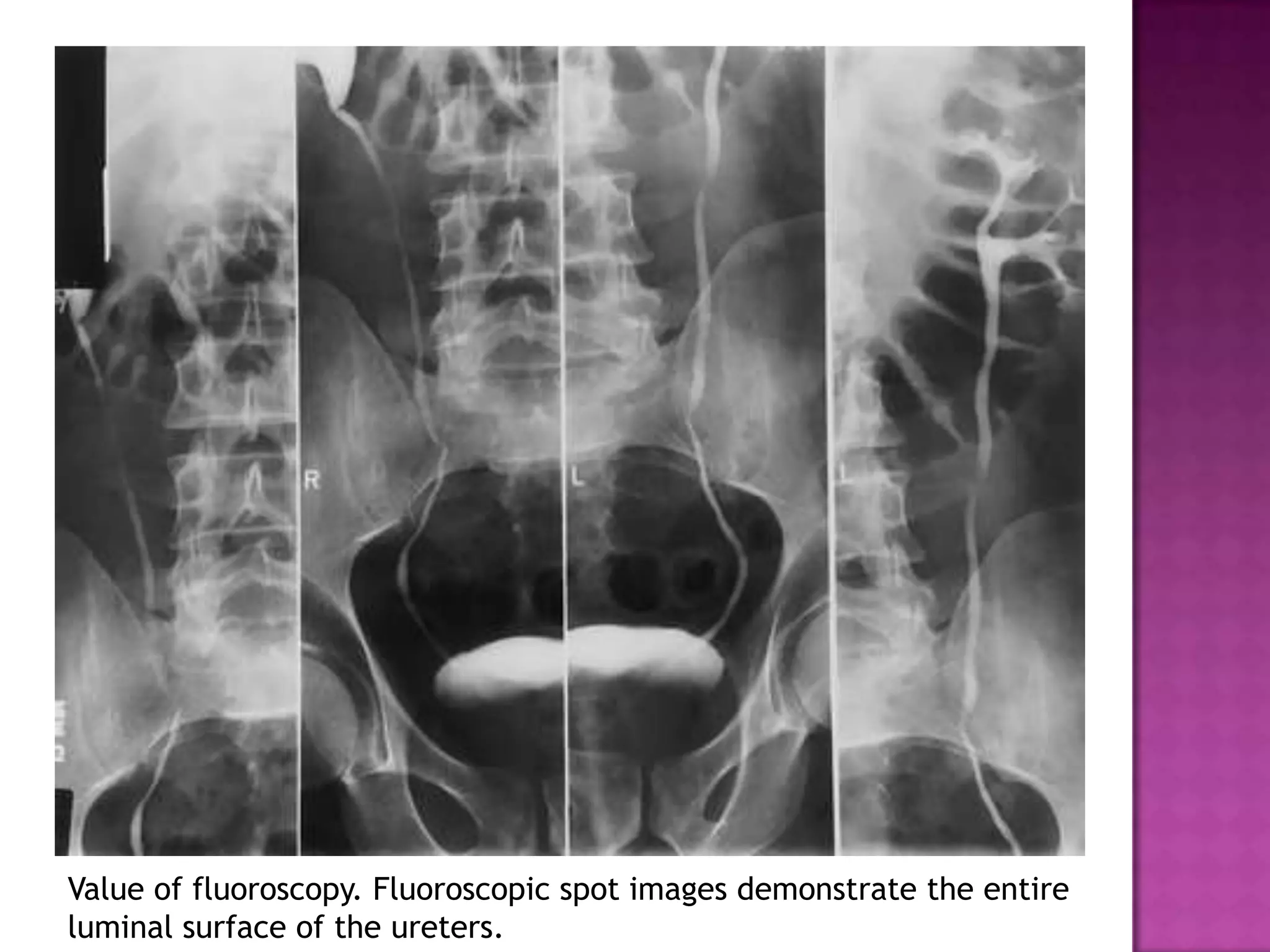
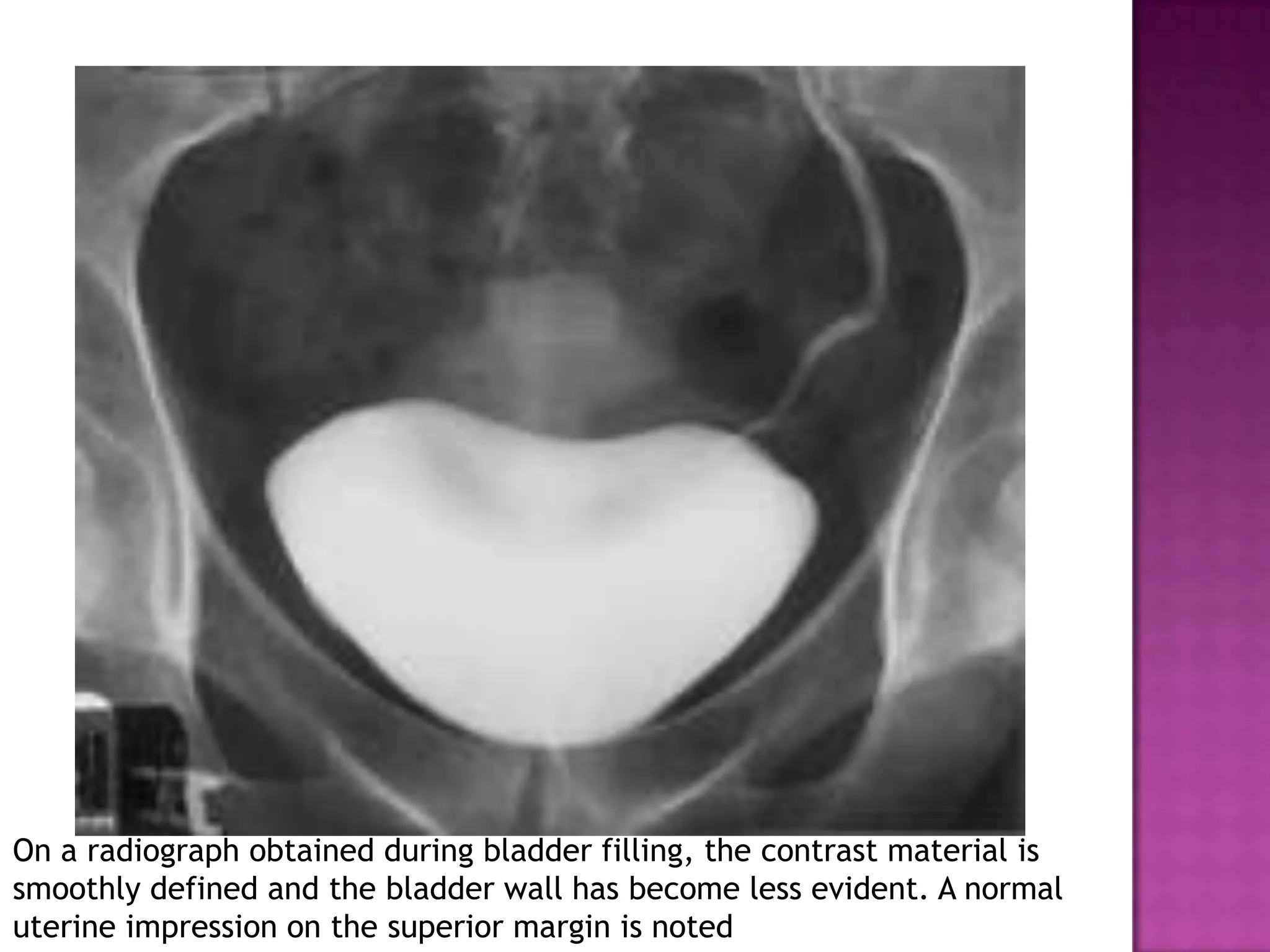
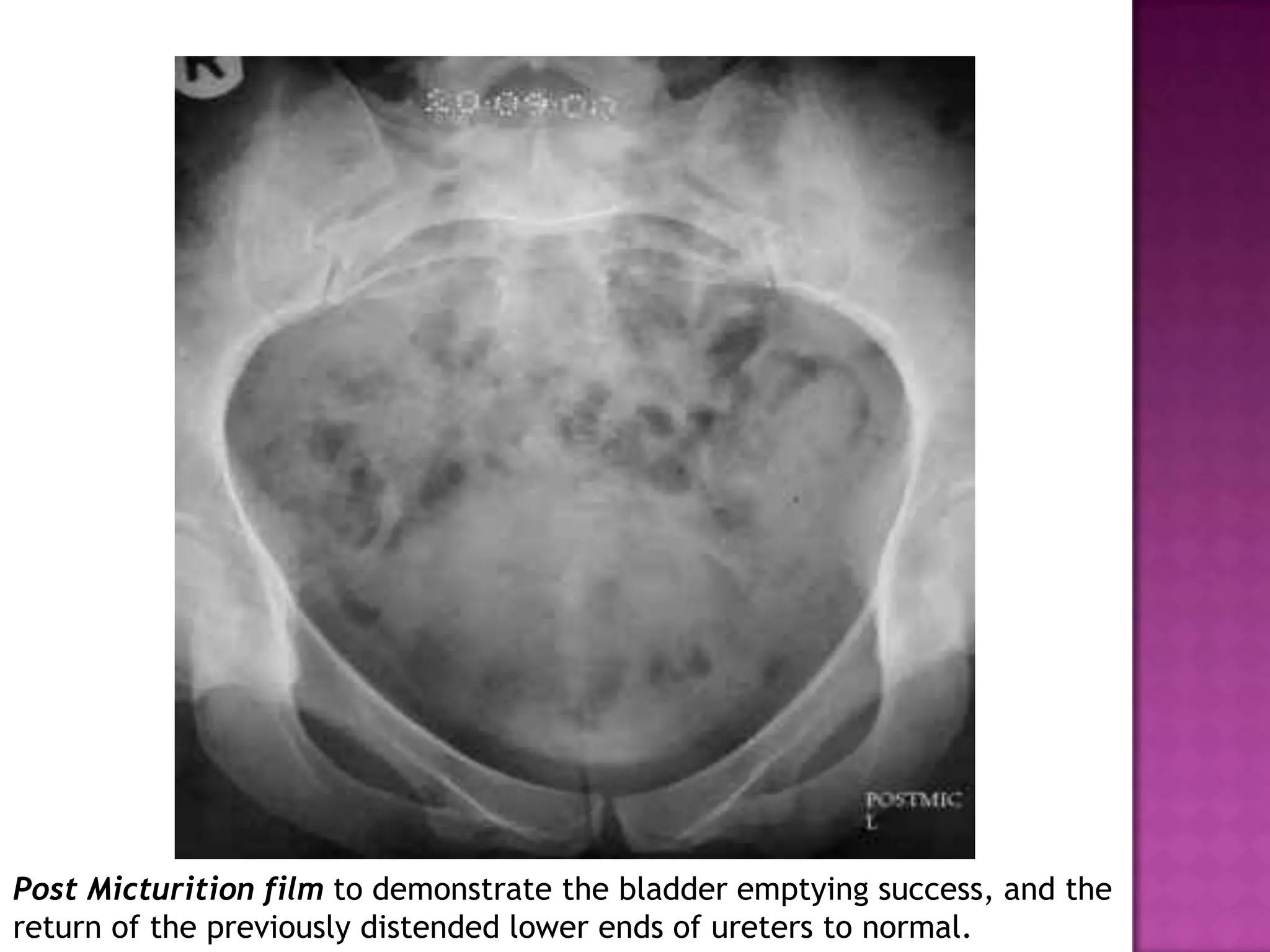
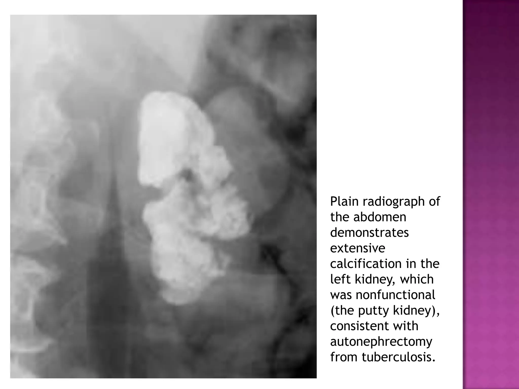
FATIMA SHAH
Structure and function of urinary system.
Ascending Urography
-
Retrograde/Cystoscopic
Descending Urography
-
Excretory/Intravenous
Timed series of radiographic
images of urinary system after
administration of IV contrast.
Suspected urinary tract pathology.
Repeated infections -focus, damage
Heamaturia
Investigation of hypertension not
controlled by medication in young
adults.
Renal colic.
Trauma.
 General contra indications to contrast
agents.
Diabetes,
Thyrotoxicosis,
Pregnancy
Raised urea creat. urography unlikely to
be successful.
Metformin therapy
Bowel prep.
Basic psychological preparation with
reassurance & explanation of technique
Bladder emptied immediately before exam.
 Hx of Previous I.V.U.
Previous experience of iodinated
contrast media.
Abdominal surgery, Allergies, drugs Hx.
Ionic or HOCM eg urograffin used.
Iodine is main element which imparts
radio-opacity.
300mg I/kg body wt. 15-25 gm of iodine
given. 20ml of 76% urograffin
Greatest single predictor of contrast
reaction is previous reaction to contrast
 MILD & TRANSIENT – NO Rx REQUIRED:
-nausea,vomiting,sensation of heat, tingling,
metallic taste,pain in arm,deire to urinate.
 ANTI-HISTAMINE & STEROID Rx:
-Skin rashes;urticaria ,diffuse erythema.
Angioneurotic edema,pruritis,sneezing&rhinorrhea
 ADRENALINE,AMINOPHYLLINE or SALBUTAMOL,O2
& STEROIDS Rx:
-Broncho spasm and layryngeal edema particularly
due to meglumine.
Scout film
 Administration of contrast
 Early nephrogram
 Tomograms
 Excretory films
Compression banding:
Aim to produce better PC distension
C/I: Recent abd surgery.
Renal trauma.
Large abd mass.
Obstruction.
If 5min film shows adequate
distension.
End of Injection, A.P. of the renal areas to show the nephrogram, i.e. the
renal parenchyma opacified by the contrast medium in the renal tubules.
Value of fluoroscopy. Fluoroscopic spot images demonstrate the entire
luminal surface of the ureters.
On a radiograph obtained during bladder filling, the contrast material is
smoothly defined and the bladder wall has become less evident. A normal
uterine impression on the superior margin is noted
Post Micturition film to demonstrate the bladder emptying success, and the
return of the previously distended lower ends of ureters to normal.
Area: supra-renal - below symphysis.
Assessment of Bones, stones, masses &
gases.
Oblique view helpful when pt symptomatic
but no cause seen on KUB.
Urethral calculus in pt with hx of severe right flank pain.Collimated Radiograph shows
calcification centered behind symphysis . CT helped confirm presence of urethral
calculus.This case shows importance of full coverage of anatomic structures at KUB.
(a) Collimated preliminary radiograph of the pelvis shows no obvious stones
Rt post oblique radiograph of pelvis shows 6mm ureteral calculus now projected onto
iliac bone.urogram (not shown)helped confirm rt ureteral obstruction 2ndry to the
stone. This case shows how a calculus can be obscured by the complex sacral anatomy.
The plain KUB
shows lumbar
spondylosis
with marked
scoliosis and
obvious
asymmetrical
thickness of
the lateral
abdominal wall
musculatures.
AP radiograph of pelvis shows parasymphyseal bone fragment along left
pubis ,mild p. symphysis diastasis, &transverse fracture of rt transverse
process .irregularity of rt SI joint space, suggestive of fracture.
Preliminary
image from
excretory
urography
demonstrates a
looped
configuration of
the distal
transverse colon
and splenic
flexure
(arrows).
50yrs female,
known diabetic
presented with
lethargy, fever
with chills and
rigors. Urine
examination
shows multiple
pus cells and
ESR is
elevatedabnor
mal gas in the
left renal
region
Another patient
aged 45yr with
similar history
and marked
suprapubic
tendernessKUB
radiograph
showing air with
in the bladder
KIDNEYS: visualised if peri-renal fat.
GUT GASES: may over lap.
Change in shape & location
displaced by compression.
CALCIFICATION OVER RENAL AREA:
-True lat/ips-ilateral post oblique views.
-Displacement with ins/exp/upright films.
EXTRA-RENAL CALCIFICATION:
-Calcified Costal Cartilage, supra-RG.
-Calcification in tail of pancreas, GB,
liver, Splenic artery.
-Phleboliths, chip fracture of TP.
On a scout image
obtained before
excretory urography,
a calculus fills nearly
the entirety of a
bifid right renal
collecting system,
giving it a branched
appearance that
resembles the
antlers of a stag.
Plain radiograph of
the abdomen
demonstrates
extensive
calcification in the
left kidney, which
was nonfunctional
(the putty kidney),
consistent with
autonephrectomy
from tuberculosis.
URETERS: not visualized.
OPACITIES:
-Intra-Luminal: ureteral stones.
-Intra-mural: schistosomiasis.
GAS SHADOWS: conform to shape of ureter.
EXTRA-URETERAL CALCIFICATION:
-mesenteric LN(mobile)
-phleboliths, calcification in arteries.
U.BLADDER & URETHRA:
- not visualised.
- visualised if calculi or foreign body.
Contrast in glomeruli & tubules.(1-3min)
Four phases:
SPONTANEOUS: Non-opacified, outlined by
RP fat on plain film.
VASCULAR: Opacification of intra-renal
blood vessels.
TOTAL BODY: “ of pre and retro renal soft
tissue + vascular nephrogram
INTRA-TUBULAR:” of intra-renal tubules.
U=([P]xGFR)/Uvol
a-extravasation of contrast
b-absent blood flow :
i-shock
ii-no glomerular filteration :
acute obstruction,infarction
Non- Visualisation:
- Insufficient dose of contrast.
- Acute, chronic renal disease
- End stage renal disease.
- Acute tubular obstruction
- Ureteral calculus.
- Renal agenesis
- Renal artery thrombosis,avulsion.
Size - Normal range-height of three vertebra.
Enlarged kidneys suggest
-polycystic disease
-acute pyelo or glomerulonephritis
Small kidneys imply chronic disease.
Shape –Cysts & tumors may cause distortion.
Orientation - disorientation may be
-intrinsic, e.g. horseshoe kidney, or
-extrinsic, i.e. pressure effect of other organs
image of a
tomographic
sequence
demonstrates
symmetric
nephrograms and
pyelograms. Renal
size is normal.
On a 10-minute
image, no pyelogram
is evident. The
nephrograms are
persistent, and the
kidneys are smaller.
With this imaging
sequence alteration,
the patient should be
evaluated
immediately for the
development of
hypotension related
to the procedure or
as a reaction to
contrast material
administration.
One-minute image
shows slight
asymmetry of the
nephrographic
opacity, with less
opacity in the right
kidney than in the
left
Image obtained
at 80 minutes
shows a
persistent, very
dense right
nephrogram, a
typical finding
in acute high-
grade
obstruction. A
2-mm stone
was discovered
at the right
ureterovesical
junction.
Enlarged kidneys in a
young patient with
early, asymmetric
findings of autosomal
dominant polycystic
kidney disease.
Nephrotomogram
shows enlarged
kidneys, the left
more so than the
right. Note the
multiple parenchymal
defects (“Swiss
cheese”
nephrogram).
Right renal
artery
stenosis for
evaluation of
renovascular
hypertension
shows a small
right kidney
with
decreased
nephrographic
density and
temporal
asymmetry of
filling of the
right
collecting
system
compared
with the left
Fifteen-minute
urographic
image helps
confirm the
asymmetric
renal size. Note
the
hyperconcentra
tion of contrast
material in the
right collecting
system
compared with
the left.
Opacification of pelvicalceal system &bladder
Filling defects include:
stone,tcc,blood clots,papillary necrosis
with sloughing of infarcted papilla.
Strictures due to :
-Post inflammation,previous stone impaction
-Post infection,TB, Shistosomiasis
-Cancer, intrinsic-tcc
extrinsic-cevical ca
Tomogram
from excretory
urography
demonstrates
absence of the
left kidney
and deviation
of the
descending
colon into the
renal fossa.
Common findings in bladder:
-Filling defects , tumors
-Trabeculated , thick walled bladder.
different patterns of excavation that can be seen with papillary necrosis:
normal (A), central excavation with ball-on-tee appearance (B), forniceal
excavation (C), lobster claw appearance (D), signet ring appearance (E), and
sloughed papilla with clubbed calix (F).
Papillary
necrosis. On an
excretory
urogram,
contrast material
fills central
excavations
(arrows) in the
papilla of the
interpolar region,
Note the
abnormal calices
in the upper and
lower poles as
well.
Papillary
necrosis.
Excavation
extending from
the caliceal
fornices (arrows)
produces the
lobster claw
deformity in
another patient.
Delayed tomographic
image from excretory
urography shows
caliceal crescents
(arrowheads)
surrounding the
dilated collecting
system. Contrast
material pools
dependently
Excretory urography
Excretory urography
Excretory urography

Excretory urography

  • 1.
  • 2.
    Structure and functionof urinary system. Ascending Urography - Retrograde/Cystoscopic Descending Urography - Excretory/Intravenous
  • 3.
    Timed series ofradiographic images of urinary system after administration of IV contrast.
  • 4.
    Suspected urinary tractpathology. Repeated infections -focus, damage Heamaturia Investigation of hypertension not controlled by medication in young adults. Renal colic. Trauma.
  • 5.
     General contraindications to contrast agents. Diabetes, Thyrotoxicosis, Pregnancy Raised urea creat. urography unlikely to be successful. Metformin therapy
  • 6.
    Bowel prep. Basic psychologicalpreparation with reassurance & explanation of technique Bladder emptied immediately before exam.  Hx of Previous I.V.U. Previous experience of iodinated contrast media. Abdominal surgery, Allergies, drugs Hx.
  • 7.
    Ionic or HOCMeg urograffin used. Iodine is main element which imparts radio-opacity. 300mg I/kg body wt. 15-25 gm of iodine given. 20ml of 76% urograffin Greatest single predictor of contrast reaction is previous reaction to contrast
  • 8.
     MILD &TRANSIENT – NO Rx REQUIRED: -nausea,vomiting,sensation of heat, tingling, metallic taste,pain in arm,deire to urinate.  ANTI-HISTAMINE & STEROID Rx: -Skin rashes;urticaria ,diffuse erythema. Angioneurotic edema,pruritis,sneezing&rhinorrhea  ADRENALINE,AMINOPHYLLINE or SALBUTAMOL,O2 & STEROIDS Rx: -Broncho spasm and layryngeal edema particularly due to meglumine.
  • 10.
    Scout film  Administrationof contrast  Early nephrogram  Tomograms  Excretory films
  • 12.
    Compression banding: Aim toproduce better PC distension C/I: Recent abd surgery. Renal trauma. Large abd mass. Obstruction. If 5min film shows adequate distension.
  • 15.
    End of Injection,A.P. of the renal areas to show the nephrogram, i.e. the renal parenchyma opacified by the contrast medium in the renal tubules.
  • 18.
    Value of fluoroscopy.Fluoroscopic spot images demonstrate the entire luminal surface of the ureters.
  • 19.
    On a radiographobtained during bladder filling, the contrast material is smoothly defined and the bladder wall has become less evident. A normal uterine impression on the superior margin is noted
  • 20.
    Post Micturition filmto demonstrate the bladder emptying success, and the return of the previously distended lower ends of ureters to normal.
  • 22.
    Area: supra-renal -below symphysis. Assessment of Bones, stones, masses & gases. Oblique view helpful when pt symptomatic but no cause seen on KUB.
  • 23.
    Urethral calculus inpt with hx of severe right flank pain.Collimated Radiograph shows calcification centered behind symphysis . CT helped confirm presence of urethral calculus.This case shows importance of full coverage of anatomic structures at KUB.
  • 24.
    (a) Collimated preliminaryradiograph of the pelvis shows no obvious stones
  • 25.
    Rt post obliqueradiograph of pelvis shows 6mm ureteral calculus now projected onto iliac bone.urogram (not shown)helped confirm rt ureteral obstruction 2ndry to the stone. This case shows how a calculus can be obscured by the complex sacral anatomy.
  • 27.
    The plain KUB showslumbar spondylosis with marked scoliosis and obvious asymmetrical thickness of the lateral abdominal wall musculatures.
  • 28.
    AP radiograph ofpelvis shows parasymphyseal bone fragment along left pubis ,mild p. symphysis diastasis, &transverse fracture of rt transverse process .irregularity of rt SI joint space, suggestive of fracture.
  • 29.
    Preliminary image from excretory urography demonstrates a looped configurationof the distal transverse colon and splenic flexure (arrows).
  • 30.
    50yrs female, known diabetic presentedwith lethargy, fever with chills and rigors. Urine examination shows multiple pus cells and ESR is elevatedabnor mal gas in the left renal region
  • 31.
    Another patient aged 45yrwith similar history and marked suprapubic tendernessKUB radiograph showing air with in the bladder
  • 32.
    KIDNEYS: visualised ifperi-renal fat. GUT GASES: may over lap. Change in shape & location displaced by compression. CALCIFICATION OVER RENAL AREA: -True lat/ips-ilateral post oblique views. -Displacement with ins/exp/upright films.
  • 33.
    EXTRA-RENAL CALCIFICATION: -Calcified CostalCartilage, supra-RG. -Calcification in tail of pancreas, GB, liver, Splenic artery. -Phleboliths, chip fracture of TP.
  • 35.
    On a scoutimage obtained before excretory urography, a calculus fills nearly the entirety of a bifid right renal collecting system, giving it a branched appearance that resembles the antlers of a stag.
  • 36.
    Plain radiograph of theabdomen demonstrates extensive calcification in the left kidney, which was nonfunctional (the putty kidney), consistent with autonephrectomy from tuberculosis.
  • 37.
    URETERS: not visualized. OPACITIES: -Intra-Luminal:ureteral stones. -Intra-mural: schistosomiasis. GAS SHADOWS: conform to shape of ureter. EXTRA-URETERAL CALCIFICATION: -mesenteric LN(mobile) -phleboliths, calcification in arteries.
  • 38.
    U.BLADDER & URETHRA: -not visualised. - visualised if calculi or foreign body.
  • 39.
    Contrast in glomeruli& tubules.(1-3min) Four phases: SPONTANEOUS: Non-opacified, outlined by RP fat on plain film. VASCULAR: Opacification of intra-renal blood vessels. TOTAL BODY: “ of pre and retro renal soft tissue + vascular nephrogram INTRA-TUBULAR:” of intra-renal tubules.
  • 40.
    U=([P]xGFR)/Uvol a-extravasation of contrast b-absentblood flow : i-shock ii-no glomerular filteration : acute obstruction,infarction
  • 41.
    Non- Visualisation: - Insufficientdose of contrast. - Acute, chronic renal disease - End stage renal disease. - Acute tubular obstruction - Ureteral calculus. - Renal agenesis - Renal artery thrombosis,avulsion.
  • 42.
    Size - Normalrange-height of three vertebra. Enlarged kidneys suggest -polycystic disease -acute pyelo or glomerulonephritis Small kidneys imply chronic disease. Shape –Cysts & tumors may cause distortion. Orientation - disorientation may be -intrinsic, e.g. horseshoe kidney, or -extrinsic, i.e. pressure effect of other organs
  • 44.
  • 45.
    On a 10-minute image,no pyelogram is evident. The nephrograms are persistent, and the kidneys are smaller. With this imaging sequence alteration, the patient should be evaluated immediately for the development of hypotension related to the procedure or as a reaction to contrast material administration.
  • 46.
    One-minute image shows slight asymmetryof the nephrographic opacity, with less opacity in the right kidney than in the left
  • 47.
    Image obtained at 80minutes shows a persistent, very dense right nephrogram, a typical finding in acute high- grade obstruction. A 2-mm stone was discovered at the right ureterovesical junction.
  • 48.
    Enlarged kidneys ina young patient with early, asymmetric findings of autosomal dominant polycystic kidney disease. Nephrotomogram shows enlarged kidneys, the left more so than the right. Note the multiple parenchymal defects (“Swiss cheese” nephrogram).
  • 49.
    Right renal artery stenosis for evaluationof renovascular hypertension shows a small right kidney with decreased nephrographic density and temporal asymmetry of filling of the right collecting system compared with the left
  • 50.
    Fifteen-minute urographic image helps confirm the asymmetric renalsize. Note the hyperconcentra tion of contrast material in the right collecting system compared with the left.
  • 51.
    Opacification of pelvicalcealsystem &bladder Filling defects include: stone,tcc,blood clots,papillary necrosis with sloughing of infarcted papilla. Strictures due to : -Post inflammation,previous stone impaction -Post infection,TB, Shistosomiasis -Cancer, intrinsic-tcc extrinsic-cevical ca
  • 52.
    Tomogram from excretory urography demonstrates absence ofthe left kidney and deviation of the descending colon into the renal fossa.
  • 53.
    Common findings inbladder: -Filling defects , tumors -Trabeculated , thick walled bladder.
  • 57.
    different patterns ofexcavation that can be seen with papillary necrosis: normal (A), central excavation with ball-on-tee appearance (B), forniceal excavation (C), lobster claw appearance (D), signet ring appearance (E), and sloughed papilla with clubbed calix (F).
  • 58.
    Papillary necrosis. On an excretory urogram, contrastmaterial fills central excavations (arrows) in the papilla of the interpolar region, Note the abnormal calices in the upper and lower poles as well.
  • 59.
    Papillary necrosis. Excavation extending from the caliceal fornices(arrows) produces the lobster claw deformity in another patient.
  • 61.
    Delayed tomographic image fromexcretory urography shows caliceal crescents (arrowheads) surrounding the dilated collecting system. Contrast material pools dependently

Editor's Notes

  • #6 the drug metformin has been required to stop 48 hours pre and post procedure, as it known to cause a reaction with the contrast agent. However the newest guidelines published by the Royal College of Radiologists suggests this is not as important for patients having <100mls of contrast, who have a normal renal function. If renal impairment is found before administration of the contrast, metformin should be stopped 48 hours before and after the procedure.[2]
  • #10 No universally accepted sequence it is tailored and modified to answer clinical queries.
  • #11 No universally accepted sequence it is tailored and modified to answer clinical queries.
  • #18 15 Minute film(On release if compression has been applied) to demonstrate the pelvicalyceal systems and the ureters.