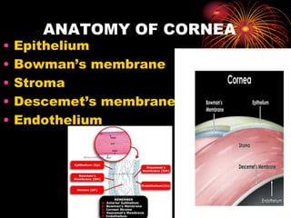
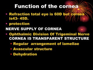
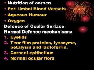
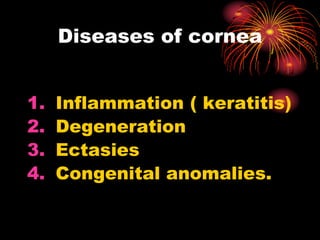
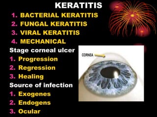
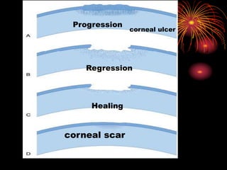
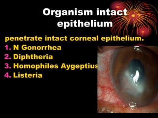
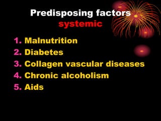
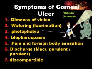
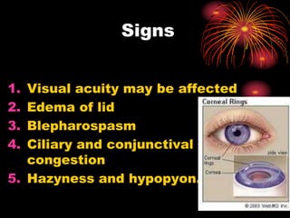
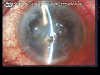
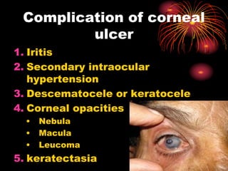
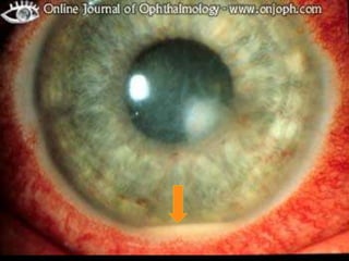
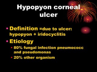
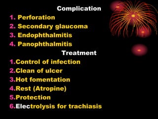
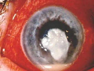
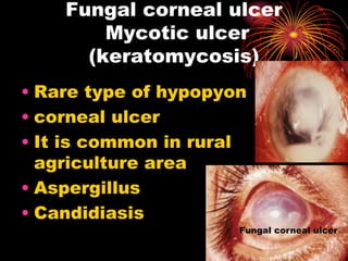
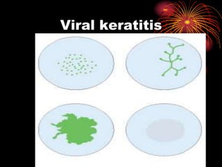
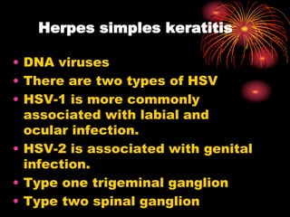
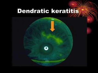
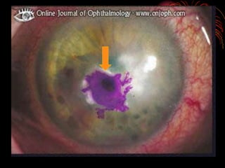
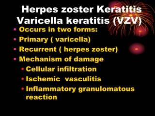
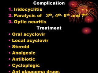
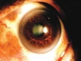
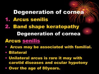
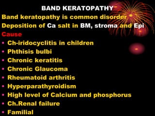
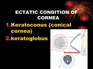
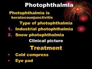
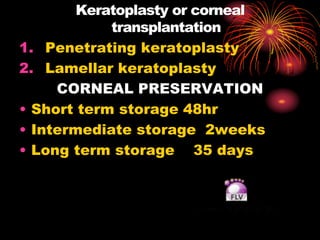
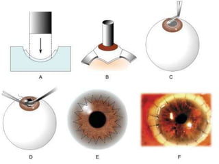
DISEASES OF CORNEA
Dr. Abdul Basir safi
Lecturer of Nangrahar Medical
faculty
head of eye department
E-mail: basirsafi123@yahoo.com
• Epithelium
• Bowman’s membrane
• Stroma
• Descemet’s membrane
• Endothelium
ANATOMY OF CORNEA
• Refraction total eye is 60D but cornea
is43- 45D.
• protection
NERVE SUPPLY OF CORNEA
• Ophthalmic Division Of Trigeminal Nerve
CORNEA IS TRANSPARENT STRUCTURE
• Regular arrangement of lamellae
• Avascular structure
• Dehydration
Function of the cornea
• Nutrition of cornea
• Peri limbal Blood Vessels
• Aqueous Humour
• Oxygen
Defence of Ocular Surface
Normal Defence mechanisms:
1. Eyelids
2. Tear film proteins, lysozyme,
betalysin and lactoferrin.
3. Corneal epithelium
4. Normal ocular flora
1. Inflammation ( keratitis)
2. Degeneration
3. Ectasies
4. Congenital anomalies.
Diseases of cornea
KERATITIS
1. BACTERIAL KERATITIS
2. FUNGAL KERATITIS
3. VIRAL KERATITIS
4. MECHANICAL
Stage corneal ulcer
1. Progression
2. Regression
3. Healing
Source of infection
1. Exogenes
2. Endogens
3. Ocular
Progression
corneal ulcer
Regression
Healing
corneal scar
penetrate intact corneal epithelium.
1. N Gonorrhea
2. Diphtheria
3. Homophiles Aygeptius
4. Listeria
Organism intact
epithelium
1. Corneal anesthesia Use of topical
anesthetic solution.
2. Local immune suppression as due
to topical corticosteroids.
3. Previous viral infection.
4. Trichiasis
5. Entropion
6. Eye lid positional
abnormality
7. Dacryo cystitis
Predisposing factors
ocular
1. Malnutrition
2. Diabetes
3. Collagen vascular diseases
4. Chronic alcoholism
5. Aids
Predisposing factors
systemic
1. Dimness of vision
2. Watering (lacrimation)
3. photophobia
4. blepharospasm
5. Pain and foreign body sensation
6. Discharge (Macu purulent /
purulent)
7. discompartible
Symptoms of Corneal
Ulcer
1. Visual acuity may be affected
2. Edema of lid
3. Blepharospasm
4. Ciliary and conjunctival
congestion
5. Hazyness and hypopyon.
Signs
1. Iritis
2. Secondary intraocular
hypertension
3. Descematocele or keratocele
4. Corneal opacities
• Nebula
• Macula
• Leucoma
5. keratectasia
Complication of corneal
ulcer
6. Synechia
7. Corneal fistula
8. Cataract
9. Staphyloma
10. Lens subluxation
11. Intra ocular hemorrhage
12. Dislocation of lens
13. Panophthalmitis
14. Endophthalmitis
Complication of corneal ulcer
Staphyloma
Synechia
LOCAL TREATMENT
• Control of infection with
appropriate antibiotic
a. based on clinical judgment
b. based on finding of smear
examination
c. based on culture and
sensitivity report
Treatment of uncomplicated
corneal ulcer
1. Control of infection (Antibiotic
drops)
2. Rest of eye (Atropine)
3. Cleanliness (cauterisition)
4. Application of hot fomentation
5. Protection of eye (black glass)
6. Support of nutrition
Treatment of uncomplicated
corneal ulcer
1. Systemic Antibiotics
2. Analgesic anti-inflammatory
3. Supportive treatment
4. Acetazolamide
Systemic Treatment
Local factors
trichiasis
exposure keratitis
chronic dacryocystitis
General factors
• Diabetes's millets
• Vitamin A deficiency
• Malnutrition
• AIDS
Non healing corneal
ulcer
• Definition =due to ulcer:
hypopyon + iridocyclitis
• Etiology
• 80% fungal infection pneumococc
and pseudomonas
• 20% other organism
Hypopyon corneal
ulcer
Clinical pictures
• Clinical picture is depends on two
factors.
• Virulence of organism
• Resistance of host
• Hypopyon in AC ( pass in AC)
• Iritis
• Synechia
• Miosis
• Secondary glaucoma
• Salagish pupil reflex
Complication
1. Perforation
2. Secondary glaucoma
3. Endophthalmitis
4. Panophthalmitis
Treatment
1.Control of infection
2.Clean of ulcer
3.Hot fomentation
4.Rest (Atropine)
5.Protection
6.Electrolysis for trachiasis
• Rare type of hypopyon
• corneal ulcer
• It is common in rural
agriculture area
• Aspergillus
• Candidiasis
Fungal corneal ulcer
Mycotic ulcer
(keratomycosis)
Fungal corneal ulcer
• Injury by vegetative material
• Injury by animal tail
• Long use of Antibiotic and
steroids
• secondary fungal ulcer
• Dry eye
• Herpes simplex keratitis
• Bullous keratopathy
Predisposing factors
Sings and symptoms
• Is sever than B ulcer.
• Massive hypopyon is present
• Irregular ulcer margin
• Dirty feature of ulcer
• Corneal abscess
treatment
• scraping and debridement of
ulcer
• Atropine
• Anti fungal drugs
• Keratoplasty
Viral keratitis
Introduction
• Viruses that cause corneal
disease are
• Herpes simplex ( HSV)
• Varicella zoster ( VZV)
• Adenovirus
• Epstein Barr virus ( EBV)
• Cytomegalovirus (CMV) can
also cause keratitis and is
more commonly associated
with AIDS
Herpes simples keratitis
• DNA viruses
• There are two types of HSV
• HSV-1 is more commonly
associated with labial and
ocular infection.
• HSV-2 is associated with genital
infection.
• Type one trigeminal ganglion
• Type two spinal ganglion
Dendratic keratitis
Treatment
1. Specific treatment
2. Non specific treatment
• Acyclovir
• Triflurothymidine drop
• Adenine arabinoside
Varicella zoster viral
Herpes zoster Keratitis
Varicella keratitis (VZV)
• Occurs in two forms:
• Primary ( varicella)
• Recurrent ( herpes zoster)
• Mechanism of damage
• Cellular infiltration
• Ischemic vasculitis
• Inflammatory granulomatous
reaction
Predisposing factors
• Old age
• Diabetes
• Alcoholic person
• Cancer
• AIDS
Complication
1. Iridocyclitis
2. Paralysis of 3th, 4th, 6th and 7th
3. Optic neuritis
Treatment
• Oral acyclovir
• Local acyclovir
• Steroid
• Analgesic
• Antibiotic
• Cycloplegic
Degeneration of cornea
1. Arcus senilis
2. Band shape keratopathy
Degeneration of cornea
Arcus senilis
• Arcus may be associated with familial.
• Bilateral
• Unilateral arcus is rare it may with
carotid diseases and ocular hypotony
• Over the age of 80years.
BAND KERATOPATHY
Band keratopathy is common disorder
Deposition of Ca salt in BM, stroma and Epi
Cause
• Ch-iridocyclitis in children
• Phthisis bulbi
• Chronic keratitis
• Chronic Glaucoma
• Rheumatoid arthritis
• Hyperparathyroidism
• High level of Calcium and phosphorus
• Ch.Renal failure
• Familial
ECTATIC CONDITION OF
CORNEA
1.Keratoconus (conical
cornea)
2.keratoglobus
keratoconus
Keratoconous
• It’s bilateral non inflammatory
condition 85%.
• Family history is not positive.
• Unilateral is rare and from
unknown causes
• Keratoconus is with Down’s
syndrome, atopic dermatitis,
Retinitis pigmentosia, aniridia,
VKC and Morffan syndrome,
osteogenesis, mitral valve
prolapse.
Munson’s sign
Keratoconous
Classification
1. Mild<48D
2. moderate (48-54D)
3. Sever >54D
PATHOLOGY
• Thinning of central cornea
• Rupture of Decement Membrane
Clinical picture
• Blurred vision
• Cone shape of cornea
• Placida's disk
• Munson’s sign
• Ophthalmoscopic sign
Placido’s disc examination
Normal
abnormal
Placido’s disc
Photophthalmia is
keratocounjunctivitis
Type of photophthalmia
1. Industrial photophthalmia
2. Snow photophthalmia
Clinical picture
Treatment
• Cold compress
• Eye pad
Photophthalmia
Keratoplasty or corneal
transplantation
1. Penetrating keratoplasty
2. Lamellar keratoplasty
CORNEAL PRESERVATION
• Short term storage 48hr
• Intermediate storage 2weeks
• Long term storage 35 days
keratomlacia
Corneal ulcer due to vit-A
deficiency
• The cornea become soft and
necrotic known as keratomlacia
• Bil cornea damage from center
• Bitot spot
• Less then 50– 70 IU
• Night blindness or nyctalopia
keratomlacia
Corneal ulcer due to vit-A
deficiency
Function of vit A
• Vision
• Mucus cell - keratin producing cell
• Anti infective vitamin
• Daily need of vitamin A in children is
1500—5000iu
• Daily need of vitamin A in adult is
3000—5000iu
Treatment
• In mild cases 30000iu per day for 1 week
• In sever cases 20000 iu / kg daily.
Treatment
Keep cornea well cover by
eye lid and bandage.
•Artificial tears and
ointment.
•Tarsorrhaphy
−Total tarsorrhaphy
−Lateral tarsorrhaphy
•Treatment of underling
causes.
‫پوښتنی‬‫؟‬

Corneal diseases by dr abdul basir safi

  • 2.
    DISEASES OF CORNEA Dr.Abdul Basir safi Lecturer of Nangrahar Medical faculty head of eye department E-mail: basirsafi123@yahoo.com
  • 3.
    • Epithelium • Bowman’smembrane • Stroma • Descemet’s membrane • Endothelium ANATOMY OF CORNEA
  • 4.
    • Refraction totaleye is 60D but cornea is43- 45D. • protection NERVE SUPPLY OF CORNEA • Ophthalmic Division Of Trigeminal Nerve CORNEA IS TRANSPARENT STRUCTURE • Regular arrangement of lamellae • Avascular structure • Dehydration Function of the cornea
  • 5.
    • Nutrition ofcornea • Peri limbal Blood Vessels • Aqueous Humour • Oxygen Defence of Ocular Surface Normal Defence mechanisms: 1. Eyelids 2. Tear film proteins, lysozyme, betalysin and lactoferrin. 3. Corneal epithelium 4. Normal ocular flora
  • 6.
    1. Inflammation (keratitis) 2. Degeneration 3. Ectasies 4. Congenital anomalies. Diseases of cornea
  • 7.
    KERATITIS 1. BACTERIAL KERATITIS 2.FUNGAL KERATITIS 3. VIRAL KERATITIS 4. MECHANICAL Stage corneal ulcer 1. Progression 2. Regression 3. Healing Source of infection 1. Exogenes 2. Endogens 3. Ocular
  • 8.
  • 9.
    penetrate intact cornealepithelium. 1. N Gonorrhea 2. Diphtheria 3. Homophiles Aygeptius 4. Listeria Organism intact epithelium
  • 10.
    1. Corneal anesthesiaUse of topical anesthetic solution. 2. Local immune suppression as due to topical corticosteroids. 3. Previous viral infection. 4. Trichiasis 5. Entropion 6. Eye lid positional abnormality 7. Dacryo cystitis Predisposing factors ocular
  • 11.
    1. Malnutrition 2. Diabetes 3.Collagen vascular diseases 4. Chronic alcoholism 5. Aids Predisposing factors systemic
  • 12.
    1. Dimness ofvision 2. Watering (lacrimation) 3. photophobia 4. blepharospasm 5. Pain and foreign body sensation 6. Discharge (Macu purulent / purulent) 7. discompartible Symptoms of Corneal Ulcer
  • 13.
    1. Visual acuitymay be affected 2. Edema of lid 3. Blepharospasm 4. Ciliary and conjunctival congestion 5. Hazyness and hypopyon. Signs
  • 15.
    1. Iritis 2. Secondaryintraocular hypertension 3. Descematocele or keratocele 4. Corneal opacities • Nebula • Macula • Leucoma 5. keratectasia Complication of corneal ulcer
  • 16.
    6. Synechia 7. Cornealfistula 8. Cataract 9. Staphyloma 10. Lens subluxation 11. Intra ocular hemorrhage 12. Dislocation of lens 13. Panophthalmitis 14. Endophthalmitis Complication of corneal ulcer Staphyloma
  • 17.
  • 18.
    LOCAL TREATMENT • Controlof infection with appropriate antibiotic a. based on clinical judgment b. based on finding of smear examination c. based on culture and sensitivity report Treatment of uncomplicated corneal ulcer
  • 19.
    1. Control ofinfection (Antibiotic drops) 2. Rest of eye (Atropine) 3. Cleanliness (cauterisition) 4. Application of hot fomentation 5. Protection of eye (black glass) 6. Support of nutrition Treatment of uncomplicated corneal ulcer
  • 20.
    1. Systemic Antibiotics 2.Analgesic anti-inflammatory 3. Supportive treatment 4. Acetazolamide Systemic Treatment
  • 21.
    Local factors trichiasis exposure keratitis chronicdacryocystitis General factors • Diabetes's millets • Vitamin A deficiency • Malnutrition • AIDS Non healing corneal ulcer
  • 23.
    • Definition =dueto ulcer: hypopyon + iridocyclitis • Etiology • 80% fungal infection pneumococc and pseudomonas • 20% other organism Hypopyon corneal ulcer
  • 24.
    Clinical pictures • Clinicalpicture is depends on two factors. • Virulence of organism • Resistance of host • Hypopyon in AC ( pass in AC) • Iritis • Synechia • Miosis • Secondary glaucoma • Salagish pupil reflex
  • 25.
    Complication 1. Perforation 2. Secondaryglaucoma 3. Endophthalmitis 4. Panophthalmitis Treatment 1.Control of infection 2.Clean of ulcer 3.Hot fomentation 4.Rest (Atropine) 5.Protection 6.Electrolysis for trachiasis
  • 27.
    • Rare typeof hypopyon • corneal ulcer • It is common in rural agriculture area • Aspergillus • Candidiasis Fungal corneal ulcer Mycotic ulcer (keratomycosis) Fungal corneal ulcer
  • 28.
    • Injury byvegetative material • Injury by animal tail • Long use of Antibiotic and steroids • secondary fungal ulcer • Dry eye • Herpes simplex keratitis • Bullous keratopathy Predisposing factors
  • 29.
    Sings and symptoms •Is sever than B ulcer. • Massive hypopyon is present • Irregular ulcer margin • Dirty feature of ulcer • Corneal abscess treatment • scraping and debridement of ulcer • Atropine • Anti fungal drugs • Keratoplasty
  • 30.
  • 31.
    Introduction • Viruses thatcause corneal disease are • Herpes simplex ( HSV) • Varicella zoster ( VZV) • Adenovirus • Epstein Barr virus ( EBV) • Cytomegalovirus (CMV) can also cause keratitis and is more commonly associated with AIDS
  • 32.
    Herpes simples keratitis •DNA viruses • There are two types of HSV • HSV-1 is more commonly associated with labial and ocular infection. • HSV-2 is associated with genital infection. • Type one trigeminal ganglion • Type two spinal ganglion
  • 33.
  • 35.
    Treatment 1. Specific treatment 2.Non specific treatment • Acyclovir • Triflurothymidine drop • Adenine arabinoside
  • 36.
  • 37.
    Herpes zoster Keratitis Varicellakeratitis (VZV) • Occurs in two forms: • Primary ( varicella) • Recurrent ( herpes zoster) • Mechanism of damage • Cellular infiltration • Ischemic vasculitis • Inflammatory granulomatous reaction
  • 38.
    Predisposing factors • Oldage • Diabetes • Alcoholic person • Cancer • AIDS
  • 39.
    Complication 1. Iridocyclitis 2. Paralysisof 3th, 4th, 6th and 7th 3. Optic neuritis Treatment • Oral acyclovir • Local acyclovir • Steroid • Analgesic • Antibiotic • Cycloplegic
  • 41.
    Degeneration of cornea 1.Arcus senilis 2. Band shape keratopathy Degeneration of cornea Arcus senilis • Arcus may be associated with familial. • Bilateral • Unilateral arcus is rare it may with carotid diseases and ocular hypotony • Over the age of 80years.
  • 43.
    BAND KERATOPATHY Band keratopathyis common disorder Deposition of Ca salt in BM, stroma and Epi Cause • Ch-iridocyclitis in children • Phthisis bulbi • Chronic keratitis • Chronic Glaucoma • Rheumatoid arthritis • Hyperparathyroidism • High level of Calcium and phosphorus • Ch.Renal failure • Familial
  • 44.
    ECTATIC CONDITION OF CORNEA 1.Keratoconus(conical cornea) 2.keratoglobus keratoconus
  • 46.
    Keratoconous • It’s bilateralnon inflammatory condition 85%. • Family history is not positive. • Unilateral is rare and from unknown causes • Keratoconus is with Down’s syndrome, atopic dermatitis, Retinitis pigmentosia, aniridia, VKC and Morffan syndrome, osteogenesis, mitral valve prolapse.
  • 47.
  • 48.
    Keratoconous Classification 1. Mild<48D 2. moderate(48-54D) 3. Sever >54D PATHOLOGY • Thinning of central cornea • Rupture of Decement Membrane Clinical picture • Blurred vision • Cone shape of cornea • Placida's disk • Munson’s sign • Ophthalmoscopic sign
  • 49.
  • 51.
    Photophthalmia is keratocounjunctivitis Type ofphotophthalmia 1. Industrial photophthalmia 2. Snow photophthalmia Clinical picture Treatment • Cold compress • Eye pad Photophthalmia
  • 52.
    Keratoplasty or corneal transplantation 1.Penetrating keratoplasty 2. Lamellar keratoplasty CORNEAL PRESERVATION • Short term storage 48hr • Intermediate storage 2weeks • Long term storage 35 days
  • 55.
    keratomlacia Corneal ulcer dueto vit-A deficiency • The cornea become soft and necrotic known as keratomlacia • Bil cornea damage from center • Bitot spot • Less then 50– 70 IU • Night blindness or nyctalopia
  • 56.
    keratomlacia Corneal ulcer dueto vit-A deficiency Function of vit A • Vision • Mucus cell - keratin producing cell • Anti infective vitamin • Daily need of vitamin A in children is 1500—5000iu • Daily need of vitamin A in adult is 3000—5000iu Treatment • In mild cases 30000iu per day for 1 week • In sever cases 20000 iu / kg daily.
  • 57.
    Treatment Keep cornea wellcover by eye lid and bandage. •Artificial tears and ointment. •Tarsorrhaphy −Total tarsorrhaphy −Lateral tarsorrhaphy •Treatment of underling causes.
  • 58.