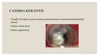
INFECTIVE CORNEALULCERS
Presented By:
◦ DEFINITION:
◦ Corneal ulcers are defects in the corneal epithelium with or without stromal infilitration.
TYPES:
A. Infectious ulcerative keratitis
B. Non infectious ulcerative keratitis.
ETIOLOGY:
Infectious
◦ bacterial & fungi
◦ Viruses
◦ Acanthamoeba
Non infectious
◦ Systemic autoimmune/ inflammatory
◦ Local toxic
INFECTIOUS NON INFECTIOUS
Pain No pain
Discharge No discharge
AC reaction present AC reaction: absent
Central Peripheral
Trauma Trauma
NON INFECTIOUS ULCERATIVE KERATITIS
Causes:
◦ Local causes;
Punctuate marginal keratitis: staphylococci, streptococci, hypersensitivity to
medications.
◦ Systemic causes:
General manifestations of systemic, immune-mediated disease.
Most common: Rheumatoid arthritis, wagner’s granulomatosis & polyarteritis
nodusa
INFECTIOUS KERATITIS
◦ A corneal ulcer is an ocular emergency that raises high stakes of questions
about diagnosis & management.
◦ When a large corneal ulcer is staring you in the face time isn’t in your side.
◦ Despite varying etiologies & presentations, as well as different treatment
approaches, cornealulcers have one thing in common: the potential to cause
devastating loss of vision.
PRINCIPLES OF MANAGEMENT OF CORNEAL DISEASE
1. Control of infection
2. Control of inflammation
3. Promotion of re-epithelialization
• Lubrication
• Lid closure
◦ Bandage soft contact lens
4. Prevention of perforation
• Tissue adhesive glue
• Conjectival flap
• Systemic immunosuppressive agents
• Corneal grafting
RISK FACTORS:
◦ Contact lens wear
◦ Trauma to the eye
◦ Reduced immunity (e.g., diabetes, immunosuppression)
SYMPTOMS:
◦ Eye pain
◦ Redness
◦ Photophobia (sensitivity to light)
◦ Blurred vision
◦ Excessive tearing
DIAGNOSIS:
◦ Clinical examination
◦ Corneal scraping for microscopy and culture
◦ Fluorescein staining
CAUSES:
◦ Bacterial causes (e.g., Staphylococcus, Streptococcus)
◦ Fungal causes (e.g., Fusarium, Aspergillus)
◦ Viral causes (e.g., Herpes simplex virus)
MICROBIAL KERATITIS (Bacterial)
Predisposing factors:
Ocular surface diseases:
◦ Post-herpetic
◦ Corneal disease,
Bullous keratopathy,
Corneal exposure,
Dry eye,
Diminished corneal sensation
Contact lens wear
Causative Organisms
80% of cases >>>> Staphylococcus aurous, Streptococuss pneumonia and
pseudomonas species
Pseudomonas aeruginosa
Is the most frequent and the most pathogenic ocular pathogen which cause
corneal perforation in just 72 hours
◦ Pathogens which can produce corneal infection in intact epithelium.
1. Neisseria gonorrhea
2. Corynebacterium diptheriae
3. Listeria
4. haemophilus
◦ Staph. Aureus and strep. Pneumoniae
oval, yellow-white, densely opaque stromal suppuration surrounded by
relatively clear cornea
MANAGEMENT
◦ History
◦ Clinical examination
◦ Corneal scrapping
DIFFERENTIATORS
Acute painful injected eye.
Profuse tearing & discharge.
Decrease visual acuity.
Large F.B
Stromal invasion with epithelial excavating edge.
TREATMENT
1. Fluroquinolones
 Every 5 mins/hour
 Hour/24 hs
 2 hour / 24 hs
2. Fortified eye drops
 Ulcer < 2 ws,
Improvement not obvious.
(N.B) Don’t miss resistant bavteria.
3. steriods
FUNGAL KERATITIS
◦ Aspergillus
◦ Fusarium
◦ Yeast candida
CLINICAL PRESENTATION
History of vegetable matter injury
Greyish-white ulcer with indistinct margins
Surrounded by feathery infiltrates
Ring infiltrate
Endothelial plaque
Hypopyon
DIFFERENTIATORS
Dull grey infiltrate
Satellite lesions
Awareness of those ulcers resembling bacterial keratitis
Awareness of those caused by yeast
Real flags
CANDIDA KERATITIS
◦ Usually develops in pre-exixting corneal disease or immunocompromised
patient
◦ Yellow-white ulcer
◦ Dense suppuration
FILAMENTOUS FUNGAL kERATITIS
◦ Firm elevated necrotic slough, “hyphate” lines that extend beyond the edge of
the ulcer into the normal cornea,
◦ Multifocal granular( or feathery) Gray-white “satellete” infiltrates in the
corneal stroma.
MANAGEMENT
Culture
Biopsy
Antifungal therapy
◦ Initially broad-spectrum econazole 1%, voriconazole 1% topically
◦ Then depending upon sensitivity Natamycin or imidazole for 6 weeks
Systemic ketoconazole
Therapeutic penetrating keratoplasty
ACANTHAMOEBA KERATITIS
Protozoan
◦ Active (trophozoite)
◦ Dormant (cystic)
Common in swimmers and
CL wearers
CLINICAL PRESENTATION
Blurred vision & disproportionate pain
Patchy anterior stromal infilterates
Perineural infilterates (radial keratoneuritis)
Infilterates coalesce- ring abscess, ulceration and
Hypopyon
White satellite lesions
MANAGEMENT
Corneal scrappings stained with calcofluor white
Corneal biopsy
Treatment with chlorhexidine,
Polyhexamethylenebiguanide drops,
Dipropamidine and propamidine.
Therapeutic penetrating keratoplasty
VIRAL KERATITIS
HERPIS SIMPLEX VIRUS
Basically it is epitheliotropic & may become neurotropic
Two types HSV-1 & HSV-2
Primary HSV-1 infection occurs most commonly in the
mucocutaneous distribution of the trigeminal nerve
CLASSIFICATION:
◦ Epithelial keratitis
◦ Disciform keratitis (localized endothelitis)
◦ Metaherpitic ulcer
◦ Necrotizing keratitis
◦ keratouvetis
DIFFERENTIATORS
◦ Dentritic ulcer
◦ Loss of corneal sensation
◦ Photophobia
TYPES OF HSV KERATITIS:
◦ Primary
◦ Recurrent
◦ Dentritic, geographic, metaherptica
◦ Diabetic foot in the eye---- Neurotrophic
MANAGEMENT
◦ Epithelial keratitis is treated with topical antivirals
Acyclovir ophthalmic ointment & Trifluridine eye drops
◦ Herpetic stromal keratitis is treated initially with
Prednisolone drops every 2 hrs accompanied by a prophylactic antiviral drug
◦ Metaherpetic ulcer treated by
Artificial tears & eye lubricants, stopping toxic medications, performing punctual occlusion,
bandage contact lens & amniotic membrane transplant
HERPES ZOSTER KERATITIS
Caused by Varicella zoster virus
Causes-varicella (chickenpox) & herpes zoster (shingles)
RISK FACTORS:
• Increasing risk (>70years)
• Neoplastic diseases
• Immunosuppressive drugs
• Organ transplant recipients
• HIV
HERPES ZOSTER OPTHAIMICUS (HZO)
Unilateral painful skin rash in one or more dermatome distributions of the fifth
cranial nerve (trigeminal nerve)
HUTCHINSON’S Sign:
Vesicles at the side or tip of the nose are potential indicators of ocular diseases
THANK YOU

corneal infection.pptx types causes management

  • 1.
  • 2.
    ◦ DEFINITION: ◦ Cornealulcers are defects in the corneal epithelium with or without stromal infilitration. TYPES: A. Infectious ulcerative keratitis B. Non infectious ulcerative keratitis. ETIOLOGY: Infectious ◦ bacterial & fungi ◦ Viruses ◦ Acanthamoeba Non infectious ◦ Systemic autoimmune/ inflammatory ◦ Local toxic
  • 3.
    INFECTIOUS NON INFECTIOUS PainNo pain Discharge No discharge AC reaction present AC reaction: absent Central Peripheral Trauma Trauma
  • 4.
    NON INFECTIOUS ULCERATIVEKERATITIS Causes: ◦ Local causes; Punctuate marginal keratitis: staphylococci, streptococci, hypersensitivity to medications. ◦ Systemic causes: General manifestations of systemic, immune-mediated disease. Most common: Rheumatoid arthritis, wagner’s granulomatosis & polyarteritis nodusa
  • 5.
    INFECTIOUS KERATITIS ◦ Acorneal ulcer is an ocular emergency that raises high stakes of questions about diagnosis & management. ◦ When a large corneal ulcer is staring you in the face time isn’t in your side. ◦ Despite varying etiologies & presentations, as well as different treatment approaches, cornealulcers have one thing in common: the potential to cause devastating loss of vision.
  • 6.
    PRINCIPLES OF MANAGEMENTOF CORNEAL DISEASE 1. Control of infection 2. Control of inflammation 3. Promotion of re-epithelialization • Lubrication • Lid closure ◦ Bandage soft contact lens 4. Prevention of perforation • Tissue adhesive glue • Conjectival flap • Systemic immunosuppressive agents • Corneal grafting
  • 7.
    RISK FACTORS: ◦ Contactlens wear ◦ Trauma to the eye ◦ Reduced immunity (e.g., diabetes, immunosuppression) SYMPTOMS: ◦ Eye pain ◦ Redness ◦ Photophobia (sensitivity to light) ◦ Blurred vision ◦ Excessive tearing
  • 8.
    DIAGNOSIS: ◦ Clinical examination ◦Corneal scraping for microscopy and culture ◦ Fluorescein staining CAUSES: ◦ Bacterial causes (e.g., Staphylococcus, Streptococcus) ◦ Fungal causes (e.g., Fusarium, Aspergillus) ◦ Viral causes (e.g., Herpes simplex virus)
  • 9.
    MICROBIAL KERATITIS (Bacterial) Predisposingfactors: Ocular surface diseases: ◦ Post-herpetic ◦ Corneal disease, Bullous keratopathy, Corneal exposure, Dry eye, Diminished corneal sensation Contact lens wear
  • 10.
    Causative Organisms 80% ofcases >>>> Staphylococcus aurous, Streptococuss pneumonia and pseudomonas species Pseudomonas aeruginosa Is the most frequent and the most pathogenic ocular pathogen which cause corneal perforation in just 72 hours ◦ Pathogens which can produce corneal infection in intact epithelium. 1. Neisseria gonorrhea 2. Corynebacterium diptheriae 3. Listeria 4. haemophilus
  • 11.
    ◦ Staph. Aureusand strep. Pneumoniae oval, yellow-white, densely opaque stromal suppuration surrounded by relatively clear cornea
  • 12.
    MANAGEMENT ◦ History ◦ Clinicalexamination ◦ Corneal scrapping
  • 13.
    DIFFERENTIATORS Acute painful injectedeye. Profuse tearing & discharge. Decrease visual acuity. Large F.B Stromal invasion with epithelial excavating edge.
  • 14.
    TREATMENT 1. Fluroquinolones  Every5 mins/hour  Hour/24 hs  2 hour / 24 hs 2. Fortified eye drops  Ulcer < 2 ws, Improvement not obvious. (N.B) Don’t miss resistant bavteria. 3. steriods
  • 15.
    FUNGAL KERATITIS ◦ Aspergillus ◦Fusarium ◦ Yeast candida
  • 16.
    CLINICAL PRESENTATION History ofvegetable matter injury Greyish-white ulcer with indistinct margins Surrounded by feathery infiltrates Ring infiltrate Endothelial plaque Hypopyon
  • 17.
    DIFFERENTIATORS Dull grey infiltrate Satellitelesions Awareness of those ulcers resembling bacterial keratitis Awareness of those caused by yeast Real flags
  • 18.
    CANDIDA KERATITIS ◦ Usuallydevelops in pre-exixting corneal disease or immunocompromised patient ◦ Yellow-white ulcer ◦ Dense suppuration
  • 19.
    FILAMENTOUS FUNGAL kERATITIS ◦Firm elevated necrotic slough, “hyphate” lines that extend beyond the edge of the ulcer into the normal cornea, ◦ Multifocal granular( or feathery) Gray-white “satellete” infiltrates in the corneal stroma.
  • 20.
    MANAGEMENT Culture Biopsy Antifungal therapy ◦ Initiallybroad-spectrum econazole 1%, voriconazole 1% topically ◦ Then depending upon sensitivity Natamycin or imidazole for 6 weeks Systemic ketoconazole Therapeutic penetrating keratoplasty
  • 21.
    ACANTHAMOEBA KERATITIS Protozoan ◦ Active(trophozoite) ◦ Dormant (cystic) Common in swimmers and CL wearers
  • 22.
    CLINICAL PRESENTATION Blurred vision& disproportionate pain Patchy anterior stromal infilterates Perineural infilterates (radial keratoneuritis) Infilterates coalesce- ring abscess, ulceration and Hypopyon White satellite lesions
  • 23.
    MANAGEMENT Corneal scrappings stainedwith calcofluor white Corneal biopsy Treatment with chlorhexidine, Polyhexamethylenebiguanide drops, Dipropamidine and propamidine. Therapeutic penetrating keratoplasty
  • 24.
    VIRAL KERATITIS HERPIS SIMPLEXVIRUS Basically it is epitheliotropic & may become neurotropic Two types HSV-1 & HSV-2 Primary HSV-1 infection occurs most commonly in the mucocutaneous distribution of the trigeminal nerve
  • 25.
    CLASSIFICATION: ◦ Epithelial keratitis ◦Disciform keratitis (localized endothelitis) ◦ Metaherpitic ulcer ◦ Necrotizing keratitis ◦ keratouvetis
  • 26.
    DIFFERENTIATORS ◦ Dentritic ulcer ◦Loss of corneal sensation ◦ Photophobia TYPES OF HSV KERATITIS: ◦ Primary ◦ Recurrent ◦ Dentritic, geographic, metaherptica ◦ Diabetic foot in the eye---- Neurotrophic
  • 27.
    MANAGEMENT ◦ Epithelial keratitisis treated with topical antivirals Acyclovir ophthalmic ointment & Trifluridine eye drops ◦ Herpetic stromal keratitis is treated initially with Prednisolone drops every 2 hrs accompanied by a prophylactic antiviral drug ◦ Metaherpetic ulcer treated by Artificial tears & eye lubricants, stopping toxic medications, performing punctual occlusion, bandage contact lens & amniotic membrane transplant
  • 28.
    HERPES ZOSTER KERATITIS Causedby Varicella zoster virus Causes-varicella (chickenpox) & herpes zoster (shingles) RISK FACTORS: • Increasing risk (>70years) • Neoplastic diseases • Immunosuppressive drugs • Organ transplant recipients • HIV
  • 29.
    HERPES ZOSTER OPTHAIMICUS(HZO) Unilateral painful skin rash in one or more dermatome distributions of the fifth cranial nerve (trigeminal nerve) HUTCHINSON’S Sign: Vesicles at the side or tip of the nose are potential indicators of ocular diseases
  • 30.