Downloaded 1,318 times

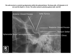
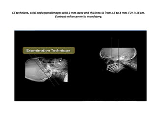
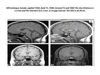
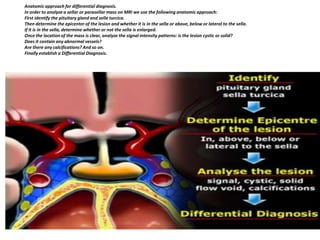

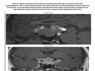
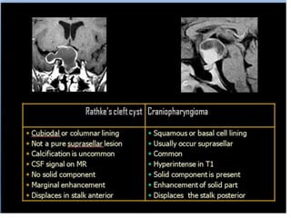
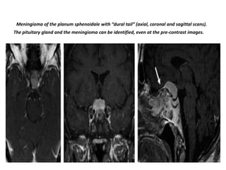
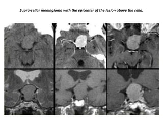

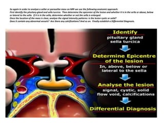
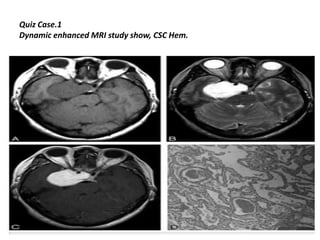
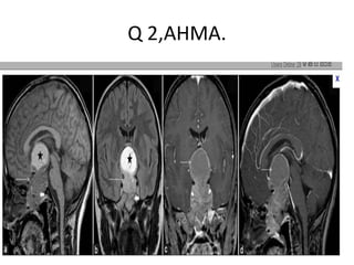
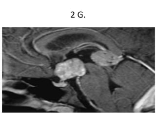

The document provides information on imaging techniques and differential diagnosis for sellar and parasellar masses. CT and MRI techniques are described for imaging the sella turcica region with details on slice thickness, field of view, and contrast usage. An anatomic approach is outlined to analyze sellar masses which involves identifying the pituitary gland, lesion location and characteristics, and establishing a differential diagnosis. Common pathologies that can occur in the sella and surrounding structures are then described individually, including the pituitary gland, stalk, optic chiasm, hypothalamus, carotid artery, cavernous sinus, and meninges. Imaging examples of lesions such as pituitary adenomas, craniopharyngiomas, and meningi


