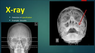
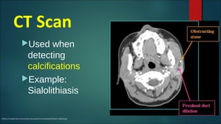
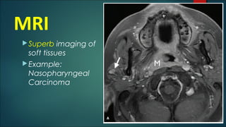
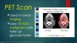
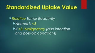
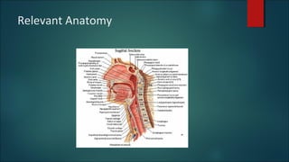
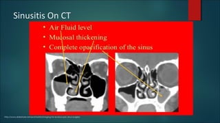
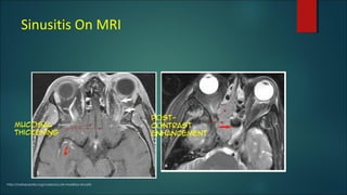
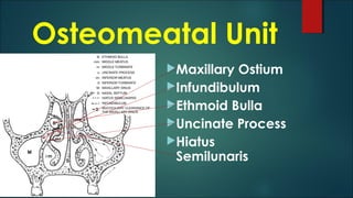
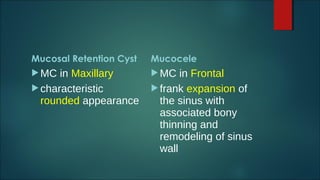
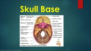
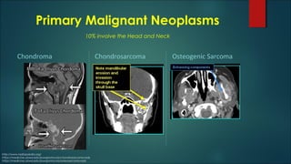
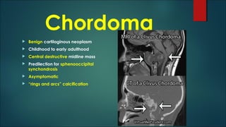
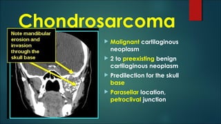
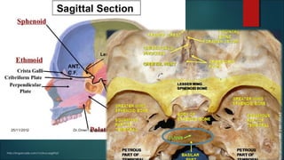
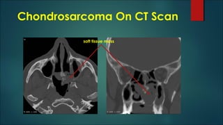
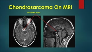
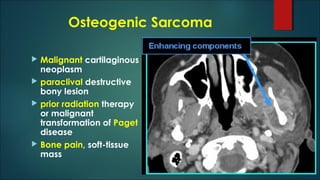
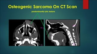
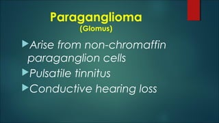
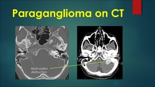
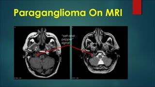
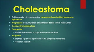
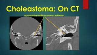
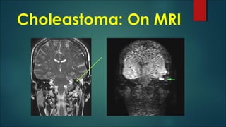
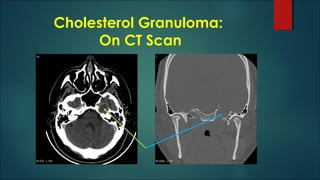
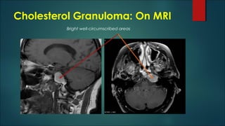
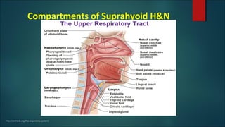
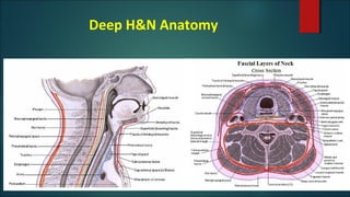
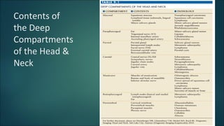
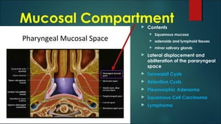
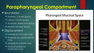
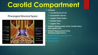
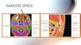
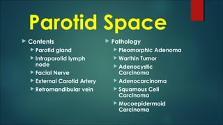
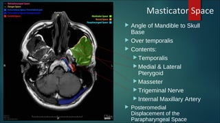
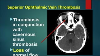
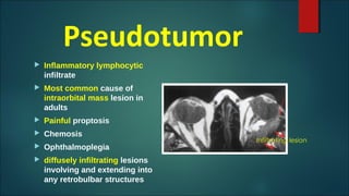
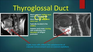
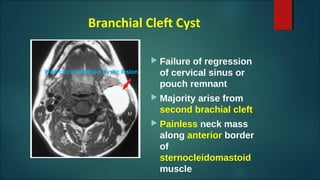
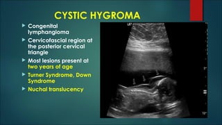
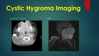
Head and Neck
Imaging
DIAGNOSTICS
Marco Alberto C. Brion, MD
Radiology Pre-Resident
Learning Objective
 Imaging Modalities for H and N
 Anatomy
 Paranasal
 Skull Base
 Suprahyoid Head and Neck
 Compartments of the head and neck
 Contents of each compartment
 Orbit
 Lymph Nodes
 Congenital Lesions
X-ray
 Detection of opacification
 Example: Sinusitis
CT Scan
Used when
detecting
calcifications
Example:
Sialolithiasis
https://medicine.uiowa.edu/iowaprotocols/sialolithiasis-radiology
MRI
Superb imaging of
soft tissues
Example:
Nasopharyngeal
Carcinoma
PET Scan
Used in tumor
staging
Uses 18 FDG –
malignant cells
take up
glucose faster
http://www.enttoday.org/article/clinicians-question-usefulness-of-multiple-post-treatment-petct-scans-for-head-and-neck-
cancer/2/
Standardized Uptake Value
Relative Tumor Reactivity
Normal is <3
If >3: Malignancy (also infection
and post-op conditions)
Relevant Anatomy
PARANASAL
SINUSES and
CAVITIES
Sinusitis
Inflammation of the sinuses
Maxillary and Ethmoid sinuses
Viral URTI
Mucosal thickening
Acute Sinusitis
air-fluid levels or foamy appearing sinus secretions
Chronic Sinusitis
mucoperiosteal thickening
osseous thickening of the sinus walls
http://shentherapies.com.au/521-2/
http://rhaudhahbinafsha.blogspot.com/2010/11/anatomy-of-sinus.html
Which modality?
Acute Sinusitis
 for better soft tissue
differentiation
 MRI
Chronic Sinusitis
 because of chronic
effects like
calcifications
 CT Scan
On X-ray
(Water’s Occipitomental view)
 Cannot assess the
extent of inflammation
and its complications
 Unreliable for children
under 1 year of age
http://slideplayer.com/slide/3936881/
Sinusitis On CT
http://www.slideshare.net/prahladnb/imaging-for-endoscopic-sinus-surgery
Sinusitis On MRI
http://radiopaedia.org/cases/acute-maxillary-sinusitis
Mucosal
thickening
post-
contrast
enhancement
Osteomeatal Unit
Maxillary Ostium
Infundibulum
Ethmoid Bulla
Uncinate Process
Hiatus
Semilunaris
Mucosal Retention Cyst
MC in Maxillary
characteristic
rounded appearance
Mucocele
MC in Frontal
frank expansion of
the sinus with
associated bony
thinning and
remodeling of sinus
wall
Skull Base
https://www.drananda.com.au/skull-base-tumours/
Primary Malignant Neoplasms
Chondroma Chondrosarcoma Osteogenic Sarcoma
http://www.radiopaedia.org/
https://medicine.uiowa.edu/iowaprotocols/chondrosarcoma-rads
https://medicine.uiowa.edu/iowaprotocols/osteosarcoma-rads
10% involve the Head and Neck
Chordoma
 Benign cartilaginous neoplasm
 Childhood to early adulthood
 Central destructive midline mass
 Predilection for sphenooccipital
synchondrosis
 Asymptomatic
 “rings and arcs” calcification
Chondrosarcoma
 Malignant cartilaginous
neoplasm
 2 to preexisting benign
cartilaginous neoplasm
 Predilection for the skull
base
 Parasellar location,
petroclival junction
http://imgarcade.com/1/clivus-sagittal/
Chondrosarcoma On CT Scan
soft tissue mass
Chondrosarcoma On MRI
Lobulated mass
Osteogenic Sarcoma
 Malignant cartilaginous
neoplasm
 paraclival destructive
bony lesion
 prior radiation therapy
or malignant
transformation of Paget
disease
 Bone pain, soft-tissue
mass
Osteogenic Sarcoma On CT Scan
predominantly lytic lesions
Paraganglioma
(Glomus)
Arise from non-chromaffin
paraganglion cells
Pulsatile tinnitus
Conductive hearing loss
Paraganglioma on CT
Moth-eaten
destruction
Paraganglioma On MRI
“salt-and-
pepper”
signal
Choleastoma
 Epidermoid cyst composed of desquamating stratified squamous
epithelium
 Progressive accumulation of epithelial debris within their lumen
 Conductive hearing loss
 Congenital
 Epithelial rests within or adjacent to temporal bone
 Acquired
 Stratified squamous epithelium of the tympanic membrane
 retraction pockets
Choleastoma: On CT
desquamating stratified squamous epithelium
Choleastoma: On MRI
Cholesterol Granuloma
 partially obstructed petrous apex air cells filled
with cholesterol debris and hemorrhagic fluid
 Hemotympanum
 Conductive hearing loss (w/ Middle Ear Effusion)
 Dizziness
 Tinnitus
 Headache
Cholesterol Granuloma:
On CT Scan
Cholesterol Granuloma: On MRI
Bright well-circumscribed areas
NECK IMAGING
http://www.fpnotebook.com/ent/Anatomy/NckAntmy.htm
Compartments of Suprahyoid H&N
http://antranik.org/the-respiratory-system/
Deep H&N Anatomy
Contents of
the Deep
Compartments
of the Head &
Neck
Mucosal Compartment Contents
 Squamous mucosa
 adenoids and lymphoid tissues
 minor salivary glands
 Lateral displacement and
obliteration of the pararyngeal
space
 Tornwaldt Cysts
 Retention Cysts
 Pleomorphic Adenoma
 Squamous Cell Carcinoma
 Lymphoma
Parapharyngeal Compartment
 Contents
 Fat
 Trigeminal Nerve
 Internal Maxillary Artery
 Ascending Pharyngeal Artery
 important landmark of
mass effect in the deep
face
Parapharyngeal Compartment
 Boundaries
 Posterior: Carotid Space
 Lateral: Parotid Space
 Anteriorly: Masticator Space
 Medial: Mucosal Space
 Displacement
 Laterally by mucosal mass
 Anteriorly by carotid sheath
mass
 Medially by parotid mass
 Posteromedially by
masticator mass
Carotid Compartment
 Contents
 Cranial Nerves IX-XII
 Sympathetic Nerves
 Jugular Chain Nodes
 Carotid Artery
 Jugular Vein
 Anterior displacement of the Carotid Artery
and Jugular Vein
 Anterior Displacement of the
Parapharyngeal Space
 Carotid Body Paragangliomas
 Schwannoma
 Neurofibroma
Parotid Space
 Contents
 Parotid gland
 Intraparotid lymph
node
 Facial Nerve
 External Carotid Artery
 Retromandibular vein
 Pathology
 Pleomorphic Adenoma
 Warthin Tumor
 Adenocystic
Carcinoma
 Adenocarcinoma
 Squamous Cell
Carcinoma
 Mucoepidermoid
Carcinoma
Signs of
Malignancy
Infiltration into deep neck structures e.g.
masticator or parapharyngeal space
Clinical involvement of the facial nerve
Masticator Space
 Angle of Mandible to Skull
Base
 Over temporalis
 Contents:
 Temporalis
 Medial & Lateral
Pterygoid
 Masseter
 Trigeminal Nerve
 Internal Maxillary Artery
 Posteromedial
Displacement of the
Parapharyngeal Space
Internal
Maxillar
y Artery
Primary Masticator Space
Malignancies
 extension of oropharyngeal or tongue base squamous cell carcinoma
 muscles of mastication
 tumor or infection from oropharyngeal or nasopharyngeal lesions
may spread along the third division of the fifth cranial nerve, allowing
the tumor to ascend through the foramen ovale into the cavernous
sinus
 extend posteriorly along the cisternal portion of the trigeminal nerve
to the brainstem
 Osteosarcoma
 Ewing Sarcoma
 Non-Hodgkin Lymphoma
Retropharyngeal Space
 Posterior to the superficial
mucosal space and
pharyngeal constrictor
muscles
 Anterior to the prevertebral
space
 posterior displacement of
the prevertebral muscles
 potential conduit for the
spread of tumor or
infection from the pharynx
to the mediastinum
Prevertebral Space
 Prevertebral fascia
 Displacement of
prevertebral muscles
anteriorly
 Contents
 Cervical vertebral
bodies
 Chordoma
 Osteomyelitis
Interspatial Disease
Secondary to lesions involving
anatomic structures normally
traversing H&N spaces
Lymphatic
Neural
Vascular
Lymph Nodes Internal Jugular Nodal Chain
 Final common pathway for
lymphatic drainage
 Jugulodigastric Lymph Node
 Highest node
 Where posterior belly of
digastric crosses near level
of hyoid bone
 Posterior to Submandibular
Gland
 Lymphatic drainage from
tonsil, oral cavity, pharynx,
submandibular nodes
Features of Malignancy
 Jugulodigastric and Submandibular normally 1.5 cm
diameter
 Other nodes abnormal if >1.0 cm
 Malignant if
 Peripheral Nodal Enhancement with Central Necrosis
 Extracapsular Spread with Infiltration
 Matted Conglomerate Mass of Nodes
 Of enlarged nodes
 70% are 2 to metastasis
 30% by benign reactive hyperplasia
Normal Lymph Node Nodal Metastasis
Hyperechoic centerHypoechoic
periphery
Increased
vascularity
http://ultrasound-images.blogspot.com/2011/11/normal-and-enlarged-lymph-nodes.html
Orbit
https://www.slideshare.net/vijayjoshi311/ocular-radiology
Optic Nerve Glioma
 Most common optic
nerve tumor
 First decade of life
 Neurofibromatosis Type I
 Bilateral
 Optic nerve sheath
enlargement
 Tubular, fusiform, kinking
Optic Sheath Meningioma
 Hemangioendothelial cells of
the arachnoid layer of the optic
nerve sheath
 Circular configuration & linear
growth along the optic nerve
 “Tram Track pattern” along
optic nerve sheath
 Invade and grow through the
dura
 Irregular asymmetric
appearance
 Extensive calcification
Cavernous Hemangioma
Most common
orbital masses in
adults
Sharply
circumscribed
rounded mass with
diffuse
enhancement or
mottled pattern
Superior Ophthalmic Vein Thrombosis
Thrombosis
in conjunction
with
cavernous
sinus
thrombosis
Loss of
Pseudotumor
 Inflammatory lymphocytic
infiltrate
 Most common cause of
intraorbital mass lesion in
adults
 Painful proptosis
 Chemosis
 Ophthalmoplegia
 diffusely infiltrating lesions
involving and extending into
any retrobulbar structures
Infiltrating lesion
Thyroglossal Duct
Cyst
Most common midline neck
mass in young patients
Most common congenital
neck cyst
Typically located in the
midline
Anterior neck mass moving
with swallowing and
elevated on tongue
protrusion
Cystic mass with uniform thin peripheral rim of
capsular enhancement and occasional septations
Branchial Cleft Cyst
 Failure of regression
of cervical sinus or
pouch remnant
 Majority arise from
second brachial cleft
 Painless neck mass
along anterior border
of
sternocleidomastoid
muscle
Well-circumscribed cystic lesion
CYSTIC HYGROMA
 Congenital
lymphangioma
 Cervicofascial region at
the posterior cervical
triangle
 Most lesions present at
two years of age
 Turner Syndrome, Down
Syndrome
 Nuchal translucency
Cystic Hygroma Imaging
Axial CT Scan MRI
References
 Fundamental of Diagnostic Radiology by Brant, MD et al (4th ed)
 Radiopaedia.org
 Harrison’s Principles of Internal Medicine by Kasper, MD et al (19th
ed)
 Clinically Oriented Anatomy by Moore et al (6th ed)

Head and Neck Imaging

Editor's Notes

  • #2 Today’s lecture will cover the different imaging modalities that will survey the head and neck region. The Head and Neck region include the sinonasal cavities, the skull base, the oral cavity, the neck, the larynx, the orbits, and temporal bone. Almost every organ system is represented, including the digestive, respiratory, nervous, osseous, and vascular systems. Because of this anatomic complexity, the head and neck region is approached with considerable trepidation.
  • #3 At the end of this lecture, the following learning objectives will be accomplished. The different imaging modalities used The anatomy of the paranasal sinuses and skull base The compartments of the suprahyoid head and neck with the contents of each compartment The imaging of the orbit Significance of the lymph nodes in tumor staging of head and neck cancers A few congenital lesions that affect the head and neck This lecture will be limited to discussing the anatomy of the head and neck. In particular, the compartments of the head and neck with their contents will be highlighted.
  • #4 As the initial imaging modality for the head and neck, conventional radiography is used to detect sinus opacification. For example, sinusitis would present on imaging with opacification of the affected sinuses, notably the maxillary sinuses.
  • #5 CT scan is used when looking for calcifications, such as in sialolithiasis.
  • #6 MRI is utilized for visualizing soft tissue pathology owing to superior discrimination. An application would be in viewing an nasopharyngeal mass.
  • #7 Aside from physical examination of the lymph nodes, newer modalities such as PET scan are used in staging tumors of the head and neck. Based on the principle of uptake of a radioisotope, 18 FDG, malignant cells would take up the glucose isotope faster compared with normal cells. Hence, they would light up when scanned.
  • #8 Standardized Uptake Value characterizes lesions found on PET scan. This value, which refers to relative reactivity of the radioisotope uptake standardized to injection dose, is normally less than 3. Malignant cells, aside from infectious and post-operative conditions, would have a value greater than 3.
  • #9 Sagittal cross section of the Head and Neck region is shown. By understanding the normal anatomy and knowing the scope of pathologic entities that occur within, an accurate assessment can be done.
  • #10 Next topic will be the study of the paranasal sinuses and cavities, focusing on the anatomy and pathology.
  • #11 Sinusitis refers to inflammation of the paranasal sinuses and the nasal cavity. Most commonly involve are the maxillary and ethmoid sinuses. The symptoms usually follow a viral upper respiratory tract infection. Mucosal thickening is noted. There are two categories according to chronicity. In Acute Sinusitis, air-fluid levels or foamy appearing sinus secretions are noted on conventional skull radiographs. In Chronic Sinusitis, noted findings include mucoperiosteal thickening and osseous thickening of the sinus walls.
  • #12 Based on chronicity, a good imaging quality can be ordered. For better soft tissue differentiation in Acute Sinusitis, MRI is preferred. To look for calcifications in Chronic Sinusitis, CT Scan is favored.
  • #13 On a conventional radiograph taken using Water’s View, noted findings include mucosal hyperplasia, complete obliteration of air spaces within the sinus cavity, and an air-fluid level due to differing densities between air and the denser fluid. To view the spread of the sinus infection to other paranasal cavities. additional studies such as Caldwell (occipitofrontal) view and Lateral view may be clinically warranted. Note that radiography by itself cannot assess the extent of inflammation and its associated complications. Radiograph is also unreliable as a diagnostic tool in infants below 1 year of age due to the underdevelopment of the sinus cavities.
  • #14 On Coronal CT scan, note the air fluid level, mucosal thickening, and complete opacification of the sinuses.
  • #15 On MRI, mucosal thickening is noted. There is also post-contrast enhancement of the inflamed mucosa opposite the fluid level.
  • #16 To understand patterns of inflammatory sinonasal disease, knowing the route of mucociliary drainage of the paranasal sinuses is critical. A major area of mucociliary drainage is the middle meatus, known as the Ostiomeatal Unit, and consists of (1) maxillary ostium, (2) infundibulum, (3) ethmoidal bulla, (4) uncinate process, and (5) hiatus semilunaris, is a common channel linking the frontal sinus, maxillary sinus, and the anterior ethmoid air cells to the middle meatus.
  • #17 Similar on radiographic appearance, both mucosal retention cyst and mucocele differ in their formation and location. Mucous retention cysts are formed due to obstruction of the mucous glands lining the sinus cavities. These lesions have a characteristic rounded appearance, with the maxillary sinus being most commonly involved. Mucocele formation is as a result of obstruction of the sinus ostium. The characteristic feature is frank expansion of the sinus with associated sinus wall bony thinning and remodeling, with the frontal sinus most commonly affected A mucopyocele is an infected mucocele.
  • #18 Five bones comprise the skull base extending from the nose anteriorly to the occipital protuberance posteriorly. These are the ethmoid, sphenoid, occipital, temporal, and frontal bones. Nerves and vessels both pass through many foramina. Any lesion from the paranasal sinuses and nasal cavity may extend to involve the skull base. They can occur either intrinsic to the skull base, or as an extrinsic lesion which may extend to involve the skull base from either above or below.
  • #19 Presented are the three most common primary malignant tumors of the skull base: Chordoma, Chondrosarcoma, and Osteogenic Sarcoma. 10% of soft tissue sarcomas arise in the head and neck region. 60% located in the extremities, with the lower extremities involved three times as often as the upper extremities. Malignant transformation of a benign soft tissue tumor is extremely rare. Several etiologic factors have been implicated in soft tissue sarcomas, including post-radiation therapy, immunodeficiency, trauma or previous surgery.
  • #20 Chordoma are relatively benign medullary cartilaginous neoplasms with benign imaging features. A bone neoplasm that arises from remnants of the primitive notochord, the lesion presents as a destructive midline mass centered in the clivus. They most frequently diagnosed in childhood to early adulthood with a peak incidence of 10-30 years. They are mostly an incidental finding with asymptomatic manifestations. They are slow growing tumors and are present due to mass effect on adjacent structures or as a mass. It is benign unless there is painful in the absence of a fracture. Radiographs will show a midline destructive bony lesion with predilection for the sphenooccipital synchondrosis. Chordomas are found along the axial skeleton and a relatively evenly distributed among three locations: sacrococcygeal: 30-50%, spheno-occipital: 30-35%, vertebral body: 15-30%. Chordoma is the most common primary malignant sacral tumor.
  • #21 Chondrosarcomas are malignant cartilaginous tumors that account for ~25% of all primary malignant bone tumors. They are most commonly found in older patients within the long bones, and can arise de novo or secondary from an existing benign cartilaginous neoplasm. On imaging these tumors have ring-and-arc chondroid matrix mineralization with aggressive features such as lytic pattern, deep endosteal scalloping and soft-tissue extension. Most common location is within the petroclival junction. Patients usually present with pain, pathological fracture, a palpable lump or local mass effect. In general chondrosarcomas are large masses at the time of diagnosis, usually &amp;gt;4 cm in diameter and &amp;gt;10 cm in 50% of cases. Note the erosion of the mandible with invasion through the skull base. Prognosis varies with both grade and location. grade grade 1: 90% 5-year survival grade 3: 29% 5-year survival location long bones have a better prognosis than axial skeleton
  • #22 Here lies the petroclival junction. The clivus is an important landmark in imaging with regards to the orientation of the patient’s head. Grossly, it is a sloping midline surface of the occipital bone located anterior to the foramen magnum.
  • #23 Note the soft tissue mass, the density of which increases with increased grade of tumor due to increased cellularity. Heterogenous contrast enhancement is also remarked.
  • #24 On MRI, a markedly lobulated mass with heterogeneous enhancement post contrast is noted.
  • #25 Osteosarcomas are malignant bone forming tumors and the second most common primary bone tumor after multiple myeloma. They account for approximately 20% of all primary bone tumors. Although plain radiography can provide a lot of information, MRI is used for local staging by assessing intraosseous tumor extension (e.g. growth plate/epiphysis) and soft-tissue-involvement. Chest CT and bone scanning have a role in distant staging. Prior radiation therapy or malignant transformation of Paget disease is a risk factor. Primary Osteosarcoma typically occurs in young patients (10-20 years) with 75% taking place before the age of 20 and a slight male predominance. This could be because the growth centers of the bone are more active during puberty/adolescence/ Secondary Osteosarcoma occurs in the elderly and are usually secondary to malignant degeneration of Paget disease, extensive bone infarcts, post-radiotherapy for other conditions, osteochondroma and osteoblastoma. They typically occur at the metadiaphysis of tubular bones in the appendicular skeleton. Common sites include: femur: ~40% (especially distal femur) tibia: ~16% (especially proximal tibia) humerus: ~15%  Patients often present with bone pain, occasionally accompanied by a soft-tissue mass or swelling. At times, the first symptoms are related to pathologic fracture. Work-up includes local staging by MRI (for skip lesions) prior to biopsy and distant staging with bone scan and chest CT. Cure, if achievable, requires aggressive surgical resection often with amputation followed by chemotherapy. If a limb-salvage procedure is feasible, a course of multidrug chemotherapy precedes surgery to downstage the tumour, followed by wide resection of the bone and insertion of an endoprosthesis. The outcome depend on different factors such as age, sex, site, size, and type but the most important predictor is the histologic degree of necrosis post induction chemotherapy; 90% histologic necrosis is associated with much better prognosis 6. Currently, the 5-year survival rate after adequate therapy is approximately 60-80% 4. The most frequent complications of conventional osteosarcoma are a pathologic fracture and the development of metastatic disease, particularly to bone, lung and regional lymph nodes.
  • #26 The role of CT is predominantly utilized in assisting biopsy and staging. CT adds little to plain radiography and MRI in the direct assessment of the tumor. The exception to this rule is predominantly lytic lesions in which small amounts of mineralized material may be inapparent on both plain film and MRI 4.
  • #27 Paragangliomas, sometimes called glomus tumors, are slow growing tumors arising from non-chromaffin paraganglion cells that are scattered throughout the body from the base of skull to the urinary bladder, but those involving the skull base, specifically the jugular foramen are referred to as a glomus jugulare. These tumors are seen in adults, typically between 40 and 60 years of age, with a moderate female predilection. These patients commonly present with pulsatile tinnitus and a conductive hearing loss.
  • #28 CT often demonstrates “moth-eaten” destruction of the bone surrounding the jugular fossa.
  • #29 MRI revealing the typical heterogeneous “salt-and-pepper” signal related to numerous flow voids.
  • #30 Cholesteatoma is histologically equivalent to an epidermoid cyst and is composed of desquamated keratinizing stratified squamous epithelium forming a mass. They usually present with conductive hearing loss. Congenital cholesteatomas originate from epithelial rests within or adjacent to the temporal bone. Acquired cholesteatomas originate from the stratified squamous epithelium of the tympanic membrane. These begin as localized tympanic membrane retraction pockets.
  • #31 A CT scan should be added in those cases where a cholesteatoma is detected with MRI. CT is required for preoperative planning (reconstruction of ossicles if needed) and to exclude perforation of the bony tegmen. Pars flaccida cholesteatoma originates in Prussak&amp;apos;s space and usually extends posteriorly. Pars tensa cholesteatoma originates in posterior mesotympanum and tends to extend posteromedially.
  • #32 Conventional non-contrast MR imaging with diffusion-weighted imaging is recommended in all patients with a suspicion of cholesteatoma. An MRI should be performed especially in patients with previous surgery for cholesteatoma since recurrence or residual tumor can be detected with great accuracy. If negative, it can obviate &amp;quot;second look&amp;quot; surgery. It is important to prepare the patient for the examination (clear the external auditory canal or the postoperative cavity) to avoid a false positive diagnosis.
  • #33 Cholesterol granuloma is a special type of middle ear granulation tissue which is particularly prone to bleeding, and is a frequent cause of a hemotympanum. Cholesterol granulomas represent the most common cystic lesion of the petrous apex.  Presentation will depend on location. At the petrous apex, patients present with conductive hearing loss, cranial nerve VI dysfunction, and tinnitus. Conductive hearing loss, dizziness, and cranial nerve VII dysfunction are present if affecting the middle ear. In the mastoid, headache is a frequent complaint. If symptomatic, surgical excision is required and this must include the cyst wall. A number of surgical approaches exist and are chosen based on both the location of the cholesterol granuloma and the degree of hearing loss. In some cases a mastoidectomy may be necessary. However high recurrence rates have been reported.
  • #34 Typically, there is an expansile well marginated lesion with thinned overlying bone. This may be dehiscent when the lesion is large. Faint peripheral enhancement post contrast may be seen. Appearance is related to location. When a cholesterol granuloma is located in the petrous apex, it is may be more aggressive in appearance, e.g. bony erosions and extension to the carotid canal or cerebellopontine angle. However, when they are located in the middle ear, associated erosion is rare. A cystic soft tissue attenuation lesion at petrous apex causing its widening and some mass effect on adjacent temporal cortex. Coronal reconstruction shows the bony expansion.
  • #35 T1: overall high signal due to cholesterol component and methemoglobin +/- low signal rim due to hemosiderin rim, and thinned adjacent bone T2 central high signal +/- peripheral low signal due to hemosiderin rim thinned adjacent bone does not attenuate on FLAIR fat suppression: remain high signal 7 T1 C + (Gd): no central enhancement although faint peripheral enhancement may be difficult to see due to intrinsic high T1 signal of lesion which is not saturated (not an adipose tissue) Bright areas are seen on MRI.
  • #36 The neck is relatively slender to allow the flexibility necessary to position the head to maximize the efficiency of its sensory organs (mainly the eyeballs but also the ears, mouth, and nose). Thus many important structures are crowded together in the neck, such as muscles, glands, arteries, veins, nerves, lymphatics, trachea, esophagus, and vertebrae. Consequently, the neck is a well-known region of vulnerability. Further, several vital structures, including the trachea, esophagus, and thyroid gland, lack the bony protection afforded other parts of the systems to which these structures belong. When a patient presents with a head and neck mass, the age of presentation is an important consideration when establishing a differential diagnostic list. In the pediatric age group, the majority of lesions (&amp;lt;90%) will be benign and consist of a variety of congenital or inflammatory entities. If a malignancy is encountered, it will most likely be a lymphoma (e.g., Burkitt lymphoma if rapid growth is noted) or rhabdomyosarcoma. In sharp contrast, when an adult presents with a head and neck mass (excluding thyroid lesions), the vast majority of lesions (&amp;gt;90%) will be malignant. In the younger adult (20 to 40 years), the most common malignancy will be lymphoma, and in adults older than 40 years, the most common neck mass will be nodal metastases.
  • #37 The suprahyoid head and neck was traditionally divided into compartments that include the nasopharynx, oropharynx, and oral cavity. An understanding of the division between these spaces is essential to accurately determine and describe the full extent of mucosal lesions.
  • #38 The deep anatomy of the head and neck is subdivided by layers of the deep cervical fascia into the following spaces: (1) superficial mucosal, (2) parapharyngeal, (3) carotid, (4) parotid, (5) masticator, (6) retropharyngeal, (7) prevertebral. When evaluating a patient with pathology in the deep head and neck, it is important to determine within which space the pathology lies. Because only a limited number of structures are located within each compartment, these are the structures from which pathology will arise. Therefore, only specific pathology will be found within these separate fascial spaces, markedly limiting the differential diagnosis.
  • #39 Presented in a table are the seven deep compartments of the head and neck with corresponding anatomic components and associated pathology. As was mentioned before, this lecture will be limited to discussing the anatomy of the head and neck.
  • #40 The first deep compartment discussed, the pharyngeal mucosal space has the following contents: squamous mucosa, adenoids and lymphoid tissues, and minor salivary glands. Lesions originating within the superficial mucosal space may invade deep to the mucosal surface, resulting first in lateral displacement and then obliteration of the parapharyngeal space. The most common benign lesions arising in the mucosal space are Tornwaldt cysts, retention cysts, and pleomorphic adenoma. Squamous cell carcinoma and lymphoma are the most common malignant lesions.
  • #41 The following contents comprise the parapharyngeal compartment. This compartment serves as an important landmark of mass effect in the deep face.
  • #42 The parapharyngeal space is surrounded by the carotid space posteriorly, the parotid space laterally, the masticator space anteriorly, and the superficial mucosal space medially. Therefore, the parapharyngeal space will be compressed on its medial surface by masses originating from the mucosal surface, displaced anteriorly by carotid sheath masses, displaced medially by parotid masses, and displaced posteriorly and medially by masses within the masticator space.
  • #43 The carotid artery, jugular vein, jugular chain nodes, sympathetic nerves, and the cranial nerves IX to XII comprise the contents of this compartment. Masses from this compartment deviate the parapharyngeal space anteriorly and will separate or anteriorly displace the carotid artery and jugular vein. Most carotid space masses are benign neoplasms that arise from nerves located within the carotid sheath. The most common lesions are paragangliomas (also called chemodectomas ) and nerve sheath tumors such as schwannomas and neurofibromas.
  • #44 The parotid space includes the parotid gland with associated lymph nodes. The parotid gland is the only salivary gland with lymph nodes contained within its capsule. Masses from the deep lobe of the parotid push the styloid process and carotid vessels posteriorly, and resulting in a characteristic widening of the stylomastoid foramen.
  • #45 Also noted are other structures such as the facial nerve, external carotid artery, and retromandibular vein. Pathology of the parotid space include benign mixed cell tumors such as Pleomorphic Adenoma and Warthin Tumor, as well as malignant tumors that account for 20% of parotid lesions, including Adenocystic Carcinoma, Adenocarcinoma, Squamous Cell Carcinoma, and Mucoepidermoid Carcinoma
  • #46 A feature predictive of malignancy is infiltration into deep neck structures, such as the masticator or parapharyngeal space. Clinical involvement of the facial nerve is another ominous finding suggestive of malignancy.
  • #47 The masticator space is formed by a superficial layer of the deep cervical fascia that surrounds the muscles of mastication and the mandible. It extends from the angle of the mandible superiorly to the skull base and over the temporalis muscle. The muscles of mastication include the temporalis, the medial and lateral pterygoid, and the masseter. In addition, branches of the trigeminal nerve and the internal maxillary artery are located within this space. Masses in the masticator space displace the parapharyngeal space medially and posteriorly.
  • #48 The Internal Maxillary Artery is located herein, between the Carotid Space laterally and the Parotid Space medially.
  • #49 Malignancies of the masticator space most often result from extension of oropharyngeal or tongue base squamous cell carcinoma to involve the muscles of mastication. In addition, tumor or infection from oropharyngeal or nasopharyngeal lesions may spread along the third division of the fifth cranial nerve, allowing the tumor to ascend through the foramen ovale into the cavernous sinus. From this location, a tumor may extend posteriorly along the cisternal portion of the trigeminal nerve to the brainstem. Primary malignancies of the masticator space include sarcomas arising from muscle, chondroid, or nerve elements. In addition, sarcomas of the bone such as osteosarcoma and Ewing sarcoma may be seen. Non-Hodgkin lymphoma will occasionally involve the mandible or extraosseous soft tissues of the masticator space.
  • #50 The retropharyngeal space is a potential space that lies posterior to the superficial mucosal space and pharyngeal constrictor muscles and anterior to the prevertebral space. A mass within this space results in characteristic posterior displacement of the prevertebral muscles. The fascial planes in this area are complex but can be considered as forming a single compartment for simplicity. This space is significant because it serves as a potential conduit for the spread of tumor or infection from the pharynx to the mediastinum.
  • #51 The prevertebral space is formed by the prevertebral fascia, which surrounds the prevertebral muscles. Masses of the prevertebral space displace the prevertebral muscles anteriorly. The structures that give rise to most pathologies in this space are the cervical vertebral bodies. Any process that involves the vertebral bodies, such as tumor (metastasis, chordoma, etc.) or osteomyelitis, may extend anteriorly to involve this space.
  • #52 Occasionally, masses may not be localized to one of the spaces described above. Such masses are often secondary to lesions involving anatomic structures that normally traverse spaces of the head and neck, e.g., lymphatics, nerves, and vessels. ( 1 ) lymphatic masses (lymphangioma); ( 2 ) neural masses (neurofibroma, schwannoma, perineural spread of tumor); ( 3 ) vascular masses (hemangioma).
  • #53 The assessment of lymph nodes is a vital part of tumor staging. There are at least 10 major lymph node groups in the head and neck. Knowledge of the location of these cervical lymph node chains and the usual modes of spread of head and neck disease is essential for successful analysis of CT and MR scans. We will focus on the principal lymph node group of the neck: the internal jugular chain. The internal jugular nodal chain serves as the final common afferent pathway for lymphatic drainage of the entire head and neck. This nodal chain follows the oblique course of the jugular vein beneath and adjacent to the anterior border of the sternocleidomastoid muscle. The jugulodigastric node is the highest node of the internal jugular chain. It is located where the posterior belly of the digastric muscle crosses this chain, near the level of the hyoid bone. The jugulodigastric lymph node is immediately posterior to the submandibular gland and provides lymphatic drainage from the tonsil, oral cavity, pharynx, and submandibular nodes.
  • #54 The jugulodigastric node and submandibular nodes may normally measure up to 1.5 cm in diameter; in contrast, all other nodes of the head and neck are considered abnormal if larger than 1.0 cm. When an enlarged node is encountered on CT or MR, differentiation between a benign reactive node and a malignant one can be difficult. Several features that suggest malignancy are ( 1 ) peripheral nodal enhancement with central necrosis, ( 2 ) extracapsular spread with infiltration of adjacent tissues ( 3 ) a matted conglomerate mass of nodes If size criteria alone are used, approximately 70% of enlarged nodes are secondary to metastatic disease and 30% are caused by benign reactive hyperplasia.
  • #55 This is a normal inguinal lymph node. Although a little prominent, it shows normal vascularity and clear differentiation between the central and peripheral regions- the central region being hyperechoic whilst the periphery shows lower echogenicity. A markedly enlarged inguinal lymph node (1.2 x 2.7 cms.) shows remarkable increase in its vascularity- a classic picture of inguinal lymphadenitis. This patient had a large infected wound in the foot Normal lymph nodes demonstrate homogeneous signal intensity, whether on precontrast or postcontrast T1WIs or T2WIs. Any heterogeneity in signal, especially in the presence of cystic change or necrosis, is consistent with metastatic disease Shape is also a differentiating feature, as a rounded shape suggests neoplastic nodal infiltration with associated nodal expansion.
  • #56 Presented are the structures that comprise the orbit on CT Scan taken in sagittal view
  • #57 Optic nerve glioma is the most common tumor of the optic nerve and typically occurs during the first decade of life. There is a high association with neurofibromatosis type 1, particularly when there is bilateral optic nerve involvement. The characteristic imaging finding is that of enlargement of the optic nerve sheath complex. The enlarged sheath complex may be tubular, fusiform, or eccentric with kinking.
  • #58 Optic sheath meningiomas arise from hemangioendothelial cells of the arachnoid layer of the optic nerve sheath. These lesions assume a circular configuration and grow in a linear fashion along the optic nerve. Optic sheath meningiomas demonstrate a characteristic “tram track” pattern of linear contrast enhancement, because the nerve sheath enhances, rather than the nerve itself. MR easily displays any tumor extension along the optic nerve sheath through the orbital apex. In contrast to optic nerve gliomas, meningiomas may invade and grow through the dura, resulting in an irregular and asymmetric appearance. Additionally, optic sheath meningiomas may be extensively calcified, whereas optic nerve gliomas rarely have any calcification.
  • #59 Cavernous hemangiomas are one of the most common orbital masses in adults. In contrast to the other vascular lesions of the orbit, hemangiomas are characterized as a sharply circumscribed, rounded mass. These lesions demonstrate diffuse enhancement, sometimes with a mottled pattern.
  • #60 Superior ophthalmic vein is well visualized on MR studies. Pathology includes thrombosis and enlargement. Thrombosis often occurs in conjunction with cavernous sinus thrombosis and presents as loss of the normal flow void related to the age of the thrombus.
  • #61 Idiopathic inflammatory pseudotumor is a poorly characterized condition that results from an inflammatory lymphocytic infiltrate. This is the most common cause of an intraorbital mass lesion in the adult population. Pseudotumor is often rapidly developing and presents with painful proptosis, chemosis, and ophthalmoplegia. Diffusely infiltrating lesions involving and extending into retrobulbar structures can be appreciated on MRI.
  • #62 Thyroglossal duct cysts are the most common congenital neck cyst. They are typically located in the midline and are the most common midline neck mass in young patients. They can be diagnosed with multiple imaging modalities, including ultrasound, CT, and MRI. Presentation is typically either as a painless rounded midline anterior neck swelling or, if infected, as a red warm painful lump. It may move with swallowing and classically elevates on tongue protrusion. On CT and MR, these lesions appear as cystic masses with a uniformly thin peripheral rim of capsular enhancement, with occasional septations Sagittal MR is ideal for determining the full extent of the lesion prior to surgery. Surgery is the treatment of choice for these lesions because they may become infected. These lesions tend to recur if incompletely resected.
  • #63 A branchial cleft cyst, sinus, or fistula may develop if there is failure of the cervical sinus or pouch remnants to regress. Although branchial abnormalities can arise from any of the pouches, the majority (95%) arise from the second branchial cleft. The course of the second branchial cleft begins at the base of the tonsillar fossa and extends between the internal and external carotid arteries. Thus, second branchial cleft cysts are typically found along this pathway, anterior to the middle portion of the sternocleidomastoid muscle and lateral to the internal jugular vein at the level of the carotid bifurcation. The usual clinical presentation is that of a painless neck mass along the anterior border of the sternocleidomastoid muscle, presenting during the first to third decade. These lesions tend to vary in size over time, often enlarging with upper respiratory tract infections. Branchial cleft cysts are readily identified on CT and MR as well-circumscribed cystic lesions. Wall thickness, irregularity, and enhancement are related to active or prior infections. With MR, the T1W signal characteristics of the cyst may be either hypointense or hyperintense. This signal variability is related to proteinaceous cyst contents, with simple fluid appearing darker on T1, and the presence of proteinaceous contents resulting in T1 shortening, that is, brighter signal on T1WIs.
  • #64 Cystic hygroma, also known as cystic or nuchal lymphangioma, refers to the cystic variety of congenital lymphangioma which, most commonly, occur in the cervicofacial regions, particularly at the posterior cervical triangle.  They usually occur in the fetal/infantile and pediatric populations with most lesions presenting by the age of two. The estimated prevalence in the fetal population is 0.2-3%.  They are most frequently associated with Turner Syndrome. Patients in the infantile or pediatric population can present with pain, dyspnea, infection, hemorrhage or respiratory compromise. Management is by surgical excision.
  • #65 They are usually well circumscribed and are of fluid density. Cystic hygromas may also have an infiltrative appearance and may be uni or multilocular. The density can also be variable with a combination of fluid, soft-tissue density and fat.  CT Commonly seen as a hypo-attenuating ill-defined neck cystic mass. MRI Reported signal characteristics include: T1: predominantly low signal unless there are haemorrhagic components T2: predominantly high signal  T1C+ (Gd): no enhancement on any component except occasional faint enhancement of rim Treatment and prognosis Management may be by surgical excision or by injection with OK-432, a preparation containing Streptococcus pyogenes antigens, which induces an inflammatory response and subsequent obliteration of the abnormal cavities. Most fetuses with cystic hygromas have a poor prognosis although it may improve in utero on its own in a tiny proportion of cases. Spontaneous remission does not necessarily exclude an abnormal karyotype.