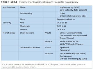
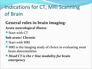
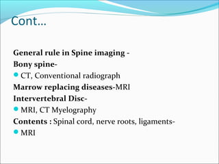
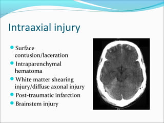
Dr. Sunil Kumar Sharma
Senior Resident
Dept. of Neurology
GMC Kota
Aim
ļ‚—To understand Indications and select the appropriate
imaging modality
ļ‚—To identify common traumatic emergency
abnormalities of brain.
ļ‚—To determine an immediate life threatening
abnormality on a CNS imaging
INTRODUCTION
ļ‚—Head trauma is the leading cause of death in people
under the age of 30.
ļ‚—Males have 2-3 x frequency of brain injury than
females
ļ‚—Due mainly to motor vehicle accidents and assaults
OUTLINE
ļ‚—Classification of traumatic brain injury
ļ‚—Clinical indications for imaging
ļ‚—Imaging technique
ļ‚—Extraaxial hemorrhage
ļ‚—Intraaxial injury
ļ‚—Brain herniations
Classification of traumatic brain
injury
Traumatic Brain Injury
Classification -
ļ‚—Primary & Secondary
ļ‚—Mechanism of injury-Penetrating, Blunt
ļ‚—Location-Intraaxial, Extraaxial
ļ‚—Clinical severity, GCS -Mild, Moderate, severe
Classification of TBI
ļ‚—Primary
ļ‚—Injury to scalp, skull fracture
ļ‚—Surface contusion/laceration
ļ‚—Intracranial hematoma
ļ‚—Diffuse axonal injury, diffuse vascular injury
Contd..
ļ‚—Secondary
ļ‚—Hypoxia-ischemia
ļ‚—swelling/edema
ļ‚—raised intracranial pressure
ļ‚—Meningitis/abscess
Imaging Modalities
ļ‚—Plain Radiography
ļ‚—Ultrasound
ļ‚—CT
ļ‚—MRI
ļ‚—Radionuclide studies
ļ‚—Angiography
Conventional X-rays
ļ‚—Uses a form of Ionizing radiation
ļ‚—Good in evaluating air containing structures, bones
and calcifications
Limitations
ļ‚—No depth information
ļ‚—Low soft tissue contrast
ļ‚—Radiation
Computed Tomography
ļ‚—Uses Ionizing radiation.
ļ‚—Cross-sectional image
ļ‚—Principle based on differential x-ray beam
attenuation by tissue
ļ‚—Image contrast, displayed as Gray scale –
Density/attenuation
Low attenuation-Darker
High Attenuation-Whiter
APPROACH TO CT BRAIN
ļ‚—Look at the scout film: ? Fracture of upper
cervical spine or skull
ļ‚—Look at bone windows to see fractures
ļ‚—Look for brain asymmetry
ļ‚—Look at sulci, Sylvian fissure and cisterns to
exclude subarachnoid hemorrhage
ļ‚—Look for subdural collection
ļ‚—Determine if mass is intraaxial (in the brain) or
extraaxial (outside)
Advantages of CT
ļ‚—High sensitivity for calcification
ļ‚—Easier and faster to perform.
ļ‚—Most sensitive for detection of acute hemorrhage
ļ‚—Increasing availability
ļ‚—No contraindications to emergency patient scanning
ļ‚—Quickest & most efficient screening technique in
acute traumatic setting
Disadvantages of CT
ļ‚—High radiation dose per examination
ļ‚—Children are more sensitive to radiation induced
cancers than adults
ļ‚—Bone marrow, thyroid, breast, and lung are at greatest
risk
ļ‚—Contrast related side effects
Factors to Consider When Determining
Need of CT in Patients with Head Injury
Indications for urgent CT scan include:
ļ‚—Evidence of skull fracture—basal, depressed,
or open
ļ‚—Abnormal results of neurologic examination
ļ‚—Seizure
ļ‚—Vomiting >1 time
ļ‚—High-risk mechanism (e.g., ejection from vehicle;
pedestrian or cyclist versus automobile)
ļ‚—Decreasing GCS score or persistently decreased GCS
score of <15.
Indications for lower threshold for CT scan
include:
ļ‚—Age >60 yr
ļ‚—Persistent anterograde amnesia
ļ‚—Retrograde amnesia >30 min
ļ‚—Coagulopathy
ļ‚—Fall >5 stairs or >3 feet
Cont..
ļ‚—Intoxication (examination unreliable)
ļ‚—LOC >30 min
ļ‚—Mechanism and location of injury
ļ‚—Social factors (e.g., abusive situation at home,
language barriers preclude accurate history)
Magnetic Resonance Imaging
Advantages of MRI
ļ‚—Uses non ionizing radiation
ļ‚—Less invasive technique
ļ‚—Multiplanar capability
ļ‚—Excellent soft tissue contrast
ļ‚—Ability to depict flowing blood without the need of IV
contrast administration
Disadvantages of MRI
ļ‚—Cost
ļ‚—Limited Availability
ļ‚—Length of the examination time
ļ‚—Physiological motion artifacts
ļ‚—Acoustic noise
Contraindications of MRI
ļ‚—Ferrous object: Oxygen tanks, wheelchairs in the scan
room is extremely dangerous
ļ‚—Pts. who have electrically &/or magnetically activated
implants: Cardiac pace makers, Implanted
defibrillators
ļ‚—Pts. with intracranial aneurysm clips
ļ‚—Metal within the eye
ļ‚—Some decorative tattoos
Indications for CT, MRI Scanning
of Brain
General rules in brain imaging-
Acute neurological illness-
ļ‚—Start with CT
Sub acute/ Chronic
ļ‚—Start with MRI
ļ‚—MRI is the imaging study of choice in evaluating most
brain abnormalities
ļ‚—Head CT is the 1st
line modality for brain
emergency
Cont…
General rule in Spine imaging -
Bony spine-
ļ‚—CT, Conventional radiograph
Marrow replacing diseases-MRI
Intervertebral Disc-
ļ‚—MRI, CT Myelography
Contents : Spinal cord, nerve roots, ligaments-
ļ‚—MRI
Extraaxial Injury
ļ‚—Skull fracture
ļ‚—Epidural hematoma
ļ‚—Subdural hematoma
ļ‚—Subarachnoid hemorrhage
Skull Fracture
ļ‚—Not predictive of intracranial injury
ļ‚—Absence does not exclude intracranial injury
ļ‚—Types-Linear, depressed, basal, comminuted
ļ‚—Risk of brain injury increases with the depth of
depression
Skull x-ray findings-
Linear fracture-
• Lucent sharply defined
line without sclerotic
margin
ļ‚—Mimics-Vascular
groove , sutures
Depressed fracture –
• Increased or double
density on x-ray
• Best evaluated by
CT
• >5mm, elevation
indicated
Epidural Hematoma
ļ‚—Blood collection within potential space between skull
inner table & dura mater
ļ‚—75 % at the temporal region
ļ‚—Usually (99%)at the coup side
ļ‚—Majority have Skull fracture 85-95% ; In children
often absent
ļ‚—Mostly arterial bleed ,Due to laceration of the middle
meningeal artery or dural veins
ļ‚—Expands and present rapidly -Tense distension
Radiological signs
ļ‚—Biconvex with a sharply defined margin
ļ‚—Does not cross suture line unless there is
fracture/diastasis
ļ‚—Mass effect +/-brain herniation
ļ‚—Displacement of sinus, falx from the skull
ļ‚—Adjacent skull fracture
MANAGEMENT OF EDH
ļ‚—EDH > 30 cm3
should be evacuated.
ļ‚—EDH < 30 cm3
and <15 mm thickness and < 5 mm
midline shift and GCS >8 may be managed
nonoperatively with serial CT
SUBDURAL HEMATOMA
ļ‚—Occurs between the dura and arachnoid
ļ‚—Can cross the sutures but not the dural reflections
ļ‚—Due to disruption of the bridging cortical veins
ļ‚—hyperdense(acute), isodense(subacute)
Hypodense(chronic),
Radiological signs
ļ‚—Crescent shape
ļ‚—The most common locations are the frontal and
parietal convexities.
ļ‚—Countercoup
ļ‚—Unlike an EDH, its spread is not limited by suture
lines; it can spread over the whole convexity, but it
almost never crosses the midline
ļ‚—Mass effect +/-brain herniation
ļ‚—Skull fracture in < 50 %
SUBDURAL HEMATOMA
MANAGEMENT OF SDH
ļ‚—Acute SDH with thickness > 10 mm or midline shift >
5mm should be evacuated
ļ‚—Patient in coma with a decrease in GCS by >2 points
with a SDH should undergo surgical evacuation.
SUBARACHNOID HEMORRAGE
ļ‚—Can originate from direct vessel injury, contused cortex
or intraventricular hemorrhage.
ļ‚—In contrast to aneurysmal SAH, the blood is superficial in
the cortex and not present in the basal cisterns.
ļ‚—Usually focal (but diffuse from aneurysm)
ļ‚—In some instances, for example, if blood is found in the
sylvian fissure, a vascular study (CTA,DSA) is needed to
rule out an aneurysm rupture.
ļ‚—Can lead to communicating hydrocephalus
Intraventricular hemorrhage
ļ‚—Most commonly due to rupture of subependymal
vessels
ļ‚—Can occur from reflux of SAH or contiguous
extension of an intracerebral hemorrhage
ļ‚—Look for blood-cerebrospinal fluid level in occipital
horns
Intraaxial injury
ļ‚—Surface
contusion/laceration
ļ‚—Intraparenchymal
hematoma
ļ‚—White matter shearing
injury/diffuse axonal injury
ļ‚—Post-traumatic infarction
ļ‚—Brainstem injury
CONTUSION/LACERATIONS
ļ‚—Most common source of traumatic SAH
ļ‚—Contusion: must involve the superficial gray
matter
ļ‚—Laceration: contusion + tear of pia-arachnoid
ļ‚—Affects the crests of gyri
ļ‚—Hemorrhage present ½ cases and occur at right
angles to the cortical surface
ļ‚—Located near the irregular bony contours: poles of
frontal lobes, temporal lobes, inferior cerebellar
hemispheres
Intraparenchymal hematoma
ļ‚—Focal collections of blood that most commonly arise
from shear-strain injury to intraparenchymal vessels.
ļ‚—Usually located in the frontotemporal white matter or
basal ganglia
ļ‚—DDx: DAI, hemorrhagic contusion
DIFFUSE AXONAL INJURYDIFFUSE AXONAL INJURY
ļ‚—Rarely detected on CT ( 20% of DAI lesions are
hemorrhagic)
ļ‚—MRI: T1, T2, T2 GRE, DWI
DAI
ļ‚—Due to acceleration/deceleration to whtie matter +
hypoxia
ļ‚—Patients have severe LOC at impact
ļ‚—Grade 1: axonal damage in WM only -67%
ļ‚—Grade 2: WM + corpus callosum (posterior > anterior)
– 21%
ļ‚—Grade 3: WM + CC + brainstem
DTI
Traumatic cerebral infarct
ļ‚—Best diagnosed by: Restricted diffusion
Location-
ļ‚— Most commonly occur in PCA vascular distribution,
ļ‚— MCA, ACA, vertebrobasilar relatively common
ļ‚— Other: Lenticulostriate, thalamoperforating;
cortical/subcortical; cerebellar
Traumatic cerebral infarct
SUBFALCIAL HERNIATION
ļ‚—Subfalcial: displacement of the cingulate gyrus under
the free edge of the falx along with the pericallosal
arteries.
ļ‚—Can lead to anterior cerebral artery infarction
DESCENDING HERNIATION
Herniation:
Ascending Transtentorial
ļ‚—Cranial shift of
vermis and parts of
superomedial
cerebellar
hemisphere through
tentorium incisura
ļ‚— Compressed
superior cerebellar,
vermian cisterns and
forth ventricle
TONSILLAR HERNIATION
ļ‚—Inferior displacement of the cerebellar tonsils
through the foramen magnum
ļ‚—Can lead to posterior cerebellar artery infarction
EXTERNAL HERNIATION
ļ‚—Due to a defect in the
skull in combination
with elevated ICP
ļ‚—Venous obstruction
can occur at the
margins of the defect.
Conclusions
ļ‚— CT = primary modality for head trauma, enough for
most parts
ļ‚— Skull x-rays still used in penetrating trauma,
suspected child abuse
ļ‚—MRI to help predicting prognosis by detection of
subtle injuries i.e., contusion and DAI
Primary vs secondary lesion.
Often, secondary lesion more important
Conclusions
ļ‚—While checking the scan, make sure to think if the
patient needs CTA or other CTs (C-spine, facial
bones,
etc)
ļ‚— Coup-contrecoup mechanism helps confirm acute
trauma nature and search for subtle lesions
Thank you
Thank you
References
ļ‚—Osborn Diagnostic Imaging Brain-2004
ļ‚—Bradley’s Neurology in Clinical Practice 6’th edition
2012
ļ‚—Slideshare.com
ļ‚—Radiopedia.com

Imaging in head trauma

  • 1.
    Dr. Sunil KumarSharma Senior Resident Dept. of Neurology GMC Kota
  • 2.
    Aim ļ‚—To understand Indicationsand select the appropriate imaging modality ļ‚—To identify common traumatic emergency abnormalities of brain. ļ‚—To determine an immediate life threatening abnormality on a CNS imaging
  • 3.
    INTRODUCTION ļ‚—Head trauma isthe leading cause of death in people under the age of 30. ļ‚—Males have 2-3 x frequency of brain injury than females ļ‚—Due mainly to motor vehicle accidents and assaults
  • 4.
    OUTLINE ļ‚—Classification of traumaticbrain injury ļ‚—Clinical indications for imaging ļ‚—Imaging technique ļ‚—Extraaxial hemorrhage ļ‚—Intraaxial injury ļ‚—Brain herniations
  • 5.
  • 6.
    Traumatic Brain Injury Classification- ļ‚—Primary & Secondary ļ‚—Mechanism of injury-Penetrating, Blunt ļ‚—Location-Intraaxial, Extraaxial ļ‚—Clinical severity, GCS -Mild, Moderate, severe
  • 7.
    Classification of TBI ļ‚—Primary ļ‚—Injuryto scalp, skull fracture ļ‚—Surface contusion/laceration ļ‚—Intracranial hematoma ļ‚—Diffuse axonal injury, diffuse vascular injury
  • 8.
  • 10.
  • 11.
    Conventional X-rays ļ‚—Uses aform of Ionizing radiation ļ‚—Good in evaluating air containing structures, bones and calcifications Limitations ļ‚—No depth information ļ‚—Low soft tissue contrast ļ‚—Radiation
  • 13.
    Computed Tomography ļ‚—Uses Ionizingradiation. ļ‚—Cross-sectional image ļ‚—Principle based on differential x-ray beam attenuation by tissue ļ‚—Image contrast, displayed as Gray scale – Density/attenuation Low attenuation-Darker High Attenuation-Whiter
  • 14.
    APPROACH TO CTBRAIN ļ‚—Look at the scout film: ? Fracture of upper cervical spine or skull ļ‚—Look at bone windows to see fractures ļ‚—Look for brain asymmetry ļ‚—Look at sulci, Sylvian fissure and cisterns to exclude subarachnoid hemorrhage ļ‚—Look for subdural collection ļ‚—Determine if mass is intraaxial (in the brain) or extraaxial (outside)
  • 15.
    Advantages of CT ļ‚—Highsensitivity for calcification ļ‚—Easier and faster to perform. ļ‚—Most sensitive for detection of acute hemorrhage ļ‚—Increasing availability ļ‚—No contraindications to emergency patient scanning ļ‚—Quickest & most efficient screening technique in acute traumatic setting
  • 16.
    Disadvantages of CT ļ‚—Highradiation dose per examination ļ‚—Children are more sensitive to radiation induced cancers than adults ļ‚—Bone marrow, thyroid, breast, and lung are at greatest risk ļ‚—Contrast related side effects
  • 17.
    Factors to ConsiderWhen Determining Need of CT in Patients with Head Injury Indications for urgent CT scan include: ļ‚—Evidence of skull fracture—basal, depressed, or open ļ‚—Abnormal results of neurologic examination ļ‚—Seizure ļ‚—Vomiting >1 time
  • 18.
    ļ‚—High-risk mechanism (e.g.,ejection from vehicle; pedestrian or cyclist versus automobile) ļ‚—Decreasing GCS score or persistently decreased GCS score of <15.
  • 19.
    Indications for lowerthreshold for CT scan include: ļ‚—Age >60 yr ļ‚—Persistent anterograde amnesia ļ‚—Retrograde amnesia >30 min ļ‚—Coagulopathy ļ‚—Fall >5 stairs or >3 feet
  • 20.
    Cont.. ļ‚—Intoxication (examination unreliable) ļ‚—LOC>30 min ļ‚—Mechanism and location of injury ļ‚—Social factors (e.g., abusive situation at home, language barriers preclude accurate history)
  • 21.
  • 22.
    Advantages of MRI ļ‚—Usesnon ionizing radiation ļ‚—Less invasive technique ļ‚—Multiplanar capability ļ‚—Excellent soft tissue contrast ļ‚—Ability to depict flowing blood without the need of IV contrast administration
  • 23.
    Disadvantages of MRI ļ‚—Cost ļ‚—LimitedAvailability ļ‚—Length of the examination time ļ‚—Physiological motion artifacts ļ‚—Acoustic noise
  • 24.
    Contraindications of MRI ļ‚—Ferrousobject: Oxygen tanks, wheelchairs in the scan room is extremely dangerous ļ‚—Pts. who have electrically &/or magnetically activated implants: Cardiac pace makers, Implanted defibrillators ļ‚—Pts. with intracranial aneurysm clips ļ‚—Metal within the eye ļ‚—Some decorative tattoos
  • 25.
    Indications for CT,MRI Scanning of Brain General rules in brain imaging- Acute neurological illness- ļ‚—Start with CT Sub acute/ Chronic ļ‚—Start with MRI ļ‚—MRI is the imaging study of choice in evaluating most brain abnormalities ļ‚—Head CT is the 1st line modality for brain emergency
  • 26.
    Cont… General rule inSpine imaging - Bony spine- ļ‚—CT, Conventional radiograph Marrow replacing diseases-MRI Intervertebral Disc- ļ‚—MRI, CT Myelography Contents : Spinal cord, nerve roots, ligaments- ļ‚—MRI
  • 27.
    Extraaxial Injury ļ‚—Skull fracture ļ‚—Epiduralhematoma ļ‚—Subdural hematoma ļ‚—Subarachnoid hemorrhage
  • 28.
    Skull Fracture ļ‚—Not predictiveof intracranial injury ļ‚—Absence does not exclude intracranial injury ļ‚—Types-Linear, depressed, basal, comminuted ļ‚—Risk of brain injury increases with the depth of depression
  • 29.
    Skull x-ray findings- Linearfracture- • Lucent sharply defined line without sclerotic margin ļ‚—Mimics-Vascular groove , sutures
  • 30.
    Depressed fracture – •Increased or double density on x-ray • Best evaluated by CT • >5mm, elevation indicated
  • 31.
    Epidural Hematoma ļ‚—Blood collectionwithin potential space between skull inner table & dura mater ļ‚—75 % at the temporal region ļ‚—Usually (99%)at the coup side ļ‚—Majority have Skull fracture 85-95% ; In children often absent ļ‚—Mostly arterial bleed ,Due to laceration of the middle meningeal artery or dural veins ļ‚—Expands and present rapidly -Tense distension
  • 32.
    Radiological signs ļ‚—Biconvex witha sharply defined margin ļ‚—Does not cross suture line unless there is fracture/diastasis ļ‚—Mass effect +/-brain herniation ļ‚—Displacement of sinus, falx from the skull ļ‚—Adjacent skull fracture
  • 34.
    MANAGEMENT OF EDH ļ‚—EDH> 30 cm3 should be evacuated. ļ‚—EDH < 30 cm3 and <15 mm thickness and < 5 mm midline shift and GCS >8 may be managed nonoperatively with serial CT
  • 35.
    SUBDURAL HEMATOMA ļ‚—Occurs betweenthe dura and arachnoid ļ‚—Can cross the sutures but not the dural reflections ļ‚—Due to disruption of the bridging cortical veins ļ‚—hyperdense(acute), isodense(subacute) Hypodense(chronic),
  • 36.
    Radiological signs ļ‚—Crescent shape ļ‚—Themost common locations are the frontal and parietal convexities. ļ‚—Countercoup ļ‚—Unlike an EDH, its spread is not limited by suture lines; it can spread over the whole convexity, but it almost never crosses the midline ļ‚—Mass effect +/-brain herniation ļ‚—Skull fracture in < 50 %
  • 37.
  • 38.
    MANAGEMENT OF SDH ļ‚—AcuteSDH with thickness > 10 mm or midline shift > 5mm should be evacuated ļ‚—Patient in coma with a decrease in GCS by >2 points with a SDH should undergo surgical evacuation.
  • 39.
    SUBARACHNOID HEMORRAGE ļ‚—Can originatefrom direct vessel injury, contused cortex or intraventricular hemorrhage. ļ‚—In contrast to aneurysmal SAH, the blood is superficial in the cortex and not present in the basal cisterns. ļ‚—Usually focal (but diffuse from aneurysm) ļ‚—In some instances, for example, if blood is found in the sylvian fissure, a vascular study (CTA,DSA) is needed to rule out an aneurysm rupture. ļ‚—Can lead to communicating hydrocephalus
  • 41.
    Intraventricular hemorrhage ļ‚—Most commonlydue to rupture of subependymal vessels ļ‚—Can occur from reflux of SAH or contiguous extension of an intracerebral hemorrhage ļ‚—Look for blood-cerebrospinal fluid level in occipital horns
  • 43.
    Intraaxial injury ļ‚—Surface contusion/laceration ļ‚—Intraparenchymal hematoma ļ‚—White mattershearing injury/diffuse axonal injury ļ‚—Post-traumatic infarction ļ‚—Brainstem injury
  • 44.
    CONTUSION/LACERATIONS ļ‚—Most common sourceof traumatic SAH ļ‚—Contusion: must involve the superficial gray matter ļ‚—Laceration: contusion + tear of pia-arachnoid ļ‚—Affects the crests of gyri ļ‚—Hemorrhage present ½ cases and occur at right angles to the cortical surface ļ‚—Located near the irregular bony contours: poles of frontal lobes, temporal lobes, inferior cerebellar hemispheres
  • 46.
    Intraparenchymal hematoma ļ‚—Focal collectionsof blood that most commonly arise from shear-strain injury to intraparenchymal vessels. ļ‚—Usually located in the frontotemporal white matter or basal ganglia ļ‚—DDx: DAI, hemorrhagic contusion
  • 48.
    DIFFUSE AXONAL INJURYDIFFUSEAXONAL INJURY ļ‚—Rarely detected on CT ( 20% of DAI lesions are hemorrhagic) ļ‚—MRI: T1, T2, T2 GRE, DWI
  • 49.
    DAI ļ‚—Due to acceleration/decelerationto whtie matter + hypoxia ļ‚—Patients have severe LOC at impact ļ‚—Grade 1: axonal damage in WM only -67% ļ‚—Grade 2: WM + corpus callosum (posterior > anterior) – 21% ļ‚—Grade 3: WM + CC + brainstem
  • 52.
  • 54.
    Traumatic cerebral infarct ļ‚—Bestdiagnosed by: Restricted diffusion Location- ļ‚— Most commonly occur in PCA vascular distribution, ļ‚— MCA, ACA, vertebrobasilar relatively common ļ‚— Other: Lenticulostriate, thalamoperforating; cortical/subcortical; cerebellar
  • 55.
  • 57.
    SUBFALCIAL HERNIATION ļ‚—Subfalcial: displacementof the cingulate gyrus under the free edge of the falx along with the pericallosal arteries. ļ‚—Can lead to anterior cerebral artery infarction
  • 58.
  • 59.
    Herniation: Ascending Transtentorial ļ‚—Cranial shiftof vermis and parts of superomedial cerebellar hemisphere through tentorium incisura ļ‚— Compressed superior cerebellar, vermian cisterns and forth ventricle
  • 61.
    TONSILLAR HERNIATION ļ‚—Inferior displacementof the cerebellar tonsils through the foramen magnum ļ‚—Can lead to posterior cerebellar artery infarction
  • 63.
    EXTERNAL HERNIATION ļ‚—Due toa defect in the skull in combination with elevated ICP ļ‚—Venous obstruction can occur at the margins of the defect.
  • 64.
    Conclusions ļ‚— CT =primary modality for head trauma, enough for most parts ļ‚— Skull x-rays still used in penetrating trauma, suspected child abuse ļ‚—MRI to help predicting prognosis by detection of subtle injuries i.e., contusion and DAI Primary vs secondary lesion. Often, secondary lesion more important
  • 65.
    Conclusions ļ‚—While checking thescan, make sure to think if the patient needs CTA or other CTs (C-spine, facial bones, etc) ļ‚— Coup-contrecoup mechanism helps confirm acute trauma nature and search for subtle lesions
  • 66.
  • 67.
    References ļ‚—Osborn Diagnostic ImagingBrain-2004 ļ‚—Bradley’s Neurology in Clinical Practice 6’th edition 2012 ļ‚—Slideshare.com ļ‚—Radiopedia.com