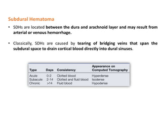
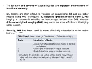
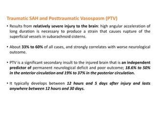
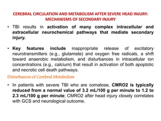
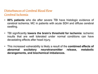
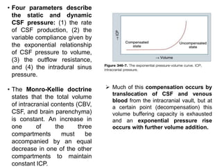
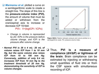
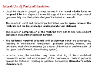
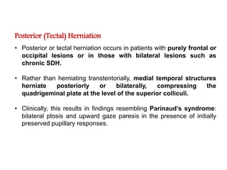
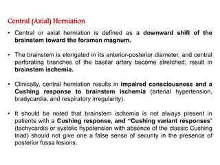
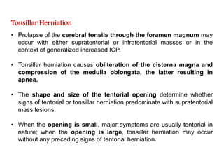
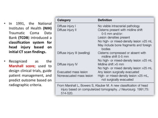
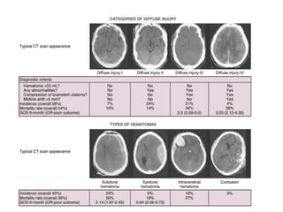
Dr. Shahnawaz Alam
MCh-Neurosurgery; SR
Traumatic Brain Injury
Moderated by:
Dr. V. C. Jha
HOD, Dept. of Neurosurgery
• The initial mechanical insult of traumatic brain injury (TBI) results in
tissue deformation that causes damage to neurons, glia, axons, and
blood vessels.
• This is followed by a more delayed phase of injury, which is
mediated by intracellular and extracellular biologic pathways.
• During this phase, many patients experience superimposed
secondary insults such as hypoxia, hypotension, cerebral swelling,
and the consequences of increased intracranial pressure (ICP).
• These secondary insults further exacerbate TBI and have a profound
negative effect on patient outcome.
• An understanding of the sequelae of TBI and secondary insults is
paramount for managing head injury.
 In a recent report from the International Mission for Prognosis and
Analysis of Clinical Trials in TBI (IMPACT) database, hypoxia and
hypotension were present on admission in 20% and 18% of TBI patients.
 Five insults consistently correlated with poor outcome in this
study: arterial hypo- tension, reduced cerebral perfusion pressure
(CPP), elevated ICP, hypoxemia, and pyrexia.
• Treating secondary injury mechanisms and preventing secondary
insults remains a focus of TBI management, and significant
advancements have been made in prehospital care, resuscitation, and
rapid radiologic diagnosis.
MECHANISMS OF BRAIN INJURY
 Bayly et al. have studied the effects of mild linear and angular head
acceleration on brain deformation in healthy volunteers. Their data suggest that
mechanical responses are mediated by divisions between brain regions (e.g.,
central sulcus), dural reflections (e.g., falx cerebri, tentorium cerebelli), and
tethering of the brain at the sella and suprasellar regions.
• Critical components include the nature of the force (contact or inertial loading),
the type of injury (rotational, translational, or angular), and the magnitude and
duration of the impact.
• Although one process may predominate, most patients with TBI experience a
combination of these mechanisms.
• Contact forces typically result in focal injuries such as coup contusions and skull
fractures.
• Inertial loading forces that are primarily translational also result in focal injuries,
such as contusions and subdural hematomas (SDHs), whereas rotational
acceleration-deceleration injuries are more likely to result in diffuse injuries
ranging from concussion to diffuse axonal injury (DAI).
• Rotational injuries are particularly concerning because they cause injury to both
the cortical surface and deep brain structures. Diffusion tensor imaging (DTI)
techniques have been used to better visualize the distribution and severity of
white matter fiber injuries after DAI.
• Angular acceleration represents a combination
of translational and rotational acceleration and
is the most common form of inertial injury.
• Because of the biomechanical properties of the
head and neck, TBI often results in deflection
of the head and neck around the middle or
lower cervical spine (the center of angular
movement).
• The resultant magnitude of rotation that occurs
with this injury depends on the distance
between the center of gravity and the center
of angulation: the smaller the distance, the
larger the rotational component of angulation.
Mechanism of angular
acceleration: interaction of
head and neck.
• The extent of injury in TBI is also determined, in large part, by the magnitude
and duration of the insult mechanism.
• A low magnitude of acceleration with a long duration results in DAI owing
to propagation of the forces deep within the brain; typically seen with motor
vehicle collisions.
• In contrast, a brief, high-velocity impact often results in tearing of
superficially located bridging veins and pial vessels, causing SDH; occurs in
falls or assaults in which the head strikes a broad, hard surface.
CLASSIFICATION OF HEAD INJURY
• Clinical condition and level of consciousness after TBI are typically described
using the Glasgow Coma Scale; Universally adopted for grading the clinical
severity of head injuries and other pathologies that impair consciousness.
• GCS plays an important role in categorizing injury severity, allowing for
standardized determination of clinical neurological status, and detecting
episodes of neurological deterioration.
• However, that the usefulness of GCS is somewhat limited with modern
therapies. For example, most patients arrive to the hospital by ambulance
unresponsive because of sedation and neuromuscular blockade.
 In a study by Gale et al., 50% of patients could not be assigned an accurate GCS
score owing to these confounding variables.
• An alternate scale for scoring clinical condition is the head injury severity
scale, but it faces similar challenges because it is largely based on the GCS
score.
• TBI can also be classified anatomically into focal or diffuse injury patterns.
• In 1991, the National
Institutes of Health (NIH)
Traumatic Coma Data
Bank (TCDB) introduced a
classification system for
head injury based on
initial CT scan findings.
• Recognized as the
Marshall score; used to
design clinical trials, guide
patient management, and
predict outcome based on
radiographic criteria.
 More recently, Maas et al.
proposed a modified version of the
Marshall score (termed the
Rotterdam score) to account for
additional radiographic criteria
that more accurately predict
survival from head injury.
 These systems describe a
strong correlation between
CT scan findings (e.g.,
compression of the basal
cisterns, presence of
subarachnoid hemorrhage
[SAH], midline shift) and
clinical course, mortality, and
functional outcome after TBI.
• Because determining an accurate post-resuscitation GCS score is extremely
difficult in the current era, it is likely that radiographic scoring systems will play a
larger role in predicting outcome and directing care in the acute period after
TBI.
FOCAL BRAIN INJURY: Brain Contusion /
Traumatic Intracranial Hematoma
Brain Contusion
• Represent focal regions of subpial hemorrhage and swelling and are present
in 22% to 31% of patients on initial CT scan.
• MC in regions that contact bony surfaces in the cranial vault during trauma:
frontal and temporal poles, orbitofrontal gyri, perisylvian cortices, and
inferolateral temporal lobe surfaces.
• Characterized by mechanism, anatomic location, or adjacent injuries.
• For example, fracture contusions/ Coup contusions / contrecoup contusions /
Gliding contusions / Intermediary contusions / Herniation contusions .
 fracture contusions result from direct contact injuries and occur immediately
adjacent to a skull fracture.
 Coup contusions refer to those that occur at the site of impact in the absence of
a fracture, whereas contrecoup contusions are those that are diametrically
opposite to the point of impact.
 Gliding contusions are focal hemorrhages involving the cortex and adjacent
white matter of the superior margins of the cerebral hemispheres; they are
caused by rotational mechanisms rather than contact forces.
 Intermediary contusions are lesions that affect deep brain structures, such as
the corpus callosum, basal ganglia, hypothalamus, and brainstem.
 Herniation contusions can occur in areas where the medial parts of the
temporal lobe contact the tentorial edge (i.e., uncal herniation) or where the
cerebellar tonsils contact the foramen magnum (i.e., tonsillar herniation).
• Contusions typically result in varying
degrees of neurological deficits; Can
cause significant mass effect owing to
surrounding edema or hemorrhagic
progression to an intracerebral
hematoma (ICH).
• Also represent a significant source of
secondary injury to adjacent tissue via
release of neurotransmitter and local
biochemical changes.
Contusions are more severe when associated
with a skull fracture, less severe in patients
with DAI, and more severe in patients who do
not experience a lucid interval.
Traumatic Intracranial Hematoma
• Approximately half of patients with severe TBI and skull fracture have a
sizeable intracranial hematoma on initial head CT.
• The three major types of traumatic intracranial hematomas are distinguished
by their location relative to the meninges: epidural, subdural, and
intracerebral.
Epidural Hematoma
• Occur in 10.6% of TBI patients admitted to the hospital and account for 5%
to 15% of fatal head injuries; MC in patients younger than 50 years,
although they do occur in all age groups.
• In adults, EDH is far less common than SDH or ICH. In pediatric patients,
however, EDH is 1.96 times more common after TBI; this is likely because of
abundant diploic and dural vascularization normally present in infants and
young children.
• The classic EDH occurs beneath a temporoparietal skull fracture as a result
of damage to the middle meningeal artery; “stripping” of the dura from the
inner table because of clot enlargement.
• They rarely occur spontaneously in patients with infections, sinusitis,
vascular anomalies, or chronic renal failure.
• EDHs can be classified by their radiographic progression into three
appearances:
1. Type I (acute or hyperacute-day 1, associated with “swirl” of unclotted
blood); occur in 58%.
2. Type II (subacute-days 2-4, solid); 31% and
3. Type III (chronic- days 7-20, mixed or lucent with contrast
enhancement); 11%.
• The classic clinical course of a patient with EDH was first described by
Jacobson in 1886; initial LOC after trauma, transient complete recovery
(“lucid interval”), then rapid progression of neurological deterioration; this
classic presentation occurs in only 14% to 21% of patients with an EDH.
• The classic lucid interval is most common in pure EDHs that are very large and
demonstrate CT signs of active bleeding (type I).
• Neurological deterioration from an expanding EDH typically results in
obtundation, contralateral hemiparesis, ipsilateral oculomotor nerve paresis,
decerebrate rigidity, arterial hypertension, cardiac arrhythmias, respiratory
disturbances, and, finally, apnea and death.
• Development of these symptoms depends on hematoma size and the
presence of associated intracranial lesions.
• Patients with pure EDHs have an excellent prognosis after surgical
evacuation, whereas those with associated intradural lesions experience good
outcome in only 44% of cases.
• Although rapid diagnosis and evacuation are critical factors, data suggest
that appropriate treatment (by a neurosurgeon, as compared with a general
surgeon in remote areas) is also important in determining patient outcome.
 Lee et al. found that patients with EDH volume >50 cc before evacuation
experience worse neurological outcome and increased mortality.
• EDHs in the posterior fossa accounting for ≈5% of all posttraumatic intracranial
mass lesions; challenging to manage because these patients may remain
conscious until late in the evolution of the hematoma, when they may suddenly
lose consciousness, become apneic, and die.
• Outcome in patients with posterior fossa EDH is generally better in children
and correlates with GCS score on admission and CT evidence of hydrocephalus
(caused by compression of the fourth ventricle).
Subdural Hematoma
• SDHs are located between the dura and arachnoid layer and may result from
arterial or venous hemorrhage.
• Classically, SDHs are caused by tearing of bridging veins that span the
subdural space to drain cortical blood directly into dural sinuses.
Acute Subdural Hematoma
• Account for 50% to 60% of all SDHs; MC after sudden head movements
that occur with assaults or falls.
• Most acute SDHs result from venous vascular injury at the brain surface,
resulting in two distinct pathologies.
1. The first type of hematoma, produced by contact forces and
associated with contusions or lacerations, results from cortical
bleeding into the adjacent subdural space and is most common at the
temporal pole. This complex of SDH and damaged and necrotic brain
is termed burst lobe.
2. The second type of SDH is located over the cerebral convexity and is
produced by inertial forces that tear bridging veins. The underlying
brain damage in this type of injury is usually milder, and primarily
caused by local ischemia from mass effect or compromised venous
outflow.
• Despite the often relatively minor underlying brain damage, prognosis is
generally poor in these patients unless the hematoma is rapidly evacuated.
• Cerebral ischemia plays a critical role in the pathology of SDH; likely
related to compressive effects of the hematoma and elevated ICP with
resultant compromised CPP.
• Timely clot evacuation (within 4 hours) generally results in significantly
improved neurological outcome. Patients with initial CT evidence of
significant hemispheric or generalized brain swelling have extremely poor
outcome with or without early surgery.
• The prognosis of SDH is still poor in many cases. It is thought that the
coexisting brain damage (DAI, contusion, laceration) is responsible for poor
neurological function after injury.
Intracerebral Hematoma
• Account for 20% to 30% of all traumatic intracranial hematomas;
Associated with extensive lobar contusions.
• By definition, an ICH is a parenchymal lesion composed of at least
two thirds blood; otherwise, the lesion is described as disrupted
tissue with areas of microscopic hemorrhage; often result from growth
and/or coalescence of smaller cerebral contusions.
• A hemorrhagic mass should be considered an ICH when there is a
homogeneous collection of blood with relatively well-defined
margins. Multiple ICHs are found in approximately 20% of TBI
patients.
• Because ICHs typically result from rupture of intrinsic cerebral
vessels; most traumatic ICHs occur in the orbitofrontal and
temporal lobes, as do most cerebral contusions.
• Deeper ICHs, such as those occurring in BG& IC , are less common
and found in approximately 2% of TBI patients.
• ICHs are most common in focal head injuries, such as missile
injuries, perforating wounds, and depressed skull fractures.
DIFFUSE BRAIN INJURY : Concussion/ DAI
Concussion
• Mildest form of diffuse injury and is thought to be caused by
rotational acceleration of the head in the absence of significant
mechanical contact.
• In its classic form, patients with concussion experience a transient
LOC f/b a rapid return to a normal state of alertness.
• The pathophysiology of concussion is poorly understood and may be
related to disturbances of consciousness from lesions of the
brainstem and diencephalon.
• DTI reveals signs of cytotoxic edema in the brain despite a normal
head CT and GCS of 15.
Diffuse Axonal Injury
 DAI was described by Strich in 1956 in his report of a series of patients
with severe posttraumatic dementia and “diffuse degeneration of
the white matter.”
• Results from severe angular and rotational acceleration and
deceleration that delivers shear and tensile forces to axons; Result in
severe impairment despite lack of gross parenchymal contusions,
lacerations, or hematomas.
• Coronal or lateral acceleration injuries produce the most severe
DAIs, whereas acceleration in the oblique or sagittal plane results in
less severe to minimal DAI.
• The histologic findings of DAI include disruption and swelling of
axons, “retraction balls” (swollen proximal ends of severed axons),
and punctate hemorrhages in the pons, midbrain, and corpus
callosum.
• The location and severity of axonal injuries are important determinants of
functional recovery.
• DAI lesions are often difficult to visualize on conventional CT and are better
imaged using MRI techniques. T2-weighted gradient-recalled echo (GRE)
imaging is particularly sensitive for hemorrhagic lesions after DAI, whereas
diffusion-weighted imaging (DWI) sequences are more effective in identifying
shear injuries.
• Recently, DTI has been used to more effectively characterize white matter
lesions after TBI.
Traumatic SAH and Posttraumatic Vasospasm (PTV)
• Results from relatively severe injury to the brain: high angular acceleration of
long duration is necessary to produce a strain that causes rupture of the
superficial vessels in subarachnoid cisterns.
• About 33% to 60% of all cases, and strongly correlates with worse neurological
outcome.
• PTV is a significant secondary insult to the injured brain that is an independent
predictor of permanent neurological deficit and poor outcome; 18.6% to 50%
in the anterior circulation and 19% to 37% in the posterior circulation.
• It typically develops between 12 hours and 5 days after injury and lasts
anywhere between 12 hours and 30 days.
Intraventricular Hemorrhage
• Less than 10% of patients with severe TBI; more likely to demonstrate
intraparenchymal and BG hemorrhages.
• Most patients with primary IVH (no significant parenchymal blood) had a
high incidence of damage to the septum pellucidum, choroid plexus, and
subependymal vein in the fornix.
• Although traumatic IVH has the potential of obstructing CSF flow, acute
hydrocephalus is an uncommon manifestation.
CEREBRAL CIRCULATION AND METABOLISM AFTER SEVERE HEAD INJURY:
MECHANISMS OF SECONDARY INJURY
• TBI results in activation of many complex intracellular and
extracellular neurochemical pathways that mediate secondary
injury.
• Key features include inappropriate release of excitatory
neurotransmitters (e.g., glutamate) and oxygen free radicals, a shift
toward anaerobic metabolism, and disturbances in intracellular ion
concentrations (e.g., calcium) that result in activation of both apoptotic
and necrotic cell death pathways.
Disturbances of Cerebral Metabolism
• In patients with severe TBI who are comatose, CMRO2 is typically
reduced from a normal value of 3.2 mL/100 g per minute to 1.2 to
2.3 mL/100 g per minute; CMRO2 after head injury closely correlates
with GCS and neurological outcome.
Disturbances of Cerebral Blood Flow
Cerebral Ischemia
• 80% patients who die after severe TBI have histologic evidence of
cerebral ischemia; MC in patients with acute SDH and diffuse cerebral
swelling.
• TBI significantly lowers the brain’s threshold for ischemia: ischemic
insults that are well tolerated under normal conditions can have
devastating effects after head injury.
• This increased vulnerability is likely a result of the combined effects of
abnormal excitatory neurotransmitter release, metabolic
derangements, and biochemical imbalances.
• In an arterial occlusion model of
ischemia, the CBF threshold at
which these changes occur is at
18 mL/100 g per minute; After
severe TBI, however, this
threshold is likely elevated to
approximately 20 mL/100 g
per minute. Neuronal
dysfunction and death depend
on both the duration and
magnitude of CBF depression
after injury.
Metabolic Autoregulation
• Although comatose patients typically experience reduction of
CMRO2 from a normal value of 3.3 mL/100 g per minute to
approximately 2.1 mL/100 g per minute, this is not always accompanied
by a proportional decrease in CBF.
• Metabolic uncoupling occurs when CBF exceeds CMRO2, a
phenomenon termed luxury perfusion or hyperemia.
 Obrist et al. defined luxury perfusion in comatose head-injured
patients as CBF above 33 mL/100 g per minute; CBF of 33 to 55
mL/100 g per minute represents relative hyperemia, and CBF above
55 mL/100 g per minute is absolute hyperemia.
• Hyperemia is a critical concept in TBI because it is strongly
associated with diffuse cerebral swelling and elevated ICP.
• In fact, in patients with acute SDH, elevated ICP, and cerebral
ischemia, CBV is half of normal.
Cerebral Blood Flow, Cerebral Blood Volume,
Arteriovenous Difference in Oxygen and Autoregulation
 Decreased cerebral metabolism results in a coupled decrease in CBF via vasoconstriction
(metabolic autoregulation): ICP decreases.
 Reduced CPP (reduction in MAP and/or elevation of ICP) results in compensatory
vasodilatation when pressure autoregulation is intact: ICP increases.
 Reduced CPP in the context of defective autoregulation results in passive decreases of
CBF and CBV: ICP decreases.
 Reduced blood viscosity, as obtained with mannitol administration, results in vasoconstriction when
viscosity autoregulation is intact: ICP decreases.
 Reduced blood viscosity in the context of impaired viscosity autoregulation, however, does not
induce a vascular response: ICP does not significantly change.
 Hyperventilation or hypocapnia normally results in vasoconstriction and reduction in CBV, causing a
reduction in ICP.
 Large-artery vasospasm decreases the diameter of the macrocirculation, resulting in reduced
perfusion pressure. This triggers compensatory vasodilatation of the microcirculation, leading to
elevated CBV in the presence of normal or reduced CBF. ICP may increase.
Elevated Intracranial Pressure
• Elevated ICP (above 20 mm Hg) is a common complication of severe
TBI that is persistent in 50% of patients with intracranial mass lesions and
33% of those with diffuse injuries; ICP above 20 is a significant
independent determinant of outcome.
• Patients whose ICP is maintained below 20 mm Hg have significantly
better outcome.
 A recent systematic review found that ICP of 20 to 40 increases
mortality by 3.5-fold and ICP above 40 increases mortality by 6.9-fold;
raised but reducible ICP increases mortality by three-fold to fourfold.
• ICP is often measured from CSF pressure, which is defined as the
pressure one must exert against a needle introduced into the CSF
space to prevent escape of fluid.
• Four parameters describe
the static and dynamic
CSF pressure: (1) the rate
of CSF production, (2) the
variable compliance given by
the exponential relationship
of CSF pressure to volume,
(3) the outflow resistance,
and (4) the intradural sinus
pressure.
• The Monro-Kellie doctrine
states that the total volume
of intracranial contents (CBV,
CSF, and brain parenchyma)
is constant. An increase in
one of the three
compartments must be
accompanied by an equal
decrease in one of the other
compartments to maintain
constant ICP.
 Much of this compensation occurs by
translocation of CSF and venous
blood from the intracranial vault, but at
a certain point (decompensation) this
volume buffering capacity is exhausted
and an exponential pressure rise
occurs with further volume addition.
 Marmarou et al. plotted a curve on
a semilogarithmic scale to create a
straight line. The slope of this line is
the pressure-volume index (PVI),
the amount of volume that must be
added or withdrawn from the
craniospinal axis to increase or
decrease ICP 10-fold:
PVI V÷(logICPi / ICPo)
 Change in volume is represented
by ΔV, ICPo is the pressure before
volume change, and ICPi is the
pressure after volume change.
• Normal PVI is 26 ± 4 mL: 26 mL of
volume raises ICP from 1 to 10 mm
Hg, but the same volume also raises
ICP from 10 to 100 mm Hg;
Conversely, addition of only 6.4 mL
increases ICP from 10 mm Hg to the
treatment threshold of 20 mm Hg,
demonstrating the sensitivity of ICP to
volume changes.
 Thus, PVI is a measure of
compliance (ΔV/ΔP) or tightness of
the brain. Brain compliance can be
estimated by injecting or withdrawing
small quantities of fluid into or from
the CSF space with simultaneous
recording of ICP.
• The following five pathways of intracranial volume increase were
described by Marmarou: (1) CSF system, (2) CBV, (3) blood-brain
barrier damage-associated edema (vasogenic edema), (4) neurotoxic
edema, and (5) ischemic edema.
• CSF components (CSF resistance to outflow and absorption) account
for approximately one third of ICP elevation.
• This component can increase substantially in patients with SAH
who experience outflow resistance owing to blockage of CSF flow
through arachnoid villi.
• The remaining two thirds of ICP elevation is attributed to a vascular
component: increased blood volume (pathway 2) and increased tissue
water (vasogenic, neurotoxic, and ischemic edema: pathways 3, 4, and
5).
• Neurotoxic and ischemic edema are thought to be of cellular origin,
whereas edema caused by blood-brain barrier damage is
extracellular in nature.
• CBV is determined by the total diameter of the cerebrovascular
bed. Most blood volume is contained in cerebral veins, with only 20
mL (one third of total CBV) located in cerebral resistance vessels (15-
300 μm).
• Autoregulatory and CO2-dependent responses result in diameter
variations between 80% and 160% of baseline, resulting in volume
changes between 64% and 256% of baseline.
• With a baseline CBV of 20 mL in the resistance vessels, CBV ranges
from 13 mL (maximal vasoconstriction) to 51 mL (maximal
vasodilatation).
 Klatzo categorized cellular edema into neurotoxic and ischemic:
 Ischemic edema is primarily caused by disturbance of cellular
osmoregulation from ionic pump failure. Neurotoxic edema is caused
by excessive release of excitatory amino acids, loss of calcium and
potassium homeostasis, and generation of free radicals.
• Any discrete, expanding
intracranial mass lesion
can lead to brain
herniation or shift of
brain tissue through
existing rigid openings
in the dura and skull,
thereby causing brain
compression and raised
ICP.
• Displacement of the brain
by expanding lesions and
raised ICP are important
mechanisms of
secondary injury.
Secondary Displacement of the
Brain
 Brain herniation occurs in five
major patterns, and each is
associated with a characteristic
clinical presentation.
Subfalcine (Cingulate) Herniation
• Mass lesions in the anterior or middle fossa may result in herniation
of the cingulate gyrus under the free edge of the falx cerebri.
• This is often asymptomatic, but in severe cases pericallosal arteries
can be compressed, resulting in unilateral or bilateral frontal infarcts.
• Clinically, this may result in lower extremity monoparesis or
paraparesis.
Lateral (Uncal) Tentorial Herniation
• Uncal herniation is caused by mass lesions in the lateral middle fossa or
temporal lobe that displace the medial edge of the uncus and hippocampal
gyrus medially over the ipsilateral edge of the tentorium cerebelli.
• This results in uncal and hippocampal herniation into the space between the
midbrain and the tentorial edge (ambient and crural cisterns).
• This results in compression of the midbrain from side to side with resultant
elongation of its anterior-posterior diameter.
• The ipsilateral cerebral peduncle and oculomotor nerve are compressed,
resulting in contralateral hemiparesis, ipsilateral pupillary dilation, and
decreased level of consciousness (as a result of distortion or deafferentation of
the upper part of the reticular-activating system).
• In some cases, the herniation may cause stretching of the contralateral
oculomotor nerve and compression of the contralateral cerebral peduncle
against the tentorium, resulting in ipsilateral hemiparesis (Kernohan’s notch
phenomenon).
Posterior (Tectal) Herniation
• Posterior or tectal herniation occurs in patients with purely frontal or
occipital lesions or in those with bilateral lesions such as
chronic SDH.
• Rather than herniating transtentorially, medial temporal structures
herniate posteriorly or bilaterally, compressing the
quadrigeminal plate at the level of the superior colliculi.
• Clinically, this results in findings resembling Parinaud’s syndrome:
bilateral ptosis and upward gaze paresis in the presence of initially
preserved pupillary responses.
Central (Axial) Herniation
• Central or axial herniation is defined as a downward shift of the
brainstem toward the foramen magnum.
• The brainstem is elongated in its anterior-posterior diameter, and central
perforating branches of the basilar artery become stretched; result in
brainstem ischemia.
• Clinically, central herniation results in impaired consciousness and a
Cushing response to brainstem ischemia (arterial hypertension,
bradycardia, and respiratory irregularity).
• It should be noted that brainstem ischemia is not always present in
patients with a Cushing response, and “Cushing variant responses”
(tachycardia or systolic hypotension with absence of the classic Cushing
triad) should not give one a false sense of security in the presence of
posterior fossa lesions.
Tonsillar Herniation
• Prolapse of the cerebral tonsils through the foramen magnum may
occur with either supratentorial or infratentorial masses or in the
context of generalized increased ICP.
• Tonsillar herniation causes obliteration of the cisterna magna and
compression of the medulla oblongata, the latter resulting in
apnea.
• The shape and size of the tentorial opening determine whether
signs of tentorial or tonsillar herniation predominate with supratentorial
mass lesions.
• When the opening is small, major symptoms are usually tentorial in
nature; when the opening is large, tonsillar herniation may occur
without any preceding signs of tentorial herniation.
CONCLUSION
• TBI is an extremely complex disease that remains a leading cause
of death and disability around the world.
• A solid understanding of cerebral metabolism and circulation
under both normal and pathologic circumstances is essential for
optimizing care for patients with severe TBI.
• Protocol-based, consistent delivery of care that is focused on a
better understanding of TBI pathophysiology likely accounts for
improved outcome in patients with severe TBI.
References:
• Youmans and Winn neurological surgery 7th edition
• Ramamurthi & Tandon's textbook of neurosurgery 3rd edition
• Internet
THANK YOU

Traumatic brain injury.pptx

  • 1.
    Dr. Shahnawaz Alam MCh-Neurosurgery;SR Traumatic Brain Injury Moderated by: Dr. V. C. Jha HOD, Dept. of Neurosurgery
  • 2.
    • The initialmechanical insult of traumatic brain injury (TBI) results in tissue deformation that causes damage to neurons, glia, axons, and blood vessels. • This is followed by a more delayed phase of injury, which is mediated by intracellular and extracellular biologic pathways. • During this phase, many patients experience superimposed secondary insults such as hypoxia, hypotension, cerebral swelling, and the consequences of increased intracranial pressure (ICP). • These secondary insults further exacerbate TBI and have a profound negative effect on patient outcome. • An understanding of the sequelae of TBI and secondary insults is paramount for managing head injury.
  • 3.
     In arecent report from the International Mission for Prognosis and Analysis of Clinical Trials in TBI (IMPACT) database, hypoxia and hypotension were present on admission in 20% and 18% of TBI patients.  Five insults consistently correlated with poor outcome in this study: arterial hypo- tension, reduced cerebral perfusion pressure (CPP), elevated ICP, hypoxemia, and pyrexia. • Treating secondary injury mechanisms and preventing secondary insults remains a focus of TBI management, and significant advancements have been made in prehospital care, resuscitation, and rapid radiologic diagnosis.
  • 4.
    MECHANISMS OF BRAININJURY  Bayly et al. have studied the effects of mild linear and angular head acceleration on brain deformation in healthy volunteers. Their data suggest that mechanical responses are mediated by divisions between brain regions (e.g., central sulcus), dural reflections (e.g., falx cerebri, tentorium cerebelli), and tethering of the brain at the sella and suprasellar regions. • Critical components include the nature of the force (contact or inertial loading), the type of injury (rotational, translational, or angular), and the magnitude and duration of the impact. • Although one process may predominate, most patients with TBI experience a combination of these mechanisms.
  • 5.
    • Contact forcestypically result in focal injuries such as coup contusions and skull fractures. • Inertial loading forces that are primarily translational also result in focal injuries, such as contusions and subdural hematomas (SDHs), whereas rotational acceleration-deceleration injuries are more likely to result in diffuse injuries ranging from concussion to diffuse axonal injury (DAI). • Rotational injuries are particularly concerning because they cause injury to both the cortical surface and deep brain structures. Diffusion tensor imaging (DTI) techniques have been used to better visualize the distribution and severity of white matter fiber injuries after DAI.
  • 6.
    • Angular accelerationrepresents a combination of translational and rotational acceleration and is the most common form of inertial injury. • Because of the biomechanical properties of the head and neck, TBI often results in deflection of the head and neck around the middle or lower cervical spine (the center of angular movement). • The resultant magnitude of rotation that occurs with this injury depends on the distance between the center of gravity and the center of angulation: the smaller the distance, the larger the rotational component of angulation. Mechanism of angular acceleration: interaction of head and neck.
  • 7.
    • The extentof injury in TBI is also determined, in large part, by the magnitude and duration of the insult mechanism. • A low magnitude of acceleration with a long duration results in DAI owing to propagation of the forces deep within the brain; typically seen with motor vehicle collisions. • In contrast, a brief, high-velocity impact often results in tearing of superficially located bridging veins and pial vessels, causing SDH; occurs in falls or assaults in which the head strikes a broad, hard surface.
  • 8.
    CLASSIFICATION OF HEADINJURY • Clinical condition and level of consciousness after TBI are typically described using the Glasgow Coma Scale; Universally adopted for grading the clinical severity of head injuries and other pathologies that impair consciousness. • GCS plays an important role in categorizing injury severity, allowing for standardized determination of clinical neurological status, and detecting episodes of neurological deterioration. • However, that the usefulness of GCS is somewhat limited with modern therapies. For example, most patients arrive to the hospital by ambulance unresponsive because of sedation and neuromuscular blockade.  In a study by Gale et al., 50% of patients could not be assigned an accurate GCS score owing to these confounding variables.
  • 9.
    • An alternatescale for scoring clinical condition is the head injury severity scale, but it faces similar challenges because it is largely based on the GCS score.
  • 10.
    • TBI canalso be classified anatomically into focal or diffuse injury patterns.
  • 11.
    • In 1991,the National Institutes of Health (NIH) Traumatic Coma Data Bank (TCDB) introduced a classification system for head injury based on initial CT scan findings. • Recognized as the Marshall score; used to design clinical trials, guide patient management, and predict outcome based on radiographic criteria.
  • 12.
     More recently,Maas et al. proposed a modified version of the Marshall score (termed the Rotterdam score) to account for additional radiographic criteria that more accurately predict survival from head injury.  These systems describe a strong correlation between CT scan findings (e.g., compression of the basal cisterns, presence of subarachnoid hemorrhage [SAH], midline shift) and clinical course, mortality, and functional outcome after TBI. • Because determining an accurate post-resuscitation GCS score is extremely difficult in the current era, it is likely that radiographic scoring systems will play a larger role in predicting outcome and directing care in the acute period after TBI.
  • 13.
    FOCAL BRAIN INJURY:Brain Contusion / Traumatic Intracranial Hematoma Brain Contusion • Represent focal regions of subpial hemorrhage and swelling and are present in 22% to 31% of patients on initial CT scan. • MC in regions that contact bony surfaces in the cranial vault during trauma: frontal and temporal poles, orbitofrontal gyri, perisylvian cortices, and inferolateral temporal lobe surfaces. • Characterized by mechanism, anatomic location, or adjacent injuries. • For example, fracture contusions/ Coup contusions / contrecoup contusions / Gliding contusions / Intermediary contusions / Herniation contusions .
  • 14.
     fracture contusionsresult from direct contact injuries and occur immediately adjacent to a skull fracture.  Coup contusions refer to those that occur at the site of impact in the absence of a fracture, whereas contrecoup contusions are those that are diametrically opposite to the point of impact.  Gliding contusions are focal hemorrhages involving the cortex and adjacent white matter of the superior margins of the cerebral hemispheres; they are caused by rotational mechanisms rather than contact forces.  Intermediary contusions are lesions that affect deep brain structures, such as the corpus callosum, basal ganglia, hypothalamus, and brainstem.  Herniation contusions can occur in areas where the medial parts of the temporal lobe contact the tentorial edge (i.e., uncal herniation) or where the cerebellar tonsils contact the foramen magnum (i.e., tonsillar herniation).
  • 15.
    • Contusions typicallyresult in varying degrees of neurological deficits; Can cause significant mass effect owing to surrounding edema or hemorrhagic progression to an intracerebral hematoma (ICH). • Also represent a significant source of secondary injury to adjacent tissue via release of neurotransmitter and local biochemical changes. Contusions are more severe when associated with a skull fracture, less severe in patients with DAI, and more severe in patients who do not experience a lucid interval.
  • 16.
    Traumatic Intracranial Hematoma •Approximately half of patients with severe TBI and skull fracture have a sizeable intracranial hematoma on initial head CT. • The three major types of traumatic intracranial hematomas are distinguished by their location relative to the meninges: epidural, subdural, and intracerebral.
  • 17.
    Epidural Hematoma • Occurin 10.6% of TBI patients admitted to the hospital and account for 5% to 15% of fatal head injuries; MC in patients younger than 50 years, although they do occur in all age groups. • In adults, EDH is far less common than SDH or ICH. In pediatric patients, however, EDH is 1.96 times more common after TBI; this is likely because of abundant diploic and dural vascularization normally present in infants and young children. • The classic EDH occurs beneath a temporoparietal skull fracture as a result of damage to the middle meningeal artery; “stripping” of the dura from the inner table because of clot enlargement. • They rarely occur spontaneously in patients with infections, sinusitis, vascular anomalies, or chronic renal failure.
  • 18.
    • EDHs canbe classified by their radiographic progression into three appearances: 1. Type I (acute or hyperacute-day 1, associated with “swirl” of unclotted blood); occur in 58%. 2. Type II (subacute-days 2-4, solid); 31% and 3. Type III (chronic- days 7-20, mixed or lucent with contrast enhancement); 11%. • The classic clinical course of a patient with EDH was first described by Jacobson in 1886; initial LOC after trauma, transient complete recovery (“lucid interval”), then rapid progression of neurological deterioration; this classic presentation occurs in only 14% to 21% of patients with an EDH. • The classic lucid interval is most common in pure EDHs that are very large and demonstrate CT signs of active bleeding (type I).
  • 19.
    • Neurological deteriorationfrom an expanding EDH typically results in obtundation, contralateral hemiparesis, ipsilateral oculomotor nerve paresis, decerebrate rigidity, arterial hypertension, cardiac arrhythmias, respiratory disturbances, and, finally, apnea and death. • Development of these symptoms depends on hematoma size and the presence of associated intracranial lesions. • Patients with pure EDHs have an excellent prognosis after surgical evacuation, whereas those with associated intradural lesions experience good outcome in only 44% of cases. • Although rapid diagnosis and evacuation are critical factors, data suggest that appropriate treatment (by a neurosurgeon, as compared with a general surgeon in remote areas) is also important in determining patient outcome.
  • 20.
     Lee etal. found that patients with EDH volume >50 cc before evacuation experience worse neurological outcome and increased mortality. • EDHs in the posterior fossa accounting for ≈5% of all posttraumatic intracranial mass lesions; challenging to manage because these patients may remain conscious until late in the evolution of the hematoma, when they may suddenly lose consciousness, become apneic, and die. • Outcome in patients with posterior fossa EDH is generally better in children and correlates with GCS score on admission and CT evidence of hydrocephalus (caused by compression of the fourth ventricle).
  • 21.
    Subdural Hematoma • SDHsare located between the dura and arachnoid layer and may result from arterial or venous hemorrhage. • Classically, SDHs are caused by tearing of bridging veins that span the subdural space to drain cortical blood directly into dural sinuses.
  • 22.
    Acute Subdural Hematoma •Account for 50% to 60% of all SDHs; MC after sudden head movements that occur with assaults or falls. • Most acute SDHs result from venous vascular injury at the brain surface, resulting in two distinct pathologies. 1. The first type of hematoma, produced by contact forces and associated with contusions or lacerations, results from cortical bleeding into the adjacent subdural space and is most common at the temporal pole. This complex of SDH and damaged and necrotic brain is termed burst lobe. 2. The second type of SDH is located over the cerebral convexity and is produced by inertial forces that tear bridging veins. The underlying brain damage in this type of injury is usually milder, and primarily caused by local ischemia from mass effect or compromised venous outflow.
  • 23.
    • Despite theoften relatively minor underlying brain damage, prognosis is generally poor in these patients unless the hematoma is rapidly evacuated. • Cerebral ischemia plays a critical role in the pathology of SDH; likely related to compressive effects of the hematoma and elevated ICP with resultant compromised CPP. • Timely clot evacuation (within 4 hours) generally results in significantly improved neurological outcome. Patients with initial CT evidence of significant hemispheric or generalized brain swelling have extremely poor outcome with or without early surgery. • The prognosis of SDH is still poor in many cases. It is thought that the coexisting brain damage (DAI, contusion, laceration) is responsible for poor neurological function after injury.
  • 24.
    Intracerebral Hematoma • Accountfor 20% to 30% of all traumatic intracranial hematomas; Associated with extensive lobar contusions. • By definition, an ICH is a parenchymal lesion composed of at least two thirds blood; otherwise, the lesion is described as disrupted tissue with areas of microscopic hemorrhage; often result from growth and/or coalescence of smaller cerebral contusions. • A hemorrhagic mass should be considered an ICH when there is a homogeneous collection of blood with relatively well-defined margins. Multiple ICHs are found in approximately 20% of TBI patients.
  • 25.
    • Because ICHstypically result from rupture of intrinsic cerebral vessels; most traumatic ICHs occur in the orbitofrontal and temporal lobes, as do most cerebral contusions. • Deeper ICHs, such as those occurring in BG& IC , are less common and found in approximately 2% of TBI patients. • ICHs are most common in focal head injuries, such as missile injuries, perforating wounds, and depressed skull fractures.
  • 26.
    DIFFUSE BRAIN INJURY: Concussion/ DAI Concussion • Mildest form of diffuse injury and is thought to be caused by rotational acceleration of the head in the absence of significant mechanical contact. • In its classic form, patients with concussion experience a transient LOC f/b a rapid return to a normal state of alertness. • The pathophysiology of concussion is poorly understood and may be related to disturbances of consciousness from lesions of the brainstem and diencephalon. • DTI reveals signs of cytotoxic edema in the brain despite a normal head CT and GCS of 15.
  • 28.
    Diffuse Axonal Injury DAI was described by Strich in 1956 in his report of a series of patients with severe posttraumatic dementia and “diffuse degeneration of the white matter.” • Results from severe angular and rotational acceleration and deceleration that delivers shear and tensile forces to axons; Result in severe impairment despite lack of gross parenchymal contusions, lacerations, or hematomas. • Coronal or lateral acceleration injuries produce the most severe DAIs, whereas acceleration in the oblique or sagittal plane results in less severe to minimal DAI. • The histologic findings of DAI include disruption and swelling of axons, “retraction balls” (swollen proximal ends of severed axons), and punctate hemorrhages in the pons, midbrain, and corpus callosum.
  • 29.
    • The locationand severity of axonal injuries are important determinants of functional recovery. • DAI lesions are often difficult to visualize on conventional CT and are better imaged using MRI techniques. T2-weighted gradient-recalled echo (GRE) imaging is particularly sensitive for hemorrhagic lesions after DAI, whereas diffusion-weighted imaging (DWI) sequences are more effective in identifying shear injuries. • Recently, DTI has been used to more effectively characterize white matter lesions after TBI.
  • 30.
    Traumatic SAH andPosttraumatic Vasospasm (PTV) • Results from relatively severe injury to the brain: high angular acceleration of long duration is necessary to produce a strain that causes rupture of the superficial vessels in subarachnoid cisterns. • About 33% to 60% of all cases, and strongly correlates with worse neurological outcome. • PTV is a significant secondary insult to the injured brain that is an independent predictor of permanent neurological deficit and poor outcome; 18.6% to 50% in the anterior circulation and 19% to 37% in the posterior circulation. • It typically develops between 12 hours and 5 days after injury and lasts anywhere between 12 hours and 30 days.
  • 31.
    Intraventricular Hemorrhage • Lessthan 10% of patients with severe TBI; more likely to demonstrate intraparenchymal and BG hemorrhages. • Most patients with primary IVH (no significant parenchymal blood) had a high incidence of damage to the septum pellucidum, choroid plexus, and subependymal vein in the fornix. • Although traumatic IVH has the potential of obstructing CSF flow, acute hydrocephalus is an uncommon manifestation.
  • 32.
    CEREBRAL CIRCULATION ANDMETABOLISM AFTER SEVERE HEAD INJURY: MECHANISMS OF SECONDARY INJURY • TBI results in activation of many complex intracellular and extracellular neurochemical pathways that mediate secondary injury. • Key features include inappropriate release of excitatory neurotransmitters (e.g., glutamate) and oxygen free radicals, a shift toward anaerobic metabolism, and disturbances in intracellular ion concentrations (e.g., calcium) that result in activation of both apoptotic and necrotic cell death pathways. Disturbances of Cerebral Metabolism • In patients with severe TBI who are comatose, CMRO2 is typically reduced from a normal value of 3.2 mL/100 g per minute to 1.2 to 2.3 mL/100 g per minute; CMRO2 after head injury closely correlates with GCS and neurological outcome.
  • 33.
    Disturbances of CerebralBlood Flow Cerebral Ischemia • 80% patients who die after severe TBI have histologic evidence of cerebral ischemia; MC in patients with acute SDH and diffuse cerebral swelling. • TBI significantly lowers the brain’s threshold for ischemia: ischemic insults that are well tolerated under normal conditions can have devastating effects after head injury. • This increased vulnerability is likely a result of the combined effects of abnormal excitatory neurotransmitter release, metabolic derangements, and biochemical imbalances.
  • 34.
    • In anarterial occlusion model of ischemia, the CBF threshold at which these changes occur is at 18 mL/100 g per minute; After severe TBI, however, this threshold is likely elevated to approximately 20 mL/100 g per minute. Neuronal dysfunction and death depend on both the duration and magnitude of CBF depression after injury.
  • 35.
    Metabolic Autoregulation • Althoughcomatose patients typically experience reduction of CMRO2 from a normal value of 3.3 mL/100 g per minute to approximately 2.1 mL/100 g per minute, this is not always accompanied by a proportional decrease in CBF. • Metabolic uncoupling occurs when CBF exceeds CMRO2, a phenomenon termed luxury perfusion or hyperemia.  Obrist et al. defined luxury perfusion in comatose head-injured patients as CBF above 33 mL/100 g per minute; CBF of 33 to 55 mL/100 g per minute represents relative hyperemia, and CBF above 55 mL/100 g per minute is absolute hyperemia. • Hyperemia is a critical concept in TBI because it is strongly associated with diffuse cerebral swelling and elevated ICP. • In fact, in patients with acute SDH, elevated ICP, and cerebral ischemia, CBV is half of normal.
  • 36.
    Cerebral Blood Flow,Cerebral Blood Volume, Arteriovenous Difference in Oxygen and Autoregulation  Decreased cerebral metabolism results in a coupled decrease in CBF via vasoconstriction (metabolic autoregulation): ICP decreases.  Reduced CPP (reduction in MAP and/or elevation of ICP) results in compensatory vasodilatation when pressure autoregulation is intact: ICP increases.  Reduced CPP in the context of defective autoregulation results in passive decreases of CBF and CBV: ICP decreases.  Reduced blood viscosity, as obtained with mannitol administration, results in vasoconstriction when viscosity autoregulation is intact: ICP decreases.  Reduced blood viscosity in the context of impaired viscosity autoregulation, however, does not induce a vascular response: ICP does not significantly change.  Hyperventilation or hypocapnia normally results in vasoconstriction and reduction in CBV, causing a reduction in ICP.  Large-artery vasospasm decreases the diameter of the macrocirculation, resulting in reduced perfusion pressure. This triggers compensatory vasodilatation of the microcirculation, leading to elevated CBV in the presence of normal or reduced CBF. ICP may increase.
  • 37.
    Elevated Intracranial Pressure •Elevated ICP (above 20 mm Hg) is a common complication of severe TBI that is persistent in 50% of patients with intracranial mass lesions and 33% of those with diffuse injuries; ICP above 20 is a significant independent determinant of outcome. • Patients whose ICP is maintained below 20 mm Hg have significantly better outcome.  A recent systematic review found that ICP of 20 to 40 increases mortality by 3.5-fold and ICP above 40 increases mortality by 6.9-fold; raised but reducible ICP increases mortality by three-fold to fourfold. • ICP is often measured from CSF pressure, which is defined as the pressure one must exert against a needle introduced into the CSF space to prevent escape of fluid.
  • 38.
    • Four parametersdescribe the static and dynamic CSF pressure: (1) the rate of CSF production, (2) the variable compliance given by the exponential relationship of CSF pressure to volume, (3) the outflow resistance, and (4) the intradural sinus pressure. • The Monro-Kellie doctrine states that the total volume of intracranial contents (CBV, CSF, and brain parenchyma) is constant. An increase in one of the three compartments must be accompanied by an equal decrease in one of the other compartments to maintain constant ICP.  Much of this compensation occurs by translocation of CSF and venous blood from the intracranial vault, but at a certain point (decompensation) this volume buffering capacity is exhausted and an exponential pressure rise occurs with further volume addition.
  • 39.
     Marmarou etal. plotted a curve on a semilogarithmic scale to create a straight line. The slope of this line is the pressure-volume index (PVI), the amount of volume that must be added or withdrawn from the craniospinal axis to increase or decrease ICP 10-fold: PVI V÷(logICPi / ICPo)  Change in volume is represented by ΔV, ICPo is the pressure before volume change, and ICPi is the pressure after volume change. • Normal PVI is 26 ± 4 mL: 26 mL of volume raises ICP from 1 to 10 mm Hg, but the same volume also raises ICP from 10 to 100 mm Hg; Conversely, addition of only 6.4 mL increases ICP from 10 mm Hg to the treatment threshold of 20 mm Hg, demonstrating the sensitivity of ICP to volume changes.  Thus, PVI is a measure of compliance (ΔV/ΔP) or tightness of the brain. Brain compliance can be estimated by injecting or withdrawing small quantities of fluid into or from the CSF space with simultaneous recording of ICP.
  • 40.
    • The followingfive pathways of intracranial volume increase were described by Marmarou: (1) CSF system, (2) CBV, (3) blood-brain barrier damage-associated edema (vasogenic edema), (4) neurotoxic edema, and (5) ischemic edema. • CSF components (CSF resistance to outflow and absorption) account for approximately one third of ICP elevation. • This component can increase substantially in patients with SAH who experience outflow resistance owing to blockage of CSF flow through arachnoid villi. • The remaining two thirds of ICP elevation is attributed to a vascular component: increased blood volume (pathway 2) and increased tissue water (vasogenic, neurotoxic, and ischemic edema: pathways 3, 4, and 5).
  • 41.
    • Neurotoxic andischemic edema are thought to be of cellular origin, whereas edema caused by blood-brain barrier damage is extracellular in nature. • CBV is determined by the total diameter of the cerebrovascular bed. Most blood volume is contained in cerebral veins, with only 20 mL (one third of total CBV) located in cerebral resistance vessels (15- 300 μm). • Autoregulatory and CO2-dependent responses result in diameter variations between 80% and 160% of baseline, resulting in volume changes between 64% and 256% of baseline. • With a baseline CBV of 20 mL in the resistance vessels, CBV ranges from 13 mL (maximal vasoconstriction) to 51 mL (maximal vasodilatation).
  • 42.
     Klatzo categorizedcellular edema into neurotoxic and ischemic:  Ischemic edema is primarily caused by disturbance of cellular osmoregulation from ionic pump failure. Neurotoxic edema is caused by excessive release of excitatory amino acids, loss of calcium and potassium homeostasis, and generation of free radicals.
  • 43.
    • Any discrete,expanding intracranial mass lesion can lead to brain herniation or shift of brain tissue through existing rigid openings in the dura and skull, thereby causing brain compression and raised ICP. • Displacement of the brain by expanding lesions and raised ICP are important mechanisms of secondary injury. Secondary Displacement of the Brain  Brain herniation occurs in five major patterns, and each is associated with a characteristic clinical presentation.
  • 45.
    Subfalcine (Cingulate) Herniation •Mass lesions in the anterior or middle fossa may result in herniation of the cingulate gyrus under the free edge of the falx cerebri. • This is often asymptomatic, but in severe cases pericallosal arteries can be compressed, resulting in unilateral or bilateral frontal infarcts. • Clinically, this may result in lower extremity monoparesis or paraparesis.
  • 46.
    Lateral (Uncal) TentorialHerniation • Uncal herniation is caused by mass lesions in the lateral middle fossa or temporal lobe that displace the medial edge of the uncus and hippocampal gyrus medially over the ipsilateral edge of the tentorium cerebelli. • This results in uncal and hippocampal herniation into the space between the midbrain and the tentorial edge (ambient and crural cisterns). • This results in compression of the midbrain from side to side with resultant elongation of its anterior-posterior diameter. • The ipsilateral cerebral peduncle and oculomotor nerve are compressed, resulting in contralateral hemiparesis, ipsilateral pupillary dilation, and decreased level of consciousness (as a result of distortion or deafferentation of the upper part of the reticular-activating system). • In some cases, the herniation may cause stretching of the contralateral oculomotor nerve and compression of the contralateral cerebral peduncle against the tentorium, resulting in ipsilateral hemiparesis (Kernohan’s notch phenomenon).
  • 47.
    Posterior (Tectal) Herniation •Posterior or tectal herniation occurs in patients with purely frontal or occipital lesions or in those with bilateral lesions such as chronic SDH. • Rather than herniating transtentorially, medial temporal structures herniate posteriorly or bilaterally, compressing the quadrigeminal plate at the level of the superior colliculi. • Clinically, this results in findings resembling Parinaud’s syndrome: bilateral ptosis and upward gaze paresis in the presence of initially preserved pupillary responses.
  • 48.
    Central (Axial) Herniation •Central or axial herniation is defined as a downward shift of the brainstem toward the foramen magnum. • The brainstem is elongated in its anterior-posterior diameter, and central perforating branches of the basilar artery become stretched; result in brainstem ischemia. • Clinically, central herniation results in impaired consciousness and a Cushing response to brainstem ischemia (arterial hypertension, bradycardia, and respiratory irregularity). • It should be noted that brainstem ischemia is not always present in patients with a Cushing response, and “Cushing variant responses” (tachycardia or systolic hypotension with absence of the classic Cushing triad) should not give one a false sense of security in the presence of posterior fossa lesions.
  • 49.
    Tonsillar Herniation • Prolapseof the cerebral tonsils through the foramen magnum may occur with either supratentorial or infratentorial masses or in the context of generalized increased ICP. • Tonsillar herniation causes obliteration of the cisterna magna and compression of the medulla oblongata, the latter resulting in apnea. • The shape and size of the tentorial opening determine whether signs of tentorial or tonsillar herniation predominate with supratentorial mass lesions. • When the opening is small, major symptoms are usually tentorial in nature; when the opening is large, tonsillar herniation may occur without any preceding signs of tentorial herniation.
  • 51.
    CONCLUSION • TBI isan extremely complex disease that remains a leading cause of death and disability around the world. • A solid understanding of cerebral metabolism and circulation under both normal and pathologic circumstances is essential for optimizing care for patients with severe TBI. • Protocol-based, consistent delivery of care that is focused on a better understanding of TBI pathophysiology likely accounts for improved outcome in patients with severe TBI.
  • 52.
    References: • Youmans andWinn neurological surgery 7th edition • Ramamurthi & Tandon's textbook of neurosurgery 3rd edition • Internet THANK YOU