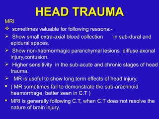
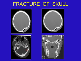
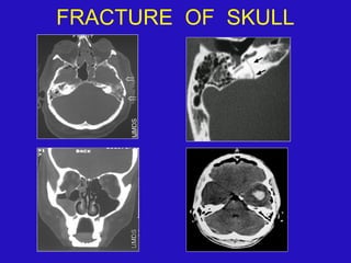
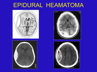
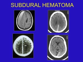
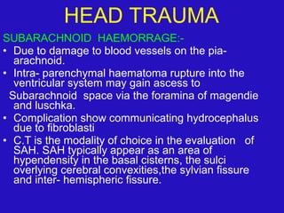
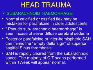
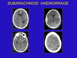
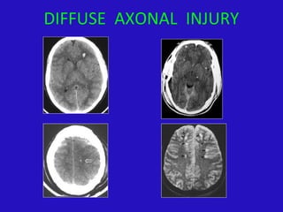
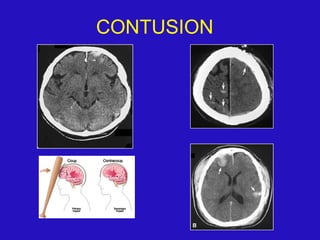
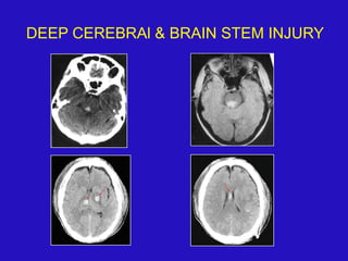
CT is the most important imaging modality for evaluating head trauma. It can detect fractures, extra-axial hemorrhages such as epidural hematomas and subdural hematomas, subarachnoid hemorrhage, intraventricular hemorrhage, and intracerebral hemorrhages. Common primary traumatic brain injuries seen on CT include contusions, diffuse axonal injury characterized by small hemorrhages, and deep cerebral and brainstem injuries. MRI can provide additional details, especially in the subacute and chronic stages.
Radiology Spotters collection by Dr Pradeep. Nice collection Radiology spotters mixed collection ppt made by or collected by Dr. Pradeep, this is a collection of confusing spotter and very important spotter commonly asked in exams, our references is radiopaedia, learning radiology and Aunt Minnie.. Thanks
Summary and illustrations of various traumatic brain injury including primary and secondary lesions as well as limited information on indications of brain imaging in trauma
Radiology Spotters mixed Bag Collection for post graduates student .PPTDr pradeep Kumar
Radiology Spotters collection by Dr Pradeep. nice collection of radiology spotter made by or collected by Dr. Pradeep, this is a collection of confusing spotter and very important spotter commonly asked in exams, our references is radiopaedia, learning radiology and Aunt Minnie.. Thanks.
A slideshow of 'Imaging of Head Trauma: Part I' describes nature, mechanism, significance of head trauma, indications and choices of imaging in patients with head trauma, and normal anatomy of the brain with emphasis on CT.
Common pathologies of the brain in comparison to CT and MRI.
Findings in trauma.
CT findings.
MRI findings.
Subarachnoid hemorrhage.
subdural hematoma.
Some Axonal injuries.
Objectives of this presentation are
Introduction to ct
Cross sectional anatomy
Common important pathologies
This presentation is aimed to educate beginers to help in ct interpretetion.
The Indian economy is classified into different sectors to simplify the analysis and understanding of economic activities. For Class 10, it's essential to grasp the sectors of the Indian economy, understand their characteristics, and recognize their importance. This guide will provide detailed notes on the Sectors of the Indian Economy Class 10, using specific long-tail keywords to enhance comprehension.
For more information, visit-www.vavaclasses.com
Instructions for Submissions thorugh G- Classroom.pptxJheel Barad
This presentation provides a briefing on how to upload submissions and documents in Google Classroom. It was prepared as part of an orientation for new Sainik School in-service teacher trainees. As a training officer, my goal is to ensure that you are comfortable and proficient with this essential tool for managing assignments and fostering student engagement.
Ethnobotany and Ethnopharmacology:
Ethnobotany in herbal drug evaluation,
Impact of Ethnobotany in traditional medicine,
New development in herbals,
Bio-prospecting tools for drug discovery,
Role of Ethnopharmacology in drug evaluation,
Reverse Pharmacology.
Welcome to TechSoup New Member Orientation and Q&A (May 2024).pdfTechSoup
In this webinar you will learn how your organization can access TechSoup's wide variety of product discount and donation programs. From hardware to software, we'll give you a tour of the tools available to help your nonprofit with productivity, collaboration, financial management, donor tracking, security, and more.
We all have good and bad thoughts from time to time and situation to situation. We are bombarded daily with spiraling thoughts(both negative and positive) creating all-consuming feel , making us difficult to manage with associated suffering. Good thoughts are like our Mob Signal (Positive thought) amidst noise(negative thought) in the atmosphere. Negative thoughts like noise outweigh positive thoughts. These thoughts often create unwanted confusion, trouble, stress and frustration in our mind as well as chaos in our physical world. Negative thoughts are also known as “distorted thinking”.
2024.06.01 Introducing a competency framework for languag learning materials ...Sandy Millin
http://sandymillin.wordpress.com/iateflwebinar2024
Published classroom materials form the basis of syllabuses, drive teacher professional development, and have a potentially huge influence on learners, teachers and education systems. All teachers also create their own materials, whether a few sentences on a blackboard, a highly-structured fully-realised online course, or anything in between. Despite this, the knowledge and skills needed to create effective language learning materials are rarely part of teacher training, and are mostly learnt by trial and error.
Knowledge and skills frameworks, generally called competency frameworks, for ELT teachers, trainers and managers have existed for a few years now. However, until I created one for my MA dissertation, there wasn’t one drawing together what we need to know and do to be able to effectively produce language learning materials.
This webinar will introduce you to my framework, highlighting the key competencies I identified from my research. It will also show how anybody involved in language teaching (any language, not just English!), teacher training, managing schools or developing language learning materials can benefit from using the framework.
Read| The latest issue of The Challenger is here! We are thrilled to announce that our school paper has qualified for the NATIONAL SCHOOLS PRESS CONFERENCE (NSPC) 2024. Thank you for your unwavering support and trust. Dive into the stories that made us stand out!
Palestine last event orientationfvgnh .pptxRaedMohamed3
An EFL lesson about the current events in Palestine. It is intended to be for intermediate students who wish to increase their listening skills through a short lesson in power point.
1. CT EVALUATION OF
HEAD TRAUMA
Dr.A.K.M.Salauddin
Consultant ,Radiology & Imaging
Bangladesh
2. leading cause of death in children and
young adults.
Peak age – 15 -24 yrs
Secondary peak > 50 years of age.
Twice as often among males compared to
females.
Generaly caused by motor vehicle
accidents, fall, assaults, violence and
sports & recreation
HEAD TRAUMAHEAD TRAUMA
3. COMPUTED TOMOGRAPHY:-
C.T is the most important step in evaluation for head
and contiguous spine injuries.
In a typical head injury windows and window level are
used:-
1) Brain window
2) Intermediate window : to assess the subdural
or epidural hematoma.
3) Bone window : to determine the presence or
absence of a bony fracture.
HEAD TRAUMAHEAD TRAUMA
4. MRI
sometimes valuable for following reasons:-
Show small extra-axial blood collection in sub-dural and
epidural spaces.
Show non-haemorrhagic paranchymal lesions diffuse axonal
injury,contusion.
Higher sensitivity in the sub-acute and chronic stages of head
trauma.
MR is useful to show long term effects of head injury.
( MR sometimes fail to demonstrate the sub-arachnoid
haemorrhage, better seen in C.T )
MRI is generally following C.T, when C.T does not resolve the
nature of brain injury.
HEAD TRAUMAHEAD TRAUMA
8. SCALP LESION
Scalp laceration
Scalp hematoma / subgaleal soft tissue swelling
Subgaleal extrusion of macerated brain through comminuted
skull fracture .
Atreiovenous fistula or pseudo-aneurysm; usually involve
superficial temporal or occipital arteries.
SKULL FRACTURES:
could be linear,depressed or diastatic and may involve cranial
vault or skull base.
Skull radiographs and C.T are effective to demonstrate
fractures..
SUTURAL DIASTASIS/ DIASTATIC FRACTURE:
Width of the suture more than 3 mm is recognized as sutural
diastasis. (normally sutures are no wider than 2 mm.)
In adult diastasis of lambdoid suture is most common,could be
bilateral.
HEAD TRAUMA
9. FRACTURE OF SKULL BASE:
• Basilar skull fracture should be sought when blood behind
tympanic membrane, otorrhoea, rinorrhoea, echymosis
surrounds the orbits without direct orbital trauma,intracranial
air,air fluid level in PNS or mastoid air cells.
• High- resolution cranial C.T with thin section is best
modalities to detect basilar fracture.
• Basilar fracture cause compression or entrapment of cranial
nertves.
• petrous bone fracture causes- ossicular chain dislocation.
• fracture of optic canal cause loss of vision.
• Sphenoidal fractures can also be associated with disruption
of intra- cavernous internal carotid artery, leading to pseudo-
aneurysm or a carotid cavernous fistula.
HEAD TRAUMA
12. EPIDURAL HEAMATOMA:
• Usually occurs within first 24 or 48 hours, delayed
development or enlargement seen
In 10 to 30%.
• Etiology - due to damage of middle meningeal artery.
- laceration of diploic veins.
- dural sinus
• located between the skull and dura with a focal
biconvex configuration. EDH may cross dural
attachments but not sutures.
• Most common site temporo-parietal area. Posterior
fossa EDH are relatively uncommon.
• On C.T scan, uncommonly EDH may be bilentricular
crescentic or irregular.
• Brain adjacent to most EDHS is severely flattened or
displaced. Secondary herniation are very common.
HEAD TRAUMA
13. EPIDURAL HEAMATOMA:.
* C.T findings Acute, Sub-acute and Chronic.
Acute epidural haematoma- 2/3 uniformly high density 1/3
homogenous in attenuation
Isodene area due to serum
Hypodense area within indicate active bleeding
Sub-acute EDH- Homogenously hyperdense – consists of
solid blood clot.
Chronic EDH – Hetrogenous or decreased attenuation as
well as enhancing membrane peripheral enhancecment
represent dura or membrane formation.
* Imaging criteria whether a epidural haematoma may be
treated conservatively are-
(1) Diameter less than 1.5 cm
(2) A minimal mid line shift of less than 2 mm
(3) The patient must be intact neurologically without focal
deficit.
HEAD TRAUMA
15. SUBDURAL HEMATOMA:
• SDHS are interposed between the dura and
arachnoid. . Post traumatic subdural collection
may be filled with CSF when there is tear in the
arachnoid then called CSF hygroma.
• Tpypically cresent shaped with concave medial
and convex lateral border, which may
extends diffusely.
• May cross suture lines but not dural
attachments.
HEAD TRAUMA
16. SUBDURAL HEMATOMA:
• COMMON SITES:
- over the fronto-parital convexities.
- middle cranial fossa
- para- falcial area.
- inter- hemispheric fissure
Adjacent to the tentorium – may simulate intra- axial lesion.
• Supra tentorial SDHS located adjacent to the tentorium
shows well-defined medial margin corresponding to the
edge of tentorium and sheet like area of increased density
that slopes laterally
• Infra tentorial SDHS located below the tentorium shows a
sharp lateral margin corresponding to the edge of tentorium
and increased density that slopes later
laterally
• Posterior fossa SDHS are rare.
HEAD TRAUMA
17. SUBDURAL HEMATOMA:
• C.T Shows as crescent shaped extra- axial
collection with a convex lateral and concave
medial border. Occasionally may be biconcave
shape.
• ACUTE SDHS- Homogenously hyperdense
- Up to 40%- mixed hyper/ hypodense
- due to unclotted blood
- serum extruded
during clot retraction
- C.S.F
- Rarely isodense- in patients with – coagulopathies
& severe anemia
HEAD TRAUMA
18. SUBACUTE SDHS:-
• Isodense within a few day to a few weeks.Difficult to
diagnosis clues -
Effacement of cortical sulci over cerebral convexity
Mid line shift
Delayed enhancement C.T scan performed after 4 to
6 hours.
CHRONIC SDHS :- Typically low attenuation in
uncomplicated SDHS.
Mixed density in 5% cases- due to recurrent haemorrage
Enhancement following contrast
Shows calcification in 0.3 to 2.7% cases
SUBDURAL HYGROMA:-
Hypodense collection equal to CSF density.
More crescentic & bilateral
At surgery show clear fluid and lack of a membrane.
HEAD TRAUMA
21. SUBARACHNOID HAEMORRAGE:-
• Due to damage to blood vessels on the pia-
arachnoid.
• Intra- parenchymal haematoma rupture into the
ventricular system may gain ascess to
Subarachnoid space via the foramina of magendie
and luschka.
• Complication show communicating hydrocephalus
due to fibroblasti
• C.T is the modality of choice in the evaluation of
SAH. SAH typically appear as an area of
hypendensity in the basal cisterns, the sulci
overlying cerebral convexities,the sylvian fissure
and inter- hemispheric fissure.
HEAD TRAUMA
22. SUBARACHNOID HAEMORRAGE:
• Normal calcified or ossified flex may be
mistaken for parafalcine in older adolescents.
• “ Pseudo sub- arachnoid haemorrhage” is
seen incase of sever diffuse cerebral oedema
• Posterior parafalcine or inter-hemispheric SAH
can mimic the “Empty delta sign’’ of superior
sagittal Sinus thrombosis.
• SAH is rapidly cleared from the subarachnoid
space. The majority of C.T scans performed
within 1Week will appear normal.
HEAD TRAUMA
24. INTRA - VENTRICULAR HAEMORRHAGEINTRA - VENTRICULAR HAEMORRHAGE
IVH are associated with other manifestations ofIVH are associated with other manifestations of
primary intra-axial brain trauma . Such as DAIprimary intra-axial brain trauma . Such as DAI
,deep cerebral gray matter and brain stem,deep cerebral gray matter and brain stem
lesions .lesions .
C.T shows high density intraventricular bloodC.T shows high density intraventricular blood
with or without a fluid level.with or without a fluid level.
Occasionally focal choroid plexux haematomaOccasionally focal choroid plexux haematoma
noted.noted.
HEAD TRAUMA
26. INTRACEREBRAl HEMATOMA:
Bilateral or multiple.
Frequently associated with SAH & SDHS
Most frequently noted at the time of injury .
may be delayed with 48 hours. .
Differentiated from hemorrhagic contusion
by sharply marginated margin, perifocal
hypodensity and mass effect.
HEAD TRAUMA
27. INTRACEREBRAl HEMATOMA:
ACUTE HEMATOMA - < 3 DAYS
C.T :-
Homogenous high density lesion (50 -70 HU) with
irregular well - efined margins.
Usually surrounded by low attenuation (oedema,
contusion ) with mass effect.
SUBCUTE HEMATOMA ( 3 – 14 DAYS )
NCCT : Gradual decrease in density from
periphery inward and becomes isodeme
to brain Parenchya (1-2 HU per day during 2nd
&
3rd
week).
CECT : Peripheral rim enhancement at inner border
of perilesional lucency.
HEAD TRAUMA
28. INTRACEREBRA HEMATOMA:
CHRONIC HEMATOMA : > 14 DAYS
C.T findings
Gradual Decreased / hypodensity .
Later show lucent hematoma (cephalomalacia due
to proteolysis and phagocytosis +surrounding
atrophy) with adjacent sulcal enlargement and
ventricular dilation with ring blush ( DDX : tumor )
HEAD TRAUMA
30. DIFFUSE AXONAL INJURY:-
Most common type of primary traumatic injury .
PATHOGENESIS Cortex and deep structures
move all different speed resulting in shearing
strain along the course of white matter tracts
especially all gray- white matter junction with
axonal tears followed by Wallerion degeneration .
Disruption of accompanying blood vessels show
numerous small hemorrhage foci .
Diffuse & Bilateral
HEAD TRAUMA
31. DIFFUSE AXONAL INJURY:-
Locations ( according to severity of trauma )
a) lobar white matter at cortico-medullary junction
more common para sagital region of frontal lobe
+ periventricular region of temporal lobe,
occasionally in parietal and occipital lobes.
b) Internal + external capsule, corona radiate,
cerebral peduncles .
c) Corpus callosum
d) Brain stem – postero lateral quadrants of mid
brain + upper pons.
HEAD TRAUMA
32. DIFFUSE AXONAL INJURY:-
C.T – Findings
- Early imaging may be subtle or normal .
- Foci of decreased density.
- May show some degrees of cerebral
swelling.
- May show small focal hemorrhage or small
petechial haemorrhage particularly at
gray-white junction and corpus callosum.
- May show extensive injury.
HEAD TRAUMA
34. CONTUSION:-
Common type of primary intra-axial lesion .
In 21% of head trauma patients.
PATH :- Tissue necrosis, capillary disruption,
petechial hemorrhage followed by
liquefaction + oedema after 4 to 7 days
.contusion may be hemorrhagic or non-
hemorrhagic.
MECHANISM :- Linear acceleration –
deceleration forces / penetrating trauma /
direct impaction.
HEAD TRAUMA
35. CONTUSION:-
1. COUP :- Direct impact to stationary brain. Injury at
the site of impaction.
2. COUNTER COUP :- Impact of moving brain on
stationary clavarium opposite to the site of the coup
and produced injury.
Contusions are typically superficial foci of punctate
or linear hemorrhages that occur
along gyral crests.
Location - Multiple bilateral lesion , temporal lobes
most frequently involved.
- Bneath an acute subdural hematoma.
HEAD TRAUMA
36. CONTUSION:-
C.T
- Initial findings may be subtle or absent.
- Early findings focal / multiple poorly defined
areas of low attenuation with irregular contour
intermixed with a few tiny areas of increased
density ( petechial hemorrhage)
- Diffuse oedema and mass effect in immediate
post-traumatic period,then gradually diminish
over time.
- Some degree of contrast enhancement.
- Isodense to brain after 2 – 3 weeks.
HEAD TRAUMA
38. DEEP CEREBRAL AND BRAIN STEM INJURY
Less common than DAI and cortical contusions.
Represent 5% to 10% of primary traumatic brain
injuries.
Most are induced by shearing forces,
Most patients have profound neurologic deficits
and a poor prognosis..
C.T. Scan - often normal initially.
- Petechial hemorrhages can sometimes be seen
in lateral brain stem, periaquedctal region region
and deep gray matter nuclei.
M.R shows brain stem lesions nicely.
HEAD TRAUMA
45. HEAD TRAUMA
SEQUELAE OF TRAUMA
1) Encephlomalacia and atrophy.
2) Pneumocephalus, Pneumatocele formation.
3) CSF Leaks and Fistulae.
4) Acquired Encephalocele or leptomeningeal cyst.
5) Cranial nerve injuries.
6) Diabetes incipidus.
7) Hydrocephalus.
8) Atrophy.
9) Subdural Hygroma.
10) Post traumatic abscess.
46. CONCLUSION :-
• C.T is important modality for evaluation of
an emergency patient with cranio-cerebral
trauma.
• Early & accurate ct evaluation cause
better patients outcomes with a decrease
morbidity & mortality statics.
& Decrease in trauma related medical
costs.
HEAD TRAUMA