
Recommended
More Related Content
What's hot
What's hot (20)
Similar to Hypersensitivity reactions
Similar to Hypersensitivity reactions (20)
More from Reshma Fathima .K
More from Reshma Fathima .K (20)
Recently uploaded
Recently uploaded (20)
Hypersensitivity reactions
- 2. Introduction • Certain human disorders are attributed to activity of the immune system. These disorders are commonly known as hypersensitivities, states of increased immune sensitivity that are mediated by antibody or cellular factors. The disorders may also involve immunodeficiencies in which failures of antibody‐mediated or cell‐mediated immunity take place. • Normally the immune system plays an important role in protecting the body from microorganisms and other foreign substances. If the activity of the immune system is excessive or overreactive, a hypersensitivity reaction develops. The consequences of a hypersensitivity reaction may be injury to the body or death. • Most injury resulting from hypersensitivities develops after an interaction has taken place between antigens and antibodies or between antigens and sensitized T- lymphocytes. The general nature of and symptoms accompanying the reaction depend upon whether antibodies or sensitized T-lymphocytes are involved.
- 3. • When antibodies are involved, the reactions fall under the heading of immediate hypersensitivity. When T-lymphocytes are involved, the reactions are characterized as delayed hypersensitivity. Immediate hypersensitivity reactions include anaphylaxis, allergic reactions, cytotoxic reactions, and immune complex reactions. Delayed hypersensitivity reactions are generally characterized as contact dermatitis or infection allergies. TYPES OF HYPERSENSITIVE REACTIONS TYPE I – IMMEDIATE, ATOPIC, ANAPHYLACTIC TYPE II – ANTIBODY DEPENDANT (CYTOTOXIC REACTIONS) TYPE III – IMMUNE COMPLEX TYPE IV – CELL MEDIATED / DELAYED TYPE OF HYPERSENSITIVITY
- 4. • Immediate hypersensitivity (TYPE 1) The reactions accompanying immediate hypersensitivity depend upon the nature of the antigen, the frequency and route of antigen contact, and the type of antibody reacting with the antigen. The initial dose of antigen is referred to as the sensitizing dose. This exposure is followed by a latent period and then a later dose of the same antigen, called the eliciting dose or shocking dose. The shocking dose sets off the hypersensitivity reaction, resulting in tissue damage. • Immediate reactions begin within minutes of contact with the eliciting dose of antigen. If antigens are introduced directly into the tissues, such as by insect sting or injection, the result is a systemic reaction such as anaphylactic shock. When the contact is a superficial one involving the epithelial tissues, the reaction is more localized, as occurs in asthma or allergic rhinitis (hay fever). These local reactions are commonly referred to as allergy. Another term used is atopy.
- 5. • The antigens eliciting an immediate hypersensitivity are called allergens, particularly when they are involved in local allergic reactions. Hapten molecules such as penicillin molecules may be involved when they are bound to larger protein molecules. Foods, feathers, pollen grains, animal dander, and dust may be allergens. Animal sera, bee venoms, and wasp venoms are also allergens. • The antibodies involved in anaphylaxis reactions are of the type IgE. In cytotoxic and immune complex reactions, IgG and IgM are involved. • Anaphylaxis. Anaphylaxis, or type I hypersensitivity, is a whole-body, immediate hypersensitivity also known as anaphylactic shock. The allergens are introduced to the body directly to the tissues in a concentrated form (intramuscular or intravenous injection, for example).
- 6. After the sensitizing dose has been administered, IgE is produced by the plasma cells. The antibodies circulate in the blood and attach at the Fc end to mast cells of the tissues and basophils in the bloodstream (Figure 1 ). This activity occurs during the latent period. When the eliciting dose of allergen is later administered, the antigens combine with antibodies on the surface of the mast cells and basophils. Figure 1 The process of anaphylaxis. (a) Allergens stimulate the production IgE antibodies, which (b) fix themselves to the surfaces of mast cells. (c) On second exposure to the allergens, a reaction occurs on the mast cell surface, and (d) the cellular granules release histamine and other stimulators of smooth muscle contraction
- 7. ANTIGEN – ANTIBODY COMBINATION Histamine and serotonin Induce spasms of the smooth muscle, such as bronchioles, small arteries and GIT lining A sudden drop in blood pressure occurs, followed by circulatory collapse and shock. Bronchospasms and edema cause constriction of the respiratory passageways, and breathing is very difficult. Facial edema occurs, and the heart rate increases due to constriction of the arteries. Swellings called “hives” develop at the site of injection and other areas of the skin. In severe cases, anaphylactic shock may result in death within several minutes to an hour. To relieve the symptoms, epinephrine is administered together with a smooth muscle relaxer, a drug such as cortisone to reduce swelling, and other drugs as appropriate the cells release a number of physiologically active substances including
- 8. • Allergic reactions. Allergic reactions (allergy) are a milder, localized form of anaphylaxis. As noted, such things as foods, pollen grains, and animal dander can induce these localized reactions. IgE, basophils, and mast cells are involved, but much less than in anaphylaxis. There appears to be a genetic basis for allergic reactions, as evidenced by their distribution in families.
- 9. • TYPE II HYPERSENSITIVITY REACTIONS Cytotoxic reactions. Cytotoxic reactions are a form of immediate hypersensitivity, sometimes referred to as type II hypersensitivity. In these reactions, IgE and IgM are produced in response to stimulation by antigens. The antibodies unite with the antigens in the bloodstream, but they also unite with analogous antigens on the surface of the human body's cells. This union sets off the complement system, and destruction of the local tissue cells ensues. An example of a cytotoxic reaction is thrombocytopenia. In this disease, antibody molecules are elicited by certain drug molecules. The antibodies unite with antigens on the surface of thrombocytes (platelets), and with complement activation, the thrombocytes are destroyed. The result is an impaired blood-clotting mechanism. Another example of the cytotoxic reaction is agranulocytosis. In this immune disorder, antibodies unite with antigens on the surface of neutrophils. As these cells are destroyed with complement activation, the capacity for phagocytosis is reduced.
- 10. The cytotoxic reaction in erythroblastosis fetalis. Cytotoxic reactions are also manifested by the transfusion reaction occurring when improper blood transfusions are performed. Another consequence is erythroblastosis fetalis, also known as hemolytic disease of the newborn, or Rh disease. In this condition, a pregnant woman produces Rh antibodies against the developing fetus, and when the Rh antibodies unite with Rh antigens on the surface of fetal red blood cells in a succeeding pregnancy, the red blood cells are destroyed (Figure 2 ).
- 11. • Immune complex disease. Immune complexes are combinations of antigen and antibody that have the ability to fix complement. The antibodies involved are IgM or IgG, and the antigens exist in fluid as soluble antigens. Proteins or nucleic acids may be involved. • An example of immune complex hypersensitivity is serum sickness. In this condition, animal serum is administered to humans, and its proteins elicit antibody production. When the antibodies and antigens unite, they form immune complexes, which activate the complement system and cause local tissue damage. The patient may display edema of the hands, face, and feet, as well as swelling of the upper respiratory tissues and impairment of normal respiration. An inflammatory response results. • Formation of immune complexes is also involved with numerous diseases includingsystemic lupus erythematosus, rheumatoid arthritis, and glomerulonephritis.Immune complex hypresensitivity is often called type III hypersensitivity.
- 12. In type 3 hypersensitivity reactions, insoluble immune complexes (aggregations of antigens and IgG and IgM antibodies) form in the blood and are deposited in various tissues (typically the skin, kidney and joints)
- 13. This deposition of the antibodies may trigger an immune response according to the classical pathway of complement activation – for eliminating cells presenting foreign antigens (which are usually, but not in this case, pathogens). There are two stages relating to the development of the complexes, firstly the complex forms when IgG and IgM antibodies are bound to an antigen, after this, the complexes can form larger ones which can be cleared by the body. It is at the first stage of this formation where clearance is not possible and the antigen-antibody complex will spread and deposit as stated above. The reaction takes hours to days to develop
- 14. Tissue damage results at the site of the immune complex with the influx of phagocytes and granuloctyes and the release of inflammatory mediators Some examples: • Immune complex glomerulonephritis • Rheumatoid arthritis • Serum sickness • Subacute bacterial endocarditis • Symptoms of malaria • Systemic lupus erythematosus • Arthus reaction • Farmer’s Lung (Arthus-type reaction)
- 15. • Arthus reaction • In immunology, the Arthus reaction is a type of local type III hypersensitivity reaction. Type III hypersensitivity reactions are immune complex mediated, and involve the deposition of an antigen/antibody complex mainly in the vascular walls, serosa (pleura, pericardium, synovium), and glomeruli.
- 16. • Delayed hypersensitivity. (TYPE IV) T-lymphocytes rather than antibodies function in cases ofdelayed hypersensitivity, also called type IV hypersensitivity. Normally these are the T-lymphocytes involved in cell- mediated immunity. The T-lymphocytes produce lymphokines, which stimulate an influx of macrophages to perform phagocytosis. In delayed hypersensitivity, the result is an exaggeration of the immune response, and the phagocytes bring about the destruction of the local tissue. • Delayed hypersensitivity (also called cellular hypersensitivity) is so named because the reaction requires a day or more to develop. One manifestation of the reaction isinfection allergy, as in the tuberculin skin test. A purified protein derivative (PPD) ofMycobacterium tuberculosis is applied to the skin superficially, and a skin reaction (swelling and redness) occurs 24 to 48 hours later if the person has had a previous exposure to the antigens of Mycobacterium tuberculosis, possibly during an episode of tuberculosis.
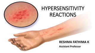
- 17. • A second manifestation of delayed hypersensitivity is contact dermatitis. In many cases, the reaction is accompanied by large, blisterlike lesions in which vesicles are surrounded by a zone of erythema (redness). Usually, the vesicles itch intensely. • Antigens involved in contact dermatitis include metals such as nickel and mercury, cosmetics, disinfectants, and plant substances such as the resins of poison ivy, poison oak, and poison sumac. The individual can be tested to determine which antigen is the cause of allergy by performing a patch test. In this procedure, a patch containing a specific antigen is attached to the skin and left in place for 48 hours to determine if a reaction will take place.