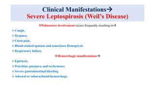
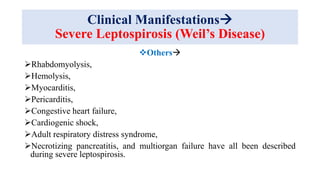
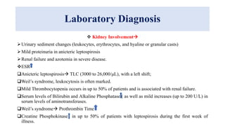
Leptospira are thin, spiral-shaped bacteria that can cause leptospirosis, a zoonotic disease. They have two flagella that allow for motility. Leptospirosis is transmitted through contact with urine from infected animals. It has varied clinical manifestations from mild flu-like symptoms to severe jaundice, kidney damage, and hemorrhaging known as Weil's disease. Diagnosis is made through microscopic agglutination testing of antibodies in serum. Proper treatment requires antibiotics.
2. Introduction
Morphology
“Leptospira” derives from the Greek leptos (thin) and Latin spira (coiled).
The leptospires are a mere 0.1 μm in diameter by 6 to 20 μm in length.
The cells have pointed ends, one or both of which is usually bent into a characteristic
hook.
Motility is conferred by the rotation of two axial flagella underlying the membrane sheath,
which are inserted at opposite ends of the cell and extend toward the central region.
Two periplasmic flagella with polar insertions are located in the periplasmic space and are
responsible for motility.
The FlaA and FlaB proteins constitute the flagellar sheath and core respectively.
Because of their small diameter, leptospires are best visualized by darkfield microscopy,
appearing as actively motile spirochetes.
These bacteria are aerobic, do not resist drought or hypertonicity, they support
alkalinization to pH 7.8.
3.
4.
5.
6. Introduction
Species & Classification
Historically, the genus Leptospira was classified into 2 Species, L. interrogans and L.
biflexa, composed of pathogenic and nonpathogenic strains, respectively.
Within each species, large numbers of serovars were differentiated using agglutinating
antibodies conferred by lipopolysaccharide (LPS) O-antigens.
More than 250 serovars of pathogenic leptospires
Antigenically related serovars were grouped into serogroups.
Leptospires are now classified into a number of species determined by DNA
reassociation.
There are currently 23 named species, including
Pathogens (e.g., L. interrogans),
Nonpathogenic saprophytes (e.g., L. biflexa), and
Species of indeterminate pathogenicity (e.g., L. inadai).
This classification is supported by 16S RNA gene sequencing.
7.
8. Epidemiology
Globally, leptospirosis is a leading zoonotic cause of morbidity and mortality
affects MORE rural subsistence farmers and urban slum dwellers.
Peak incidence occurs in the rainy season in tropical regions and the late
summer to early fall in temperate regions.
In developing countries with poor urban housing standards, leptospirosis
outbreaks occur regularly after heavy rainfall and flooding.
Morbidity and mortality are greatest in the poorest regions of the world and in
areas where surveillance is not routinely performed.
Leptospirosis patients are commonly misdiagnosed with dengue, malaria, and
other infections.
More important than incidence, estimates suggest that globally ~2.90 million of
disability adjusted life-years (DALYs) are lost per annum from the
approximately 1.03 million cases.
9. Epidemiology
Leptospirosis is maintained in nature by chronic renal infection of carrier animals.
The most important reservoirs are rodents and other small mammals, but livestock and
companion animals are also significant sources of human infection.
Infection of carrier animals usually occurs during infancy, and once infected, animals
may shed leptospires in their urine intermittently or continuously throughout life.
Infection occurs through direct or indirect contact with urine or tissues of infected
animals.
Direct contact is important in transmission to
Veterinarians,
Workers in milking sheds on dairy farms,
Abattoir workers, butchers, hunters, and animal handlers;
Transmission to children handling puppies and to dog handlers has been reported.
10. Epidemiology
Males are predominantly affected, with approximately 80% of the total
burden.
Indirect contact is more common and is responsible for disease after
exposure to wet soil or water.
The great majority of cases are acquired by this route in the tropics, either
through occupational exposure to water, as in rice or taro farming, or
through exposure to damp soil and water during avocational activities.
Recreational exposures have become relatively more important, often in
association with adventure tourism to tropical endemic areas.
Several large point-source waterborne outbreaks involving attack rates as
high as 42% have occurred recently after athletic events
11.
12. The map shows,
through different
colors, Leptospira
isolation from
different animal
orders. colors,
Leptospira isolation
from different
animal orders. from
different animal
orders.
13. Pathogenesis
The pathogenesis of leptospirosis is incompletely understood.
Leptospires enter the host through abrasions in the skin or through intact mucous
membranes, especially the conjunctiva and the lining of the oro- and nasopharynx.
Drinking of contaminated water may introduce leptospires through the mouth, throat, or
esophagus.
After entry of the organisms, leptospiremia develops, with subsequent spread to all
organs.
Multiplication takes place in blood and in tissues, and leptospires can be isolated from
blood and cerebrospinal fluid (CSF) during the first 4–10 days of illness.
All forms of leptospires can damage the wall of small blood vessels vasculitis with
leakage and extravasation of cells, including hemorrhages.
The most important known pathogenic properties of leptospires are adhesion to cell
surfaces and cellular toxicity.
14. Pathogenesis
Vasculitis is responsible for the most important manifestations of the disease.
Leptospires mainly infect the kidneys and liver but other organs may be
affected.
In the kidney, leptospires migrate to the interstitium, renal tubules, and tubular
lumen, causing interstitial nephritis and tubular necrosis.
Hypovolemia due to dehydration or altered capillary permeability may contribute
to the development of renal failure.
In the liver, centrilobular necrosis with proliferation of Kupffer’s cells may be
found.
However, severe hepatocellular necrosis is not a feature of leptospirosis.
Pulmonary involvement is the result of hemorrhage and not of inflammation.
15. Pathogenesis
Skeletal muscle Swelling, vacuolation of the myofibrils, and focal necrosis.
In severe leptospirosis, vasculitis may ultimately impair the microcirculation and
increase capillary permeability, resulting in fluid leakage and hypovolemia.
When antibodies are formed, leptospires are eliminated from all sites in the host except
the eye, the proximal renal tubules, and perhaps the brain, where they may persist
for weeks or months.
The persistence of leptospires in the aqueous humor occasionally causes chronic or
recurrent uveitis.
The systemic immune response may also produce symptomatic inflammatory reactions.
A rise in antibody titer coincides with the development of meningitis.
After the start of antimicrobial treatment for leptospirosis, a Jarisch-Herxheimer
reaction similar to that seen in other spirochetal diseases may develop.
16.
17.
18.
19. Pathogenesis
Comparison of models of Leptospira infection in resistant and susceptible
hosts suggests the importance of the inflammatory response in influencing
the disease outcome.
In Leptospira-infected mice, the inflammatory response is rapid and strictly
regulated. Consequently, homeostasis is maintained as bacteria are rapidly
cleared from the blood and no or only mild symptoms develop, which relate
to mild leptospirosis observed in 90% of human infections.
In Leptospira-infected hamsters, cytokine production is delayed and
sustained, which could trigger the onset of a cytokine storm and impair
pathogen clearance from the blood.
The resulting sepsis-like phenotype could be associated to increased
bacterial load, organ failures and hemorrhages found in infected hamsters as
well as in severe forms of leptospirosis in humans.
20. Clinical Manifestations
Many Leptospira-infected persons remain asymptomatic.
In symptomatic cases of leptospirosis, clinical manifestations vary from
mild to serious or even fatal.
More than 90% of symptomatic persons have the relatively mild and usually
anicteric form of leptospirosis, with or without meningitis.
Severe leptospirosis with profound jaundice (Weil’s syndrome) develops
in 5–10% of infected individuals.
Some serovars tend to cause more severe disease than others.
The incubation period is usually 1–2 weeks but ranges from 2 to 20 days.
Typically, an acute leptospiremic phase is followed by an immune
leptospiruric phase.
21. Clinical Manifestations
Anicteric Leptospirosis
Leptospirosis may present as an acute influenza-like illness
Fever,
Chills,
Severe headache (frontal or retroorbital) and sometimes develops
photophobia,
Nausea,
Vomiting, and myalgias.
Muscle pain, which especially affects the calves, back, and abdomen
Less common features include sore throat and rash.
Mental confusion may be evident.
Pulmonary Cough and chest pain and in a few cases by hemoptysis.
22. Clinical Manifestations
Anicteric Leptospirosis
The most common finding on physical examination
Fever with conjunctival suffusion.
Less common findings include
Muscle tenderness,
Lymphadenopathy,
Pharyngeal injection,
Rash,
Hepatomegaly, and splenomegaly.
The rash may be macular, maculopapular, erythematous, urticarial, or
hemorrhagic.
Mild jaundice may be present.
23.
24. Clinical Manifestations
Anicteric Leptospirosis
Most patients become asymptomatic within 1 week.
After an interval of 1–3 days, the illness recurs in a number of cases. The start of
this second (immune) phase coincides with the development of antibodies.
Usually the symptoms last for only a few days, but occasionally they persist for
weeks.
Often the fever is less pronounced and the myalgias are less severe than in the
leptospiremic phase.
An important event during the immune phase is the development of aseptic
meningitis.
Although no more than 15% of all patients have symptoms and signs of
meningitis, many patients have CSF pleocytosis.
Meningeal symptoms usually disappear within a few days but may persist for
weeks.
25. Clinical Manifestations
Anicteric Leptospirosis
Aseptic meningitis is more common among children than among
adults.
Iritis, iridocyclitis, and chorioretinitis—late complications that may
persist for years—can become apparent as early as the third week but
often present several months after the initial illness.
Mortality rates in anicteric leptospirosis are low, although death as a
result of pulmonary hemorrhage occurred in 2.4% of cases in a
Chinese outbreak.
26. Clinical Manifestations
Severe Leptospirosis (Weil’s Disease)
Weil’s syndrome is characterized by
Jaundice,
Renal dysfunction, and
Hemorrhagic diathesis; by pulmonary involvement in many cases
Mortality rates of 5–15%.
In Europe, this syndrome is frequently but not exclusively associated with
infection due to serovar Icterohaemorrhagiae/ Copenhageni.
The onset of illness is no different from that of less severe leptospirosis;
however,
After 4–9 days, jaundice as well as renal and vascular dysfunction
generally develop.
27. Clinical Manifestations
Severe Leptospirosis (Weil’s Disease)
A biphasic disease pattern like that seen in anicteric leptospirosis
is lacking.
The jaundice of Weil’s syndrome, which can be profound and give an
orange cast to the skin.
Hepatomegaly and tenderness in the right upper quadrant are usually
detected.
Splenomegaly is found in 20% of cases.
Renal failure may develop, often during the second week of illness.
Hypovolemia and decreased renal perfusion contribute to the
development of Acute Tubular Necrosis with oliguria or anuria.
28. Clinical Manifestations
Severe Leptospirosis (Weil’s Disease)
Pulmonary involvement occurs frequently resulting in
Cough,
Dyspnea,
Chest pain,
Blood-stained sputum and sometimes Hemoptysis
Respiratory failure.
Hemorrhagic manifestations
Epistaxis,
Petechiae, purpura, and ecchymoses
Severe gastrointestinal bleeding
Adrenal or subarachnoid hemorrhage.
29. Clinical Manifestations
Severe Leptospirosis (Weil’s Disease)
Others
Rhabdomyolysis,
Hemolysis,
Myocarditis,
Pericarditis,
Congestive heart failure,
Cardiogenic shock,
Adult respiratory distress syndrome,
Necrotizing pancreatitis, and multiorgan failure have all been described
during severe leptospirosis.
30. Laboratory Diagnosis
Kidney Involvement
Urinary sediment changes (leukocytes, erythrocytes, and hyaline or granular casts)
Mild proteinuria in anicteric leptospirosis
Renal failure and azotemia in severe disease.
ESR
Anicteric leptospirosis TLC (3000 to 26,000/μL), with a left shift;
Weil’s syndrome, leukocytosis is often marked.
Mild Thrombocytopenia occurs in up to 50% of patients and is associated with renal failure.
Serum levels of Bilirubin and Alkaline Phosphatase ( as well as mild increases (up to 200 U/L) in
serum levels of aminotransferases.
Weil’s syndrome Prothrombin Time
Creatine Phosphokinase in up to 50% of patients with leptospirosis during the first week of
illness.
31. Laboratory Diagnosis
When a meningeal reaction develops, polymorphonuclear leukocytes
predominate initially and the number of mononuclear cells increases later.
The protein concentration in the CSF may be elevated; CSF glucose
levels are normal.
In severe leptospirosis, pulmonary radiographic abnormalities are common.
These abnormalities most frequently develop 3–9 days after the onset of
illness.
The most common radiographic finding is a patchy alveolar pattern that
corresponds to scattered alveolar hemorrhage.
Radiographic abnormalities most often affect the lower lobes in the
periphery of the lung fields.
32. Laboratory Diagnosis
Microscopic Agglutination Test (MAT)
Leptospires have a
Very long doubling times in culture
Culture takes weeks to grow,
Diagnosis of leptospirosis mostly depends on serological results.
MAT has been widely used for the diagnosis of leptospirosis through detection of
antibodies produced against the antigens of Leptospira serovars.
This technique utilizes live bacterial cultures and is routinely performed by
incubating patient’s serum with various serovars of Leptospira.
MAT titre is obtained by testing various serum dilutions with a positive serovar. A
four-fold rise of MAT antibody titre is a definite evidence of Leptospira infection.
Regarded as the gold standard for all diagnostic techniques, this assay has a
high sensitivity and allows for the detection of group-specific antibodies.
33. Laboratory Diagnosis
Microscopic Agglutination Test (MAT)-Test Limitations
In regions where leptospirosis is common, there may be a substantial proportion
of the population with elevated titres of MAT.
The serum from patients may react with a different serovar than the infected
one.
In case of many numbers of samples, performing MAT would be very difficult as
it is a complicated test.
Diagnostic laboratories are also required to have all the circulating types of
Leptospira serovars, which may be costly.
It would not be useful during the early stages of the disease as the antibodies
against the leptospires are usually not present, or if at all present, it will be at an
extremely low level in the cerebrospinal fluid.
35. Laboratory Diagnosis
Microsphere Immunoassay (MIA)Luminex xMap
Technology
The MIA test is carried out by preparing antigens from pure Leptospira cultures and
preparing immunoassays for IgG and IgM.
Briefly, the technique relies on magnetic-coated polystyrene beads filled with bi-coloured
fluorescent dyes in different ratios resulting in 500 distinct bead sets.
Each bead set may be coated with a different antigen to allow simultaneous measurement
of antibody response to up to 500 different antigens.
ADVANTAGES
Able to positively diagnose samples from those that were previously deemed non-reactive.
Capable of differentiating between IgM and IgG antibodies against Leptospira.
High throughput screening system processing of high numbers of patient samples per
day (Bulk Sampling).
Identify antibody types as well as the reactivity of antigens.
Significant cost-reduction
36.
37. Laboratory Diagnosis
ELISA
ELISA can also be used to diagnose leptospirosis by utilizing
leptospiral-specific IgM and IgG from sera of patients infected with
different leptospiral serovars.
Subjects with leptospirosis produced specific IgM and IgG antibodies
that are detectable by ELISA, even with low titre of antigens in their
serum.
ELISA anti-IgM technique is a suitable method for detecting
leptospiral antibodies in sera for diagnostic and epidemiological
purposes.
Antibody levels are generally low or absent during early phases of the
infection could easily lead to false negative diagnoses.
38. Laboratory Diagnosis
Indirect Haemagglutination Assay (IHA)
IHA also detects IgM and IgG produced against Leptospira antigens in the
body after bacterial entry (within 4-6 days).
Commercial kits for this assay are widely available in the market.
IA has a diagnostic sensitivity of 92% and a specificity of 95%.
This assay can be utilized as an initial diagnostic tool for patients who are
clinically suspected to be having acute leptospirosis.
It is advantageous due to its relatively low cost, requires no specialized
equipment or any strict incubation conditions.
One significant disadvantage of using IHA is that the results may not be
interpretable when there is non specific haemagglutination.
IHA is significantly less sensitive than ELISA for the diagnosis of
leptospirosis.
39.
40.
41. Laboratory Diagnosis
Dipstick Assay (ICT)
Dipstick assay is an easy and robust technique that allows for rapid
screening and diagnosis of patients suspected of having leptospirosis.
The LEPTO dipstick test is a rapid field test for leptospirosis that does not
require special laboratory equipment or well trained personnel.
This assay demonstrated high sensitivity, specificity and predictive values.
Assesses the samples using a dipstick which contains two horizontal bands
namely, the lower band consisting of broadly reactive specific antigens and
the upper band which acts as an internal control as it consists of antihuman
IgM antibodies.
Bound IgM antibodies are detected in non-enzymatic reactions with a
stabilized anti-human IgM dye conjugate.
42.
43. Laboratory Diagnosis (Direct Methods)
Microscopy
This technique is particularly useful for
Observing leptospires in culture,
Particularly when they are present in large numbers,
Observing the agglutination formed via MAT (microscopic agglutination
test).
Leptospires present in patient samples can be concentrated using
centrifugation.
Disadvantage Direct microscopic observation of leptospires in urine
(leptouria test) may have a low specificity since the presence of fibrin and
protein in the urine samples can be mistaken for leptospires.
44. Laboratory Diagnosis (Direct Methods)
Microscopy
Phase contrast microscopy is useful for visualizing leptospires.
Leptospires can be easily detected under dark field microscopy as thin,
coiled, motile organisms in blood and urine samples of patients with
leptospirosis.
Positivity of dark field microscopy decreases from 100% to 90.9% as
the duration of infection increases for more than one week.
Another disadvantage both false positive and false negative
diagnosis can be easily made.
45.
46. Laboratory Diagnosis (Direct Methods)
Staining Techniques
The Warthin-Starry stain is widely in use now sometimes confer false negative
results, as the leptospirosis burden in tissue biopsies (such as kidneys) may not be
significant.
Immunohistochemical assay and immunoglobulin fluorescent staining are also
documented to be useful diagnostic tools for leptospirosis.
Immunoglobulin staining is usually done on tissues with positive immunoreactivity
towards leptospiral antigens.
This technique has the advantage of being useful with formalin-fixed tissue.
Detect leptospires even when their numbers are significantly low, or when there are
materials that precludes the use of dark field microscopy.
It may be not be advantageous in early infections.
48. Laboratory Diagnosis (Direct Methods)
Culture
Samples from a suspected patient, usually urine and/ or blood sample, are taken
and streaked onto a culture flask containing fluid media (generally used for
primary culture).
Oleic acid-albumin media of EMJH is the most commonly used media for this
purpose.
Antibiotics such as rifampicin, neomycin, actidione can be added to the media
for selective isolation of bacteria from contaminated samples.
Leptospires can be cultured from blood or cerebrospinal fluid samples during the
acute phases of the infection (lasts for about 10 days).
Highly accurate results but tedious and long process culture can take almost 3
months to grow.
As leptospires are highly infectious organisms, they need to be handled with
utmost care risk of laboratory-acquired infections with this technique.