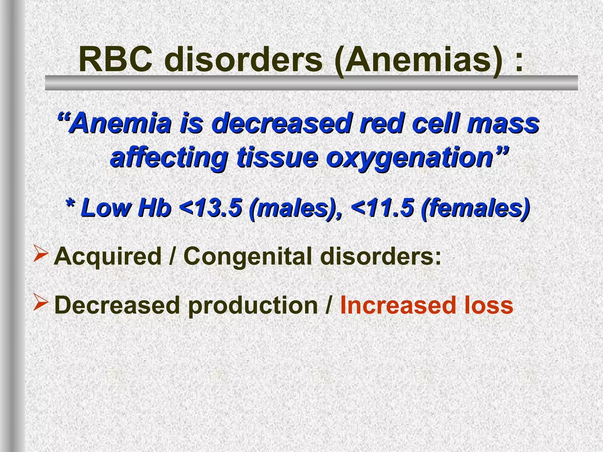
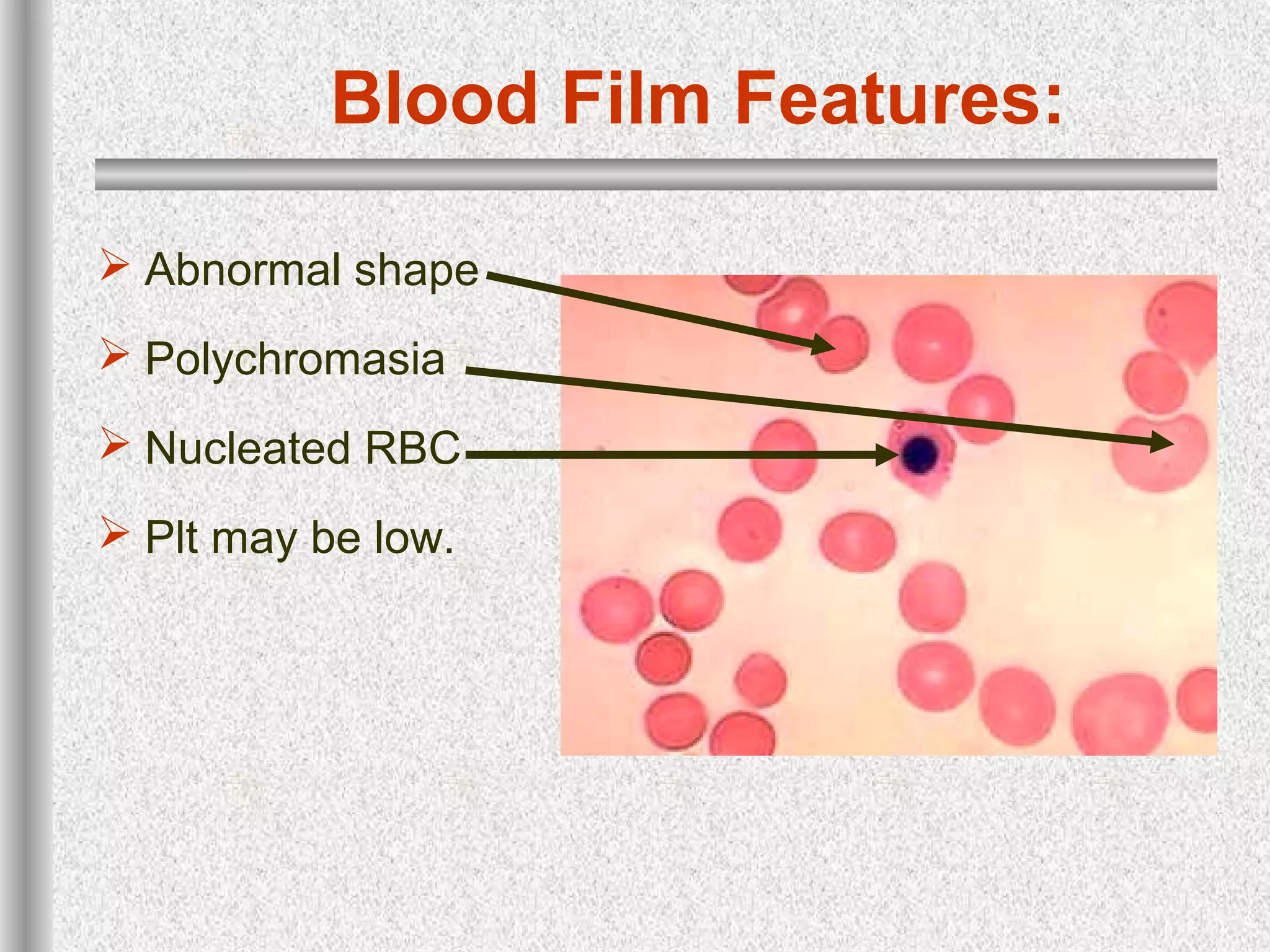
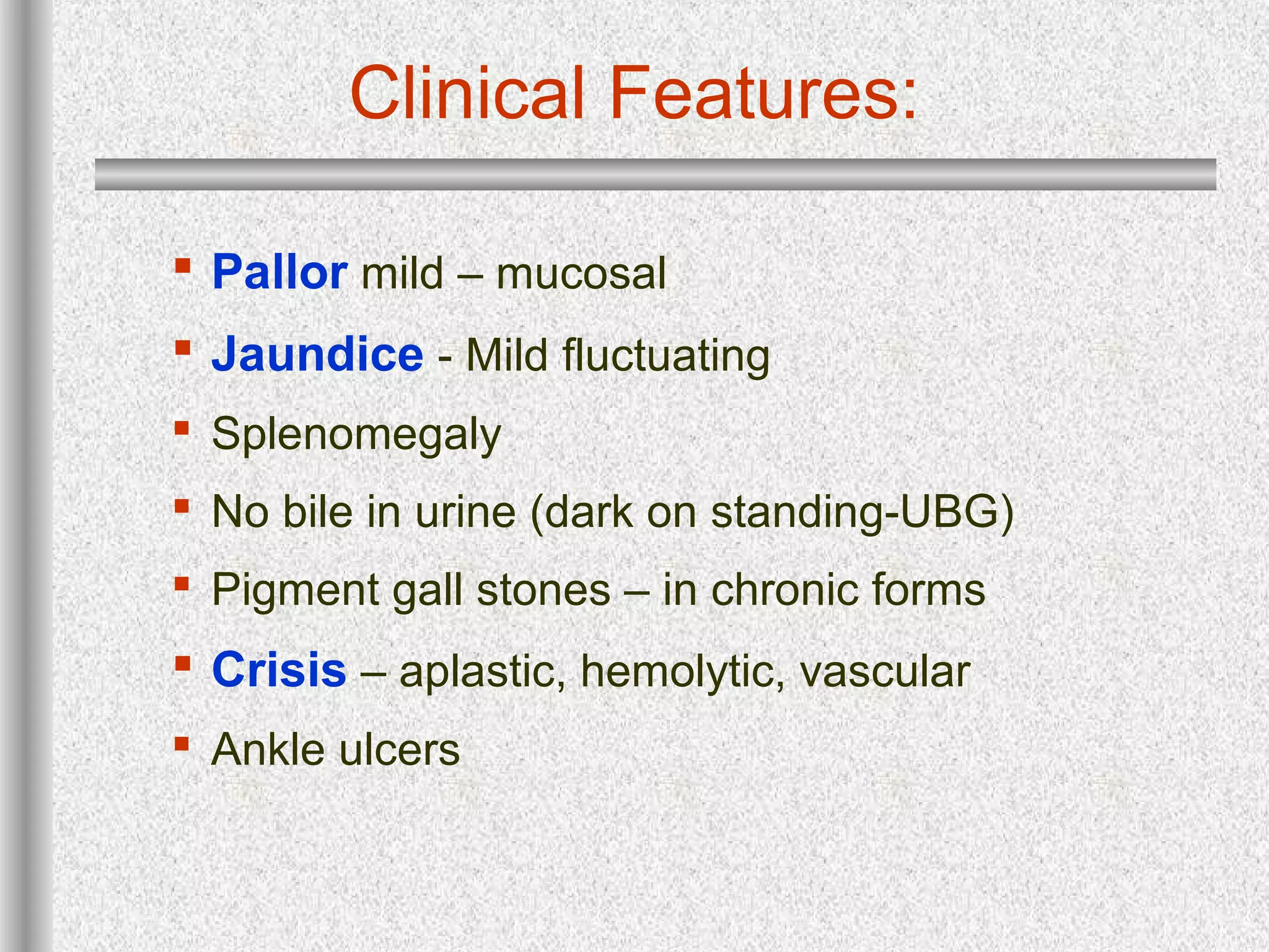
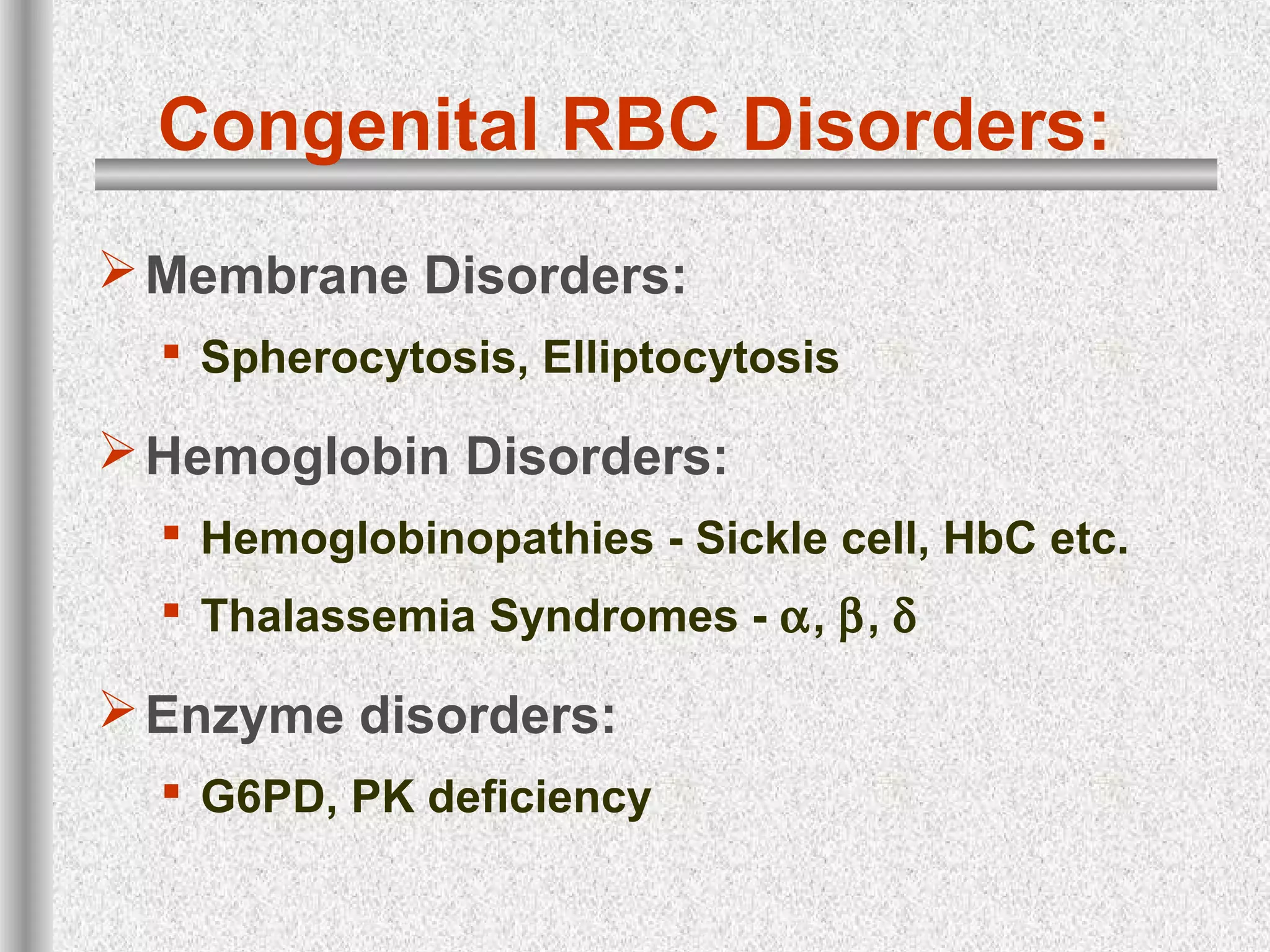
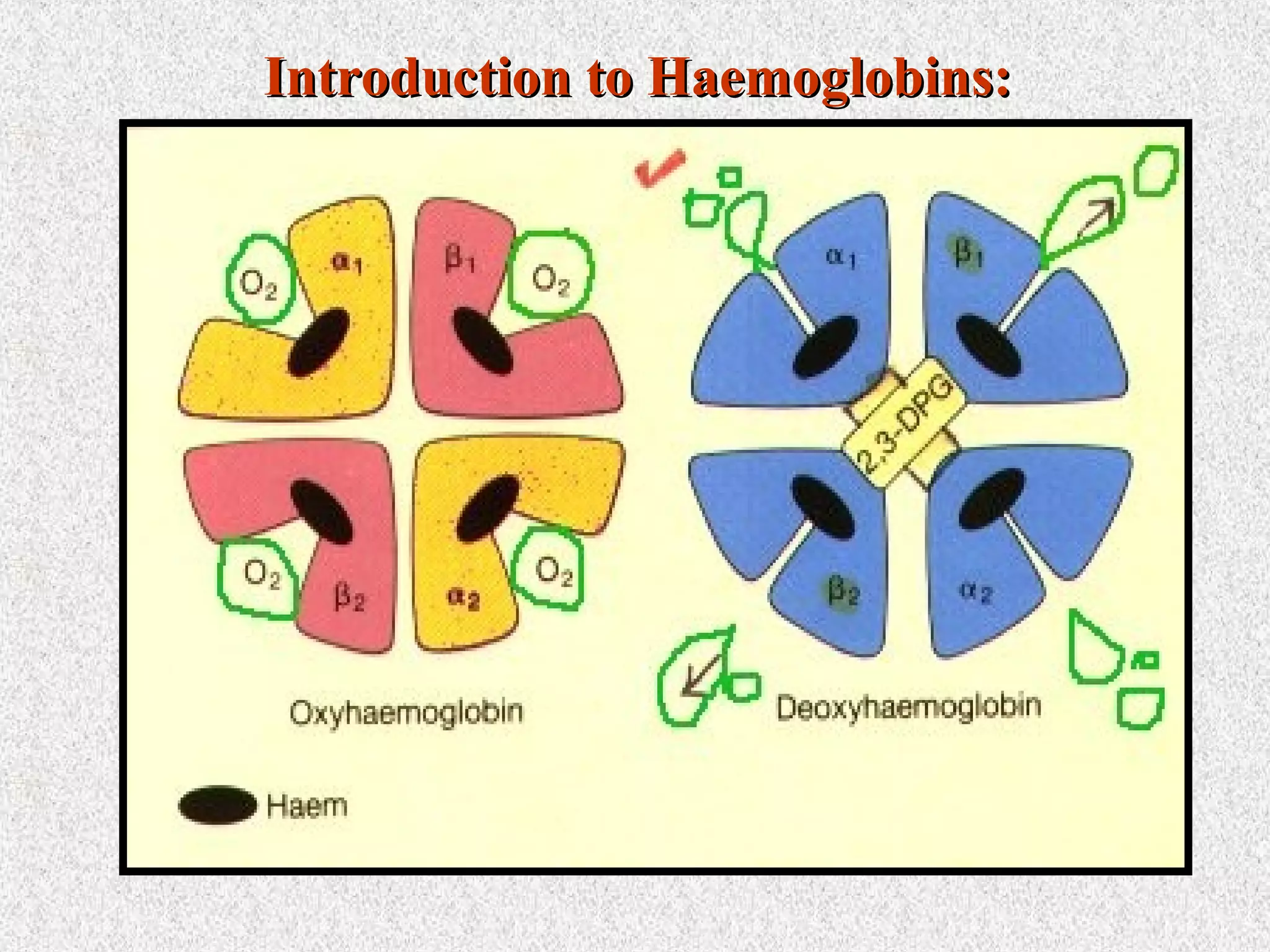
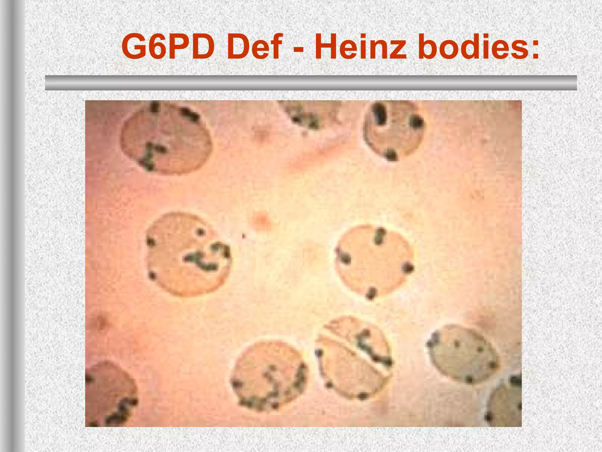
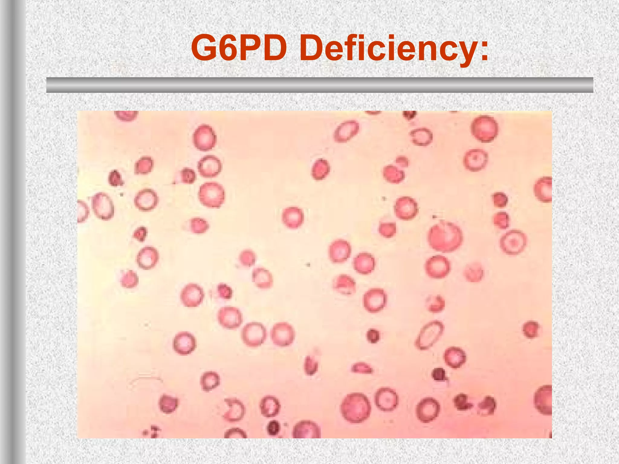
HEMATOLOGY
Hemolytic Anemia
Dr. Rafi Ahmed Ghori
FCPS
Professor Medicine
"Pleasure in the job"Pleasure in the job
putsputs
perfection in the work."perfection in the work."
-- Aristotle-- Aristotle
Blood Smear - Normal
C.B.C / FBC / Hemogram
 Haemoglobin - 15±2.5, 14 ±2.5 - g/dl
 PCV - 0.47 ±0.07, 0.42 ±0.05 - l/l (%)
 Haematocrit, Total RBC volume - better
 RBC count - 5.5 ±1, 4.8 ± 1 x10¹²/l
 MCHC - Hb/PCV - 30-36 - g/dl
 Hb synthesis within RBC
 MCH - Hb/RBC - 29.5 ± 2.5 pg/l
 Average Hb in RBC
 MCV - PCV/RBC 85 ± 8 – fl
 RBC Maturation
RBC disorders (Anemias) :
““Anemia is decreased red cell massAnemia is decreased red cell mass
affecting tissue oxygenation”affecting tissue oxygenation”
* Low Hb <13.5 (males), <11.5 (females)* Low Hb <13.5 (males), <11.5 (females)
Acquired / Congenital disorders:
Decreased production / Increased loss
Haemolytic An. Introduction
Anemia due to Increased RBC destruction
Decreased life span (<120d)
Breakdown  ↑Bilirubin (Unconj)  Jaundice
Increased RBC production - ↑ reticulocytes
Low Haptoglobins – Hb carrier proteins.
Ketabolism of Hb:
Polychromasia - Hemolytic
An.
Blood Film Features:
 Abnormal shape
 Polychromasia
 Nucleated RBC
 Plt may be low.
Clinical Features:
 Pallor mild – mucosal
 Jaundice - Mild fluctuating
 Splenomegaly
 No bile in urine (dark on standing-UBG)
 Pigment gall stones – in chronic forms
 Crisis – aplastic, hemolytic, vascular
 Ankle ulcers
Hemolytic Anemia - Types:
 Immune lysis
• Warm & Cold Ab, Auto & Allo immune
 Mechanical Damage
• Valve, Microangiopathy (DIC), prosthesis, march
 Hereditary Defects
• Membrane, Hb & Enzyme defect
 Infection induced
• Clostridia, malaria, septicemia
Clinical
Features
of Sickle
Cell
Disease
Congenital RBC Disorders:
Membrane Disorders:
 Spherocytosis, Elliptocytosis
Hemoglobin Disorders:
 Hemoglobinopathies - Sickle cell, HbC etc.
 Thalassemia Syndromes - α, β, δ
Enzyme disorders:
 G6PD, PK deficiency
Laboratory Evaluation:
 Features of RBC breakdown:
 Hyperbilirubinemia
 Increased Urine UBG & Faecal stercobilinogen.
 Low or absent Haptoglobins
 Features of increased RBC Production:
 Reticulocytosis
 Marrow erythroid hyperplasia – bone changes
 Damaged RBC
 Morphology, Osmotic Fragility
 Decreased RBC survival – 51
Cr labelling.
 Hemoglobin electrophoresis, enzyme abnormality
Laboratory Evaluation:
Intravascular Haemolysis:
 Haemoglobinaemia, Haemoglobinuria
 Haemosiderinuria – Renal tubular cells
 Methhaemalbuminaemia – Schumm’s test.
Molecular genetics
 Hb Electrophoresis
 Globin synthesis studies.
CBC Analyzer Report
Blood Smear Interpretation:
A B C D
E F G H
I J
A. Normal
B. Micro/hypo
C. Macro
D. Target
E. Sphero
F. Heinz body
G. Schistocyte
H. nRBC
I. Polychrom
J. Teardrop
Hb Electrophoresis – New born
_
S
F
A
Bart
s
+
C
S
A
F
pH 6.3pH 8.6
““Seeing much, suffering muchSeeing much, suffering much
and studying much are theand studying much are the
three pillars of learning.”three pillars of learning.”
–Benjamin Disraeli
MOLECULAR PATHOLOGY :
 Normal adult blood contain 3 types of Hb.
 The major component is HbA - α2
ß2
.
 The minor component fetal Hb (α2
γ2
) and Hb
A2 (α2
δ2
)
Structure & Synthesis of Haemoglobin:
Hb in adult Hb A Hb F Hb A2
Structure α2
ß 2
α2
γ2
α2
δ2
Normal % 96-98 0.5-0.8 1.5-3.2
Introduction to Haemoglobins:Introduction to Haemoglobins:
GENE CLUSTER & HB SWITCH
EFFECTS OF EXCESS α CHAINS
Thalassemia Syndromes:
Group of disorders with decreased production
of α or β chains.
Features:
 Low Hb – depending on type.
 Microcytic Hypochromic RBC
 Unlike IDA, uniform - low RDW
 Target forms typical. No pencil forms.
 Heinz bodies – globin deposits
 HBH inclusions – Golf ball cells
Thalassemia Syndromes:
Etiologically α, β, δβ thalassemia, HbH dis.
Clinically classified into
 Hydrops fetalis(α) – IU death
 Thalassemia major (β) – transfusion dep
 Thalassemia intermedia (αβ) –
spleenomegaly, Fe
 Thalassemia minor (αβ) - symptomless.
α-Thalassemia:
Decreased production of α chains.
Classification
 α0
– (four gene deletion) - Hydrops fetalis
 α+
2/3 gene deletion – Thal. Intermedia, HbH dis.
 α0
trait, α+
trait – Thal Minor, HPFH
No α thalassemia major.
HbH inclusions can be demonstrated in some
cases. (less in trait, more in intermedia)
Hydrops Fetalis:
α-Thalassemia:
Blood Smear & HbH PreparationBlood Smear & HbH Preparation
Thalassemia Trait:
Thalassemia Major:
ß Thalassemia Major:
Sickle Cell Disease:
G6PD Def - Heinz bodies:
Her. Spherocytosis:
Hereditary Elliptocytosis:
G6PD Deficiency:
G6PD Deficiency Anemia:
If you don'tIf you don't
stand for something,stand for something,
you will fall for anything…!you will fall for anything…!
Hb Barts levels in Cord blood inHb Barts levels in Cord blood in αα thalassemiathalassemia
Phenotype Equivalent No
of Functional
Genes
% Barts
Normal 4 0
α thal trait
(mild)
3 0-1
α thal trait
(severe)
2 2-8*
Hb H disease 1 10-40
Hb Barts Hydrops 0 ~80
Higgs DR et al Blood 73, 1081, 1989
* Some references mention upto 15%

hemolytic anemia 1

  • 1.
    HEMATOLOGY Hemolytic Anemia Dr. RafiAhmed Ghori FCPS Professor Medicine
  • 2.
    "Pleasure in thejob"Pleasure in the job putsputs perfection in the work."perfection in the work." -- Aristotle-- Aristotle
  • 3.
  • 4.
    C.B.C / FBC/ Hemogram  Haemoglobin - 15±2.5, 14 ±2.5 - g/dl  PCV - 0.47 ±0.07, 0.42 ±0.05 - l/l (%)  Haematocrit, Total RBC volume - better  RBC count - 5.5 ±1, 4.8 ± 1 x10¹²/l  MCHC - Hb/PCV - 30-36 - g/dl  Hb synthesis within RBC  MCH - Hb/RBC - 29.5 ± 2.5 pg/l  Average Hb in RBC  MCV - PCV/RBC 85 ± 8 – fl  RBC Maturation
  • 5.
    RBC disorders (Anemias): ““Anemia is decreased red cell massAnemia is decreased red cell mass affecting tissue oxygenation”affecting tissue oxygenation” * Low Hb <13.5 (males), <11.5 (females)* Low Hb <13.5 (males), <11.5 (females) Acquired / Congenital disorders: Decreased production / Increased loss
  • 6.
    Haemolytic An. Introduction Anemiadue to Increased RBC destruction Decreased life span (<120d) Breakdown  ↑Bilirubin (Unconj)  Jaundice Increased RBC production - ↑ reticulocytes Low Haptoglobins – Hb carrier proteins.
  • 7.
  • 8.
  • 9.
    Blood Film Features: Abnormal shape  Polychromasia  Nucleated RBC  Plt may be low.
  • 10.
    Clinical Features:  Pallormild – mucosal  Jaundice - Mild fluctuating  Splenomegaly  No bile in urine (dark on standing-UBG)  Pigment gall stones – in chronic forms  Crisis – aplastic, hemolytic, vascular  Ankle ulcers
  • 11.
    Hemolytic Anemia -Types:  Immune lysis • Warm & Cold Ab, Auto & Allo immune  Mechanical Damage • Valve, Microangiopathy (DIC), prosthesis, march  Hereditary Defects • Membrane, Hb & Enzyme defect  Infection induced • Clostridia, malaria, septicemia
  • 12.
  • 13.
    Congenital RBC Disorders: MembraneDisorders:  Spherocytosis, Elliptocytosis Hemoglobin Disorders:  Hemoglobinopathies - Sickle cell, HbC etc.  Thalassemia Syndromes - α, β, δ Enzyme disorders:  G6PD, PK deficiency
  • 14.
    Laboratory Evaluation:  Featuresof RBC breakdown:  Hyperbilirubinemia  Increased Urine UBG & Faecal stercobilinogen.  Low or absent Haptoglobins  Features of increased RBC Production:  Reticulocytosis  Marrow erythroid hyperplasia – bone changes  Damaged RBC  Morphology, Osmotic Fragility  Decreased RBC survival – 51 Cr labelling.  Hemoglobin electrophoresis, enzyme abnormality
  • 15.
    Laboratory Evaluation: Intravascular Haemolysis: Haemoglobinaemia, Haemoglobinuria  Haemosiderinuria – Renal tubular cells  Methhaemalbuminaemia – Schumm’s test. Molecular genetics  Hb Electrophoresis  Globin synthesis studies.
  • 16.
  • 17.
    Blood Smear Interpretation: AB C D E F G H I J A. Normal B. Micro/hypo C. Macro D. Target E. Sphero F. Heinz body G. Schistocyte H. nRBC I. Polychrom J. Teardrop
  • 18.
    Hb Electrophoresis –New born _ S F A Bart s + C S A F pH 6.3pH 8.6
  • 19.
    ““Seeing much, sufferingmuchSeeing much, suffering much and studying much are theand studying much are the three pillars of learning.”three pillars of learning.” –Benjamin Disraeli
  • 20.
    MOLECULAR PATHOLOGY : Normal adult blood contain 3 types of Hb.  The major component is HbA - α2 ß2 .  The minor component fetal Hb (α2 γ2 ) and Hb A2 (α2 δ2 ) Structure & Synthesis of Haemoglobin: Hb in adult Hb A Hb F Hb A2 Structure α2 ß 2 α2 γ2 α2 δ2 Normal % 96-98 0.5-0.8 1.5-3.2
  • 21.
  • 22.
    GENE CLUSTER &HB SWITCH
  • 23.
  • 24.
    Thalassemia Syndromes: Group ofdisorders with decreased production of α or β chains. Features:  Low Hb – depending on type.  Microcytic Hypochromic RBC  Unlike IDA, uniform - low RDW  Target forms typical. No pencil forms.  Heinz bodies – globin deposits  HBH inclusions – Golf ball cells
  • 25.
    Thalassemia Syndromes: Etiologically α,β, δβ thalassemia, HbH dis. Clinically classified into  Hydrops fetalis(α) – IU death  Thalassemia major (β) – transfusion dep  Thalassemia intermedia (αβ) – spleenomegaly, Fe  Thalassemia minor (αβ) - symptomless.
  • 26.
    α-Thalassemia: Decreased production ofα chains. Classification  α0 – (four gene deletion) - Hydrops fetalis  α+ 2/3 gene deletion – Thal. Intermedia, HbH dis.  α0 trait, α+ trait – Thal Minor, HPFH No α thalassemia major. HbH inclusions can be demonstrated in some cases. (less in trait, more in intermedia)
  • 27.
  • 28.
  • 29.
    Blood Smear &HbH PreparationBlood Smear & HbH Preparation
  • 30.
  • 31.
  • 32.
  • 33.
  • 34.
    G6PD Def -Heinz bodies:
  • 35.
  • 36.
  • 37.
  • 38.
  • 39.
    If you don'tIfyou don't stand for something,stand for something, you will fall for anything…!you will fall for anything…!
  • 40.
    Hb Barts levelsin Cord blood inHb Barts levels in Cord blood in αα thalassemiathalassemia Phenotype Equivalent No of Functional Genes % Barts Normal 4 0 α thal trait (mild) 3 0-1 α thal trait (severe) 2 2-8* Hb H disease 1 10-40 Hb Barts Hydrops 0 ~80 Higgs DR et al Blood 73, 1081, 1989 * Some references mention upto 15%