SlideShare a Scribd company logo
By Ali S. Mayali
Anemia
DEFINITION
• any condition in which the number of red cells or the
amount of hemoglobin in the blood is less than
normal.
• pathophysiological condition in which the body
cannot meet its demands for oxygen.
BLOOD REFERENCE VALUES
• Hemoglobin (M: 13.0 – 17.0 g/dl and F: 12.0 – 15.0 g/dl)
• RBC count RBC count Males range 5.5 x 10¹²/L, Females 4.5 x 10¹²/L.
• Mean cell volume MCV (80 – 100 fl)
• Mean cell hemoglobin (27 – 32 pg)? the average mass of hemoglobin per red
blood cell in a sample of blood. MCH value is diminished in hypochromic
anemias.
• Mean cell hemoglobin concentration MCHC (30-35 g/dl).
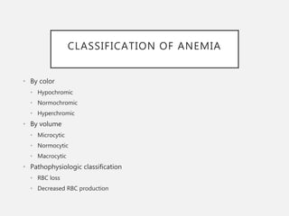
CLASSIFICATION OF ANEMIA
• By color
• Hypochromic
• Normochromic
• Hyperchromic
• By volume
• Microcytic
• Normocytic
• Macrocytic
• Pathophysiologic classification
• RBC loss
• Decreased RBC production
CLASSIFICATION BY VOLUME
I. microcytic anemia (MCV <80 fl)
1. iron deficiency anemia
2. thalassemia syndromes
3. anemia of chronic disease
4. sideroblastic anemia
II. normocytic anemia (MCV 80-100 fl)
1. anemia of blood loss
2. hemolytic anemia
III. macrocytic anemia (MCV >100 fl)
1. megaloblastic anemia
PATHOPHYSIOLOGIC
CLASSIFICATION
I RBC loss
• Blood loss
• Increased RBC destruction
a. intrinsic abnormality
b. extrinsic abnormality
II Decreased RBC production
• Stem cell abnormality
• erythroblast abnormality
• Unknown/multiple mechanism
PATHOPHYSIOLOGIC
CLASSIFICATION
I RBC loss
• Blood loss
• acute : trauma, massive hemorrhage
• chronic : GI lesion, GYN lesion
• Increased RBC destruction
a. intrinsic abnormality
b. extrinsic abnormality
PATHOPHYSIOLOGIC
CLASSIFICATION
I RBC loss
• Blood loss
• Increased RBC destruction
a. intrinsic abnormality
i. membrane disorder (e.g. hereditary spherocytosis)
ii. enzyme disorder (e.g. G6PD deficiency & PKD)
iii. Hgb synthesis disorder (e.g. Thalassemia & sickle cell disease)
iv. acquired membrane defect
a. extrinsic abnormality
PATHOPHYSIOLOGIC
CLASSIFICATION
I RBC loss
• Blood loss
• Increased RBC destruction
a. intrinsic abnormality
b. extrinsic abnormality
a. mechanical trauma (e.g. TTP/HUS, DIC)
b. chemical injury (e.g. lead poisoning)
c. Infection (e.g. malaria)
d. immunologic injury (e.g. autoimmune hemolytic anemia)
PATHOPHYSIOLOGIC
CLASSIFICATION
I RBC loss
• Blood loss
• Increased RBC destruction
a. intrinsic abnormality
b. extrinsic abnormality
II Decreased RBC production
• Stem cell abnormality
• erythroblast abnormality
• Unknown/multiple mechanism
PATHOPHYSIOLOGIC
CLASSIFICATION
I RBC loss
• Blood loss
• Increased RBC destruction
a. intrinsic abnormality
b. extrinsic abnormality
II Decreased RBC production
• Stem cell abnormality (aplastic anemia)
• erythroblast abnormality (megaloblastic anemia & IDA)
• Unknown/multiple mechanism (sideroblastic anemia)
PATHOPHYSIOLOGIC
CLASSIFICATION
I RBC loss
• Blood loss
• Increased RBC destruction
a. intrinsic abnormality
i. membrane disorder (hereditary spherocytosis & elliptocytosis)
ii. enzyme disorder (e.g. G6PD deficiency & PKD)
iii. Hgb synthesis disorder (Thalassemia & sickle cell disease)
iv. acquired membrane defect (PNH)
a. extrinsic abnormality
HEREDITARY SPHEROCYTOSIS
• Deficiency of Beta Spectrin or Ankyrin  Loss of membrane surface
area becomes more spherical Destruction in Spleen
• Clinical findings:
2. splenomegaly
3. marked compensatory erythroid hyperplasia in BM
4. jaundice, pigment cholelithiasis (↑ bilirubin)
• Laboratory findings : normal MCV with increased MCHC
• Treatment: splenectomy
HEREDITARY ELLIPTOCYTOSIS
• Abnormally large number of patient’s erythrocytes are
elliptical rather than typical biconcave shape.
• Inherited disease (autosomal dominant)
• Impaired aggregation of spectrin.
• majority require no treatment.
PATHOPHYSIOLOGIC
CLASSIFICATION
I RBC loss
• Blood loss
• Increased RBC destruction
a. intrinsic abnormality
i. membrane disorder (hereditary spherocytosis & elliptocytosis)
ii. enzyme disorder (e.g. G6PD deficiency & PKD)
iii. Hgb synthesis disorder (Thalassemia & sickle cell disease)
iv. acquired membrane defect (PNH)
a. extrinsic abnormality
G6PD DEFICIENCY
• G6PD deficiency is an X-linked inherited condition in which the body
doesn't have enough of the enzyme glucose-6-phosphate
dehydrogenase
• Function? Pivotal enzyme in HMP Shunt & produces NADPH to
protect RBC against oxidative stress
• Most common enzymopathy -10% world’s population
• Neonatal jaundice
• Hemolysis after exposure to oxidant stress
• Drugs
• Fava beans
G6PD DEFICIENCY
G6PD DEFICIENCY
• Lab findings:
• Heinz bodies : precipitates of denatured hemoglobin material
• “bite cells”? When a macrophage in the spleen identifies a RBC with
a Heinz body, it removes the precipitate and a small piece of the
membrane, leading to characteristic "bite cells“
• Treatment:
• Stop the precipitating drug or treat the infection
• Acute transfusions if required
G6PD DEFICIENCY
PYRUVATE KINASE DEFICIENCY
• also called erythrocyte pyruvate kinase deficiency, is an inherited metabolic
disorder of the enzyme pyruvate kinase which affects the survival of red blood cells
due to inability to maintain normal ATP levels.
• Splenomegaly
• Diagnosis: 1. fluorescent spot test 2. enzyme assay
• patients tolerate anemia rather well ( result in: high levels of 2,3-DPG)
phosphoenol pyruvate pyruvate lactat
e
P
K
ADP ATP *NADH NAD
PATHOPHYSIOLOGIC
CLASSIFICATION
I RBC loss
• Blood loss
• Increased RBC destruction
a. intrinsic abnormality
i. membrane disorder (hereditary spherocytosis & elliptocytosis)
ii. enzyme disorder (G6PD deficiency & PKD)
iii. Hgb synthesis disorder (Thalassemia & sickle cell disease)
iv. acquired membrane defect (PNH)
a. extrinsic abnormality
THALASSEMIA SYNDROMES
• heterogeneous hemolytic disorders characterized by
quantitative abnormalities of hemoglobin synthesis.
• genetic defect in globin production > inadequate Hgb
formation > hypochromic anemia (low hemoglobin in
RBC)
• Types:
• α-thalassemia : α-chain deficiency
• β-thalassemia : β-chain deficiency
THALASSEMIA SYNDROMES
• There are many forms of thalassemia. Each type has
many different subtypes. Both alpha and beta
thalassemia include the following two forms:
• Thalassemia major (gene defect from both parents)
• Thalassemia minor (faulty gene from only one parent)
• α thalassemia major causes stillbirth
• Children born with β thalassemia major (Cooley's
anemia) are normal at birth, but develop severe
anemia after 6 months where HbF must be replaced
with HbA .
α-Thalassemia
• There are a total of 4 alpha globin genes, alpha
thalassemia result from the deletions of some or all
of the genes.Disease Defect Hb A % HbH % MCV,fl
Silent carrier (asymptomatic) One deletion 97 0 90
Thalassemia traits Two deletions 90-95 rare 70-80
HbH (B4) Three deletions 70-95 5-30 60-70
Hb Bart (hydrops fetalis) Four deletions
Death in utero
0 5-10
β Thalassemia Major
• Severe, homozygous
• Childhood, growth delay, chipmunk face.
• Severe anemia , hepatosplenomegaly
• Treatment:
• Require transfusion once to twice a month
• Treat iron overload (due to chronic transfusion, give deferasirox or
deferoxamine)
• splenectomy reduce hemolysis
• Bone marrow tranplantation might be used.
• Diagnosis:
• P. Smear- severe microcytosis, target cells
• Hb electrophoresis: HbF - 90-96 % HbA2- 3.5 %- 5.5% HbA - 0 %
β Thalassemia Intermedia
• Similar stigmata like major
• Survive without transfusion
• Less severe than major
• Moderate anemia, microcytosis, hypochromia
• Diagnosis:
• P. Smear- severe microcytosis, target cells
• Hb electrophoresis: HbF - 20% - 100% HbA2 3.5 % -
5.5% HbA 0% - 30%
β Thalassemia Minor
• Most of the time, they do not have symptoms.
SICKLE CELL DISEASE
• Severe hereditary form of anemia (recessive) in which
an abnormal hemoglobin (hemoglobin S) leads to
chronic hemolytic anemia, pain and organ failure.
• Structurally abnormal hemoglobin (hemoglobin S)
result from a point mutation in the β chain of
hemoglobin molecule (glutamic acid  valine).
• If only one parent has sickle cell; their child would have
sickle cell trait. People with sickle cell trait do not have
the symptoms of sickle cell anemia.
• Symptoms appears after 8 to 10 weeks of age, when
the HbF has been replaced by HbS.
SICKLE CELL DISEASE
• There are two major consequences of RBC sickling
• Chronic hemolytic anemia.
• Blood vessels occlusion.
• Premature destruction of RBC in the spleen causes
hemolysis and anemia.
• Vessels occlusion disrupt blood flow causing tissue
ischemia and pain crisis.
• Other symptoms: Fatigue, paleness, rapid heart rate and
shortness of breath
• Splenic infraction may lead to splenic dysfunction which
predispose the patient to infections.
SICKLE CELL DISEASE
• Neonatal diagnosis of sickle cell disease is made on
the basis of clinical findings and hemoglobin
electrophoresis.
• The goal of treatment is to manage and control
symptoms, and to limit the number of crises.
PATHOPHYSIOLOGIC
CLASSIFICATION
I RBC loss
• Blood loss
• Increased RBC destruction
a. intrinsic abnormality
i. membrane disorder (e.g. hereditary spherocytosis)
ii. enzyme disorder (G6PD deficiency & PKD)
iii. Hgb synthesis disorder (Thalassemia & sickle cell disease)
iv. Acquired membrane defect (PNH)
a. extrinsic abnormality
PAROXYSMAL NOCTURNAL
HEMOGLOBINURIA
• Is a rare disease in which red blood cells break down
earlier than normal.
• Persons with this disease have blood cells that are
missing a gene called PIG-A. This gene allows a substance
called glycosyl-phosphatidylinositol (GPI) to help certain
proteins stick to cells.
• Without PIG-A, important proteins cannot connect to the
cell surface and protect the cell from substances in the
blood called complement. As a result, red blood cells
break down too early. The red cells leak hemoglobin into
the blood, which can pass into the urine. This can happen
at any time, but is more likely to occur during the night or
early morning.
• Risk Factors: Unknown!
PATHOPHYSIOLOGIC
CLASSIFICATION
I RBC loss
• Blood loss
• Increased RBC destruction
a. intrinsic abnormality
b. extrinsic abnormality
a. mechanical trauma (e.g. TTP/HUS, DIC)
b. chemical injury (e.g. lead poisoning)
c. Infection (e.g. malaria)
d. immunologic injury (e.g. autoimmune hemolytic anemia)
MECHANICAL TRAUMA
• Mechanical heart valves, Arterial grafts: cause
shear stress damage
• March hemoglobinuria: Red cell damage in
capillaries of feet
• Thermal injury: burns
• Microangiopathic hemolytic anemia (MAHA):
by passage of RBC through fibrin strands
deposited in small vessels  disruption of RBC
PATHOPHYSIOLOGIC
CLASSIFICATION
I RBC loss
• Blood loss
• Increased RBC destruction
a. intrinsic abnormality
b. extrinsic abnormality
a. mechanical trauma (e.g. TTP/HUS, DIC)
b. chemical injury (e.g. lead poisoning)
c. Infection (e.g. malaria)
d. immunologic injury (e.g. autoimmune hemolytic anemia)
LEAD POISONING
• inhibition of 5’-nucleotidase and
sodium-potassium pump  decreased
RBC survival.
• Other chemical injury by drugs: oxidant
denaturation of hemoglobin e.g:
Dapsone, sulphasalazine, Arsenic gas,
Cu, Nitrates & Nitrobenzene.
PATHOPHYSIOLOGIC
CLASSIFICATION
I RBC loss
• Blood loss
• Increased RBC destruction
a. intrinsic abnormality
b. extrinsic abnormality
a. mechanical trauma (e.g. TTP/HUS, DIC)
b. chemical injury (e.g. lead poisoning)
c. Infection (e.g. malaria)
d. immunologic injury (e.g. autoimmune hemolytic anemia)
MALARIA
• intracellular protozoan parasite
(Plasmodium)
• intravascular hemolysis: severe called
‘Blackwater fever’
• worldwide infection affecting 100 million
people 1 to 1.5 million deaths each year.
PATHOPHYSIOLOGIC
CLASSIFICATION
I RBC loss
• Blood loss
• Increased RBC destruction
a. intrinsic abnormality
b. extrinsic abnormality
a. mechanical trauma (e.g. TTP/HUS, DIC)
b. chemical injury (e.g. lead poisoning)
c. Infection (e.g. malaria)
d. immunologic injury (e.g. autoimmune hemolytic anemia)
AUTOIMMUNE HEMOLYTIC
ANEMIA
• Result from RBC destruction due to RBC
autoantibodies: Ig G, M, E, A
• Most commonly-idiopathic
• Classification
• Warm AI hemolysis: Ab binds at 37degree
Celsius
• Cold AI Hemolysis: Ab binds at 4 degree
Celsius
Warm Al Hemolysis
• Can occurs at all age groups
• F > M
• Causes:
• 50% Idiopathic
• Rest - secondary causes: Lymphoid neoplasm, Solid
Tumors, Autoimmune and drugs like Alpha methyl
DOPA.
• Investigations:
• Normocytic, Increased LDH, Reticulocytosis, decreased
haptoglobin
• Increased indirect Bilirubin.
• Direct Coomb’s Test / Antiglobulin test
Direct antiglobulin test
demonstrating the presence of
autoantibodies (shown here) or
complement on the surface of
the red blood cell.
complement
Warm Al Hemolysis
•Treatment:
• Correct the underlying cause
• Prednisolone 1mg/kg orally until Hb reaches 10mg/dl then
taper slowly and stop
• Transfusion: for life threatening problems
• If no response to steroids > Splenectomy or
immunosuppressive: Azathioprine, Cyclophosphamide
Cold AI Hemolysis
• Usually IgM
• Acute or Chronic form
• Chronic:
• C/F:
• Elderly patients
• Cold , painful & often blue fingers, toes, ears, or nose ( Acrocyanosis)
• Investigations:
• e/o hemolysis
• P Smear: Microspherocytosis
• Ig M with specificity to I or I Ag
Cold AI Hemolysis
• Treatment:
• Treatment of the underlying
cause
• Keep extremities warm
• Steroids treatment
• Blood transfusion
Anemia

More Related Content

What's hot

Child with pallor & jaundice (hemolytic anemia)
Child with pallor & jaundice (hemolytic anemia)Child with pallor & jaundice (hemolytic anemia)
Child with pallor & jaundice (hemolytic anemia)
Safia Sky
 
Hemolytic anemia - Approach and Management
Hemolytic anemia - Approach and ManagementHemolytic anemia - Approach and Management
Hemolytic anemia - Approach and Management
Chetan Ganteppanavar
 
Thalassemia
ThalassemiaThalassemia
Thalassemia
Anamika Dev
 
Thalassemia By IPMS-KUM Peshawar
Thalassemia By IPMS-KUM PeshawarThalassemia By IPMS-KUM Peshawar
Thalassemia By IPMS-KUM Peshawar
Asif Zeb
 
lab work up for hemolytic anemia
lab work up for hemolytic anemialab work up for hemolytic anemia
lab work up for hemolytic anemia
farzana fathima
 
anemia approach
anemia approachanemia approach
anemia approach
Nishant Prabhakar
 
An Overview of Hemolytic anemia
An Overview of Hemolytic anemiaAn Overview of Hemolytic anemia
An Overview of Hemolytic anemia
Prakash Poudel
 
Thalassemia- Introduction and Diagnosis
Thalassemia- Introduction and DiagnosisThalassemia- Introduction and Diagnosis
Thalassemia- Introduction and Diagnosis
Dr. Amita Yadav
 
Hemolytic anemia ppt presentation
Hemolytic anemia ppt presentationHemolytic anemia ppt presentation
Hemolytic anemia ppt presentation
National Academy of Young Scientists
 
Hemolytic anemia akk
Hemolytic anemia akkHemolytic anemia akk
Hemolytic anemia akk
Askin Kaplan
 
Approach to hemolytic anemia naglaa
Approach to hemolytic anemia naglaaApproach to hemolytic anemia naglaa
Approach to hemolytic anemia naglaaNaglaa Makram
 
Medicine 5th year, 8th lecture/part one (Dr. Sabir)
Medicine 5th year, 8th lecture/part one (Dr. Sabir)Medicine 5th year, 8th lecture/part one (Dr. Sabir)
Medicine 5th year, 8th lecture/part one (Dr. Sabir)
College of Medicine, Sulaymaniyah
 
Presentation Mdc Hematology
Presentation Mdc HematologyPresentation Mdc Hematology
Presentation Mdc Hematology
Miami Dade
 
Non transfusion dependent thalassemias beta-thalassemia intermedia and hb e b...
Non transfusion dependent thalassemias beta-thalassemia intermedia and hb e b...Non transfusion dependent thalassemias beta-thalassemia intermedia and hb e b...
Non transfusion dependent thalassemias beta-thalassemia intermedia and hb e b...
Thalassaemia International Federation
 
Thalassaemia
ThalassaemiaThalassaemia
Thalassaemia
Syed Nayyer Alvi
 
Thalassemia
ThalassemiaThalassemia
Thalassemia
Abdullah Almazyad
 
Diagnosis of iron deficiency anemia
Diagnosis of iron deficiency anemia Diagnosis of iron deficiency anemia
Diagnosis of iron deficiency anemia
Seth g s medical college and kem hospital mumbai
 

What's hot (20)

Child with pallor & jaundice (hemolytic anemia)
Child with pallor & jaundice (hemolytic anemia)Child with pallor & jaundice (hemolytic anemia)
Child with pallor & jaundice (hemolytic anemia)
 
Hemolytic anemia - Approach and Management
Hemolytic anemia - Approach and ManagementHemolytic anemia - Approach and Management
Hemolytic anemia - Approach and Management
 
Rbc Patho B
Rbc  Patho BRbc  Patho B
Rbc Patho B
 
4a..hemolytic anemia
4a..hemolytic anemia4a..hemolytic anemia
4a..hemolytic anemia
 
Thalassemia
ThalassemiaThalassemia
Thalassemia
 
Thalassemia By IPMS-KUM Peshawar
Thalassemia By IPMS-KUM PeshawarThalassemia By IPMS-KUM Peshawar
Thalassemia By IPMS-KUM Peshawar
 
lab work up for hemolytic anemia
lab work up for hemolytic anemialab work up for hemolytic anemia
lab work up for hemolytic anemia
 
anemia approach
anemia approachanemia approach
anemia approach
 
An Overview of Hemolytic anemia
An Overview of Hemolytic anemiaAn Overview of Hemolytic anemia
An Overview of Hemolytic anemia
 
Thalassemia- Introduction and Diagnosis
Thalassemia- Introduction and DiagnosisThalassemia- Introduction and Diagnosis
Thalassemia- Introduction and Diagnosis
 
Hemolytic anemia ppt presentation
Hemolytic anemia ppt presentationHemolytic anemia ppt presentation
Hemolytic anemia ppt presentation
 
Hemolytic anemia akk
Hemolytic anemia akkHemolytic anemia akk
Hemolytic anemia akk
 
Approach to hemolytic anemia naglaa
Approach to hemolytic anemia naglaaApproach to hemolytic anemia naglaa
Approach to hemolytic anemia naglaa
 
Medicine 5th year, 8th lecture/part one (Dr. Sabir)
Medicine 5th year, 8th lecture/part one (Dr. Sabir)Medicine 5th year, 8th lecture/part one (Dr. Sabir)
Medicine 5th year, 8th lecture/part one (Dr. Sabir)
 
Presentation Mdc Hematology
Presentation Mdc HematologyPresentation Mdc Hematology
Presentation Mdc Hematology
 
Non transfusion dependent thalassemias beta-thalassemia intermedia and hb e b...
Non transfusion dependent thalassemias beta-thalassemia intermedia and hb e b...Non transfusion dependent thalassemias beta-thalassemia intermedia and hb e b...
Non transfusion dependent thalassemias beta-thalassemia intermedia and hb e b...
 
Thalassaemia
ThalassaemiaThalassaemia
Thalassaemia
 
Thalassemia
ThalassemiaThalassemia
Thalassemia
 
Thalassemia55
Thalassemia55Thalassemia55
Thalassemia55
 
Diagnosis of iron deficiency anemia
Diagnosis of iron deficiency anemia Diagnosis of iron deficiency anemia
Diagnosis of iron deficiency anemia
 

Viewers also liked

Layers of the neck
Layers of the neckLayers of the neck
Layers of the neck
Ali S. Mayali
 
Mastoid Process
Mastoid ProcessMastoid Process
Mastoid Process
Ali S. Mayali
 
Pharmacokinetics
PharmacokineticsPharmacokinetics
Pharmacokinetics
Ali S. Mayali
 
Diagnosis and treatment of acid base disorders(1)
Diagnosis and treatment of acid base disorders(1)Diagnosis and treatment of acid base disorders(1)
Diagnosis and treatment of acid base disorders(1)
aparna jayara
 
Acid base disorders
Acid base disordersAcid base disorders
Acid base disorders
Leonardo Paskah S
 
Interpretation of the Arterial Blood Gas analysis
Interpretation of the Arterial Blood Gas analysisInterpretation of the Arterial Blood Gas analysis
Interpretation of the Arterial Blood Gas analysisVishal Golay
 
Arterial Blood Bas (ABG) Procedure and Interpretation
Arterial Blood Bas (ABG) Procedure and InterpretationArterial Blood Bas (ABG) Procedure and Interpretation
Arterial Blood Bas (ABG) Procedure and Interpretation
Louie Ray
 
Abg interpretation
Abg interpretationAbg interpretation
Abg interpretation
Ali S. Mayali
 

Viewers also liked (9)

Layers of the neck
Layers of the neckLayers of the neck
Layers of the neck
 
Mastoid Process
Mastoid ProcessMastoid Process
Mastoid Process
 
Pharmacokinetics
PharmacokineticsPharmacokinetics
Pharmacokinetics
 
Diagnosis and treatment of acid base disorders(1)
Diagnosis and treatment of acid base disorders(1)Diagnosis and treatment of acid base disorders(1)
Diagnosis and treatment of acid base disorders(1)
 
Acid base disorders
Acid base disordersAcid base disorders
Acid base disorders
 
Interpretation of the Arterial Blood Gas analysis
Interpretation of the Arterial Blood Gas analysisInterpretation of the Arterial Blood Gas analysis
Interpretation of the Arterial Blood Gas analysis
 
Arterial Blood Bas (ABG) Procedure and Interpretation
Arterial Blood Bas (ABG) Procedure and InterpretationArterial Blood Bas (ABG) Procedure and Interpretation
Arterial Blood Bas (ABG) Procedure and Interpretation
 
Blood Gas Interpretation
Blood Gas InterpretationBlood Gas Interpretation
Blood Gas Interpretation
 
Abg interpretation
Abg interpretationAbg interpretation
Abg interpretation
 

Similar to Anemia

Anemia in Child
Anemia in ChildAnemia in Child
Anemia in Child
ShaliniShal11
 
Anaemia pathology ppt
Anaemia pathology pptAnaemia pathology ppt
Anaemia pathology ppt
Nicholaus Kapunga
 
Anemia
AnemiaAnemia
Anemia; causes, types, laboratory investigations
Anemia; causes, types, laboratory investigationsAnemia; causes, types, laboratory investigations
Anemia; causes, types, laboratory investigations
Nawsherwan Mohammad
 
Hemolytic anemia.pptx
Hemolytic anemia.pptxHemolytic anemia.pptx
Hemolytic anemia.pptx
SunilMulgund1
 
Hemolytic-Anemia.pptx
Hemolytic-Anemia.pptxHemolytic-Anemia.pptx
Hemolytic-Anemia.pptx
Amrit Agarwal
 
Hemolytic-Anemia - a review of Hemolytic-Anemia
Hemolytic-Anemia - a  review of Hemolytic-AnemiaHemolytic-Anemia - a  review of Hemolytic-Anemia
Hemolytic-Anemia - a review of Hemolytic-Anemia
bhavyarkrishnan2000
 
Anemia.pptx
Anemia.pptxAnemia.pptx
Anemia.pptx
Dr. Irtaza Rehman
 
Making the diagnosis in hematology
Making the diagnosis in hematologyMaking the diagnosis in hematology
Making the diagnosis in hematologyfracpractice
 
laboratorydiagnosisofanemiaugs-170319165804 (1).pptx
laboratorydiagnosisofanemiaugs-170319165804 (1).pptxlaboratorydiagnosisofanemiaugs-170319165804 (1).pptx
laboratorydiagnosisofanemiaugs-170319165804 (1).pptx
DebdattaMandal3
 
Causes of Anemia in childhood, etiology investigation and mangement
Causes of Anemia in childhood, etiology investigation and mangementCauses of Anemia in childhood, etiology investigation and mangement
Causes of Anemia in childhood, etiology investigation and mangement
MariamMahmoud73
 
Rbc disorders-3
Rbc disorders-3Rbc disorders-3
Rbc disorders-3
Prasad CSBR
 
Laboratory diagnosis of anemia
Laboratory diagnosis of anemiaLaboratory diagnosis of anemia
Laboratory diagnosis of anemia
Dr. Varughese George
 
Lab. Diagnosis of Anemia.pptx
Lab. Diagnosis of Anemia.pptxLab. Diagnosis of Anemia.pptx
Lab. Diagnosis of Anemia.pptx
John Doe
 
Anemia.pdf
Anemia.pdfAnemia.pdf
Anemia.pdf
AritraHalder14
 
ANEMIA
ANEMIAANEMIA
Anemia seminar
Anemia seminarAnemia seminar
Anemia seminar
Ismail Abduljabar
 
is-Anemia.pptx
is-Anemia.pptxis-Anemia.pptx
is-Anemia.pptx
ShamsaBariyow
 
Anemia presentation for medical students
Anemia presentation for medical studentsAnemia presentation for medical students
Anemia presentation for medical students
IbrahimKargbo13
 

Similar to Anemia (20)

Anemia in Child
Anemia in ChildAnemia in Child
Anemia in Child
 
Anaemia pathology ppt
Anaemia pathology pptAnaemia pathology ppt
Anaemia pathology ppt
 
Anemia
AnemiaAnemia
Anemia
 
Anemia; causes, types, laboratory investigations
Anemia; causes, types, laboratory investigationsAnemia; causes, types, laboratory investigations
Anemia; causes, types, laboratory investigations
 
Hemolytic anemia.pptx
Hemolytic anemia.pptxHemolytic anemia.pptx
Hemolytic anemia.pptx
 
Hemolytic-Anemia.pptx
Hemolytic-Anemia.pptxHemolytic-Anemia.pptx
Hemolytic-Anemia.pptx
 
Hemolytic-Anemia - a review of Hemolytic-Anemia
Hemolytic-Anemia - a  review of Hemolytic-AnemiaHemolytic-Anemia - a  review of Hemolytic-Anemia
Hemolytic-Anemia - a review of Hemolytic-Anemia
 
Anemia.pptx
Anemia.pptxAnemia.pptx
Anemia.pptx
 
Anemia
AnemiaAnemia
Anemia
 
Making the diagnosis in hematology
Making the diagnosis in hematologyMaking the diagnosis in hematology
Making the diagnosis in hematology
 
laboratorydiagnosisofanemiaugs-170319165804 (1).pptx
laboratorydiagnosisofanemiaugs-170319165804 (1).pptxlaboratorydiagnosisofanemiaugs-170319165804 (1).pptx
laboratorydiagnosisofanemiaugs-170319165804 (1).pptx
 
Causes of Anemia in childhood, etiology investigation and mangement
Causes of Anemia in childhood, etiology investigation and mangementCauses of Anemia in childhood, etiology investigation and mangement
Causes of Anemia in childhood, etiology investigation and mangement
 
Rbc disorders-3
Rbc disorders-3Rbc disorders-3
Rbc disorders-3
 
Laboratory diagnosis of anemia
Laboratory diagnosis of anemiaLaboratory diagnosis of anemia
Laboratory diagnosis of anemia
 
Lab. Diagnosis of Anemia.pptx
Lab. Diagnosis of Anemia.pptxLab. Diagnosis of Anemia.pptx
Lab. Diagnosis of Anemia.pptx
 
Anemia.pdf
Anemia.pdfAnemia.pdf
Anemia.pdf
 
ANEMIA
ANEMIAANEMIA
ANEMIA
 
Anemia seminar
Anemia seminarAnemia seminar
Anemia seminar
 
is-Anemia.pptx
is-Anemia.pptxis-Anemia.pptx
is-Anemia.pptx
 
Anemia presentation for medical students
Anemia presentation for medical studentsAnemia presentation for medical students
Anemia presentation for medical students
 

Recently uploaded

Maxilla, Mandible & Hyoid Bone & Clinical Correlations by Dr. RIG.pptx
Maxilla, Mandible & Hyoid Bone & Clinical Correlations by Dr. RIG.pptxMaxilla, Mandible & Hyoid Bone & Clinical Correlations by Dr. RIG.pptx
Maxilla, Mandible & Hyoid Bone & Clinical Correlations by Dr. RIG.pptx
Dr. Rabia Inam Gandapore
 
Tom Selleck Health: A Comprehensive Look at the Iconic Actor’s Wellness Journey
Tom Selleck Health: A Comprehensive Look at the Iconic Actor’s Wellness JourneyTom Selleck Health: A Comprehensive Look at the Iconic Actor’s Wellness Journey
Tom Selleck Health: A Comprehensive Look at the Iconic Actor’s Wellness Journey
greendigital
 
For Better Surat #ℂall #Girl Service ❤85270-49040❤ Surat #ℂall #Girls
For Better Surat #ℂall #Girl Service ❤85270-49040❤ Surat #ℂall #GirlsFor Better Surat #ℂall #Girl Service ❤85270-49040❤ Surat #ℂall #Girls
For Better Surat #ℂall #Girl Service ❤85270-49040❤ Surat #ℂall #Girls
Savita Shen $i11
 
Physiology of Special Chemical Sensation of Taste
Physiology of Special Chemical Sensation of TastePhysiology of Special Chemical Sensation of Taste
Physiology of Special Chemical Sensation of Taste
MedicoseAcademics
 
BRACHYTHERAPY OVERVIEW AND APPLICATORS
BRACHYTHERAPY OVERVIEW  AND  APPLICATORSBRACHYTHERAPY OVERVIEW  AND  APPLICATORS
BRACHYTHERAPY OVERVIEW AND APPLICATORS
Krishan Murari
 
Thyroid Gland- Gross Anatomy by Dr. Rabia Inam Gandapore.pptx
Thyroid Gland- Gross Anatomy by Dr. Rabia Inam Gandapore.pptxThyroid Gland- Gross Anatomy by Dr. Rabia Inam Gandapore.pptx
Thyroid Gland- Gross Anatomy by Dr. Rabia Inam Gandapore.pptx
Dr. Rabia Inam Gandapore
 
Report Back from SGO 2024: What’s the Latest in Cervical Cancer?
Report Back from SGO 2024: What’s the Latest in Cervical Cancer?Report Back from SGO 2024: What’s the Latest in Cervical Cancer?
Report Back from SGO 2024: What’s the Latest in Cervical Cancer?
bkling
 
KDIGO 2024 guidelines for diabetologists
KDIGO 2024 guidelines for diabetologistsKDIGO 2024 guidelines for diabetologists
KDIGO 2024 guidelines for diabetologists
د.محمود نجيب
 
Novas diretrizes da OMS para os cuidados perinatais de mais qualidade
Novas diretrizes da OMS para os cuidados perinatais de mais qualidadeNovas diretrizes da OMS para os cuidados perinatais de mais qualidade
Novas diretrizes da OMS para os cuidados perinatais de mais qualidade
Prof. Marcus Renato de Carvalho
 
Light House Retreats: Plant Medicine Retreat Europe
Light House Retreats: Plant Medicine Retreat EuropeLight House Retreats: Plant Medicine Retreat Europe
Light House Retreats: Plant Medicine Retreat Europe
Lighthouse Retreat
 
planning for change nursing Management ppt
planning for change nursing Management pptplanning for change nursing Management ppt
planning for change nursing Management ppt
Thangamjayarani
 
Pharynx and Clinical Correlations BY Dr.Rabia Inam Gandapore.pptx
Pharynx and Clinical Correlations BY Dr.Rabia Inam Gandapore.pptxPharynx and Clinical Correlations BY Dr.Rabia Inam Gandapore.pptx
Pharynx and Clinical Correlations BY Dr.Rabia Inam Gandapore.pptx
Dr. Rabia Inam Gandapore
 
Ophthalmology Clinical Tests for OSCE exam
Ophthalmology Clinical Tests for OSCE examOphthalmology Clinical Tests for OSCE exam
Ophthalmology Clinical Tests for OSCE exam
KafrELShiekh University
 
Cervical & Brachial Plexus By Dr. RIG.pptx
Cervical & Brachial Plexus By Dr. RIG.pptxCervical & Brachial Plexus By Dr. RIG.pptx
Cervical & Brachial Plexus By Dr. RIG.pptx
Dr. Rabia Inam Gandapore
 
Basavarajeeyam - Ayurvedic heritage book of Andhra pradesh
Basavarajeeyam - Ayurvedic heritage book of Andhra pradeshBasavarajeeyam - Ayurvedic heritage book of Andhra pradesh
Basavarajeeyam - Ayurvedic heritage book of Andhra pradesh
Dr. Madduru Muni Haritha
 
ARTIFICIAL INTELLIGENCE IN HEALTHCARE.pdf
ARTIFICIAL INTELLIGENCE IN  HEALTHCARE.pdfARTIFICIAL INTELLIGENCE IN  HEALTHCARE.pdf
ARTIFICIAL INTELLIGENCE IN HEALTHCARE.pdf
Anujkumaranit
 
263778731218 Abortion Clinic /Pills In Harare ,
263778731218 Abortion Clinic /Pills In Harare ,263778731218 Abortion Clinic /Pills In Harare ,
263778731218 Abortion Clinic /Pills In Harare ,
sisternakatoto
 
New Directions in Targeted Therapeutic Approaches for Older Adults With Mantl...
New Directions in Targeted Therapeutic Approaches for Older Adults With Mantl...New Directions in Targeted Therapeutic Approaches for Older Adults With Mantl...
New Directions in Targeted Therapeutic Approaches for Older Adults With Mantl...
i3 Health
 
Non-respiratory Functions of the Lungs.pdf
Non-respiratory Functions of the Lungs.pdfNon-respiratory Functions of the Lungs.pdf
Non-respiratory Functions of the Lungs.pdf
MedicoseAcademics
 
ARTHROLOGY PPT NCISM SYLLABUS AYURVEDA STUDENTS
ARTHROLOGY PPT NCISM SYLLABUS AYURVEDA STUDENTSARTHROLOGY PPT NCISM SYLLABUS AYURVEDA STUDENTS
ARTHROLOGY PPT NCISM SYLLABUS AYURVEDA STUDENTS
Dr. Vinay Pareek
 

Recently uploaded (20)

Maxilla, Mandible & Hyoid Bone & Clinical Correlations by Dr. RIG.pptx
Maxilla, Mandible & Hyoid Bone & Clinical Correlations by Dr. RIG.pptxMaxilla, Mandible & Hyoid Bone & Clinical Correlations by Dr. RIG.pptx
Maxilla, Mandible & Hyoid Bone & Clinical Correlations by Dr. RIG.pptx
 
Tom Selleck Health: A Comprehensive Look at the Iconic Actor’s Wellness Journey
Tom Selleck Health: A Comprehensive Look at the Iconic Actor’s Wellness JourneyTom Selleck Health: A Comprehensive Look at the Iconic Actor’s Wellness Journey
Tom Selleck Health: A Comprehensive Look at the Iconic Actor’s Wellness Journey
 
For Better Surat #ℂall #Girl Service ❤85270-49040❤ Surat #ℂall #Girls
For Better Surat #ℂall #Girl Service ❤85270-49040❤ Surat #ℂall #GirlsFor Better Surat #ℂall #Girl Service ❤85270-49040❤ Surat #ℂall #Girls
For Better Surat #ℂall #Girl Service ❤85270-49040❤ Surat #ℂall #Girls
 
Physiology of Special Chemical Sensation of Taste
Physiology of Special Chemical Sensation of TastePhysiology of Special Chemical Sensation of Taste
Physiology of Special Chemical Sensation of Taste
 
BRACHYTHERAPY OVERVIEW AND APPLICATORS
BRACHYTHERAPY OVERVIEW  AND  APPLICATORSBRACHYTHERAPY OVERVIEW  AND  APPLICATORS
BRACHYTHERAPY OVERVIEW AND APPLICATORS
 
Thyroid Gland- Gross Anatomy by Dr. Rabia Inam Gandapore.pptx
Thyroid Gland- Gross Anatomy by Dr. Rabia Inam Gandapore.pptxThyroid Gland- Gross Anatomy by Dr. Rabia Inam Gandapore.pptx
Thyroid Gland- Gross Anatomy by Dr. Rabia Inam Gandapore.pptx
 
Report Back from SGO 2024: What’s the Latest in Cervical Cancer?
Report Back from SGO 2024: What’s the Latest in Cervical Cancer?Report Back from SGO 2024: What’s the Latest in Cervical Cancer?
Report Back from SGO 2024: What’s the Latest in Cervical Cancer?
 
KDIGO 2024 guidelines for diabetologists
KDIGO 2024 guidelines for diabetologistsKDIGO 2024 guidelines for diabetologists
KDIGO 2024 guidelines for diabetologists
 
Novas diretrizes da OMS para os cuidados perinatais de mais qualidade
Novas diretrizes da OMS para os cuidados perinatais de mais qualidadeNovas diretrizes da OMS para os cuidados perinatais de mais qualidade
Novas diretrizes da OMS para os cuidados perinatais de mais qualidade
 
Light House Retreats: Plant Medicine Retreat Europe
Light House Retreats: Plant Medicine Retreat EuropeLight House Retreats: Plant Medicine Retreat Europe
Light House Retreats: Plant Medicine Retreat Europe
 
planning for change nursing Management ppt
planning for change nursing Management pptplanning for change nursing Management ppt
planning for change nursing Management ppt
 
Pharynx and Clinical Correlations BY Dr.Rabia Inam Gandapore.pptx
Pharynx and Clinical Correlations BY Dr.Rabia Inam Gandapore.pptxPharynx and Clinical Correlations BY Dr.Rabia Inam Gandapore.pptx
Pharynx and Clinical Correlations BY Dr.Rabia Inam Gandapore.pptx
 
Ophthalmology Clinical Tests for OSCE exam
Ophthalmology Clinical Tests for OSCE examOphthalmology Clinical Tests for OSCE exam
Ophthalmology Clinical Tests for OSCE exam
 
Cervical & Brachial Plexus By Dr. RIG.pptx
Cervical & Brachial Plexus By Dr. RIG.pptxCervical & Brachial Plexus By Dr. RIG.pptx
Cervical & Brachial Plexus By Dr. RIG.pptx
 
Basavarajeeyam - Ayurvedic heritage book of Andhra pradesh
Basavarajeeyam - Ayurvedic heritage book of Andhra pradeshBasavarajeeyam - Ayurvedic heritage book of Andhra pradesh
Basavarajeeyam - Ayurvedic heritage book of Andhra pradesh
 
ARTIFICIAL INTELLIGENCE IN HEALTHCARE.pdf
ARTIFICIAL INTELLIGENCE IN  HEALTHCARE.pdfARTIFICIAL INTELLIGENCE IN  HEALTHCARE.pdf
ARTIFICIAL INTELLIGENCE IN HEALTHCARE.pdf
 
263778731218 Abortion Clinic /Pills In Harare ,
263778731218 Abortion Clinic /Pills In Harare ,263778731218 Abortion Clinic /Pills In Harare ,
263778731218 Abortion Clinic /Pills In Harare ,
 
New Directions in Targeted Therapeutic Approaches for Older Adults With Mantl...
New Directions in Targeted Therapeutic Approaches for Older Adults With Mantl...New Directions in Targeted Therapeutic Approaches for Older Adults With Mantl...
New Directions in Targeted Therapeutic Approaches for Older Adults With Mantl...
 
Non-respiratory Functions of the Lungs.pdf
Non-respiratory Functions of the Lungs.pdfNon-respiratory Functions of the Lungs.pdf
Non-respiratory Functions of the Lungs.pdf
 
ARTHROLOGY PPT NCISM SYLLABUS AYURVEDA STUDENTS
ARTHROLOGY PPT NCISM SYLLABUS AYURVEDA STUDENTSARTHROLOGY PPT NCISM SYLLABUS AYURVEDA STUDENTS
ARTHROLOGY PPT NCISM SYLLABUS AYURVEDA STUDENTS
 

Anemia

  • 1. By Ali S. Mayali Anemia
  • 2. DEFINITION • any condition in which the number of red cells or the amount of hemoglobin in the blood is less than normal. • pathophysiological condition in which the body cannot meet its demands for oxygen.
  • 3. BLOOD REFERENCE VALUES • Hemoglobin (M: 13.0 – 17.0 g/dl and F: 12.0 – 15.0 g/dl) • RBC count RBC count Males range 5.5 x 10¹²/L, Females 4.5 x 10¹²/L. • Mean cell volume MCV (80 – 100 fl) • Mean cell hemoglobin (27 – 32 pg)? the average mass of hemoglobin per red blood cell in a sample of blood. MCH value is diminished in hypochromic anemias. • Mean cell hemoglobin concentration MCHC (30-35 g/dl).
  • 4. CLASSIFICATION OF ANEMIA • By color • Hypochromic • Normochromic • Hyperchromic • By volume • Microcytic • Normocytic • Macrocytic • Pathophysiologic classification • RBC loss • Decreased RBC production
  • 5. CLASSIFICATION BY VOLUME I. microcytic anemia (MCV <80 fl) 1. iron deficiency anemia 2. thalassemia syndromes 3. anemia of chronic disease 4. sideroblastic anemia II. normocytic anemia (MCV 80-100 fl) 1. anemia of blood loss 2. hemolytic anemia III. macrocytic anemia (MCV >100 fl) 1. megaloblastic anemia
  • 6. PATHOPHYSIOLOGIC CLASSIFICATION I RBC loss • Blood loss • Increased RBC destruction a. intrinsic abnormality b. extrinsic abnormality II Decreased RBC production • Stem cell abnormality • erythroblast abnormality • Unknown/multiple mechanism
  • 7. PATHOPHYSIOLOGIC CLASSIFICATION I RBC loss • Blood loss • acute : trauma, massive hemorrhage • chronic : GI lesion, GYN lesion • Increased RBC destruction a. intrinsic abnormality b. extrinsic abnormality
  • 8. PATHOPHYSIOLOGIC CLASSIFICATION I RBC loss • Blood loss • Increased RBC destruction a. intrinsic abnormality i. membrane disorder (e.g. hereditary spherocytosis) ii. enzyme disorder (e.g. G6PD deficiency & PKD) iii. Hgb synthesis disorder (e.g. Thalassemia & sickle cell disease) iv. acquired membrane defect a. extrinsic abnormality
  • 9. PATHOPHYSIOLOGIC CLASSIFICATION I RBC loss • Blood loss • Increased RBC destruction a. intrinsic abnormality b. extrinsic abnormality a. mechanical trauma (e.g. TTP/HUS, DIC) b. chemical injury (e.g. lead poisoning) c. Infection (e.g. malaria) d. immunologic injury (e.g. autoimmune hemolytic anemia)
  • 10. PATHOPHYSIOLOGIC CLASSIFICATION I RBC loss • Blood loss • Increased RBC destruction a. intrinsic abnormality b. extrinsic abnormality II Decreased RBC production • Stem cell abnormality • erythroblast abnormality • Unknown/multiple mechanism
  • 11. PATHOPHYSIOLOGIC CLASSIFICATION I RBC loss • Blood loss • Increased RBC destruction a. intrinsic abnormality b. extrinsic abnormality II Decreased RBC production • Stem cell abnormality (aplastic anemia) • erythroblast abnormality (megaloblastic anemia & IDA) • Unknown/multiple mechanism (sideroblastic anemia)
  • 12. PATHOPHYSIOLOGIC CLASSIFICATION I RBC loss • Blood loss • Increased RBC destruction a. intrinsic abnormality i. membrane disorder (hereditary spherocytosis & elliptocytosis) ii. enzyme disorder (e.g. G6PD deficiency & PKD) iii. Hgb synthesis disorder (Thalassemia & sickle cell disease) iv. acquired membrane defect (PNH) a. extrinsic abnormality
  • 13. HEREDITARY SPHEROCYTOSIS • Deficiency of Beta Spectrin or Ankyrin  Loss of membrane surface area becomes more spherical Destruction in Spleen • Clinical findings: 2. splenomegaly 3. marked compensatory erythroid hyperplasia in BM 4. jaundice, pigment cholelithiasis (↑ bilirubin) • Laboratory findings : normal MCV with increased MCHC • Treatment: splenectomy
  • 14. HEREDITARY ELLIPTOCYTOSIS • Abnormally large number of patient’s erythrocytes are elliptical rather than typical biconcave shape. • Inherited disease (autosomal dominant) • Impaired aggregation of spectrin. • majority require no treatment.
  • 15. PATHOPHYSIOLOGIC CLASSIFICATION I RBC loss • Blood loss • Increased RBC destruction a. intrinsic abnormality i. membrane disorder (hereditary spherocytosis & elliptocytosis) ii. enzyme disorder (e.g. G6PD deficiency & PKD) iii. Hgb synthesis disorder (Thalassemia & sickle cell disease) iv. acquired membrane defect (PNH) a. extrinsic abnormality
  • 16. G6PD DEFICIENCY • G6PD deficiency is an X-linked inherited condition in which the body doesn't have enough of the enzyme glucose-6-phosphate dehydrogenase • Function? Pivotal enzyme in HMP Shunt & produces NADPH to protect RBC against oxidative stress • Most common enzymopathy -10% world’s population • Neonatal jaundice • Hemolysis after exposure to oxidant stress • Drugs • Fava beans
  • 18. G6PD DEFICIENCY • Lab findings: • Heinz bodies : precipitates of denatured hemoglobin material • “bite cells”? When a macrophage in the spleen identifies a RBC with a Heinz body, it removes the precipitate and a small piece of the membrane, leading to characteristic "bite cells“ • Treatment: • Stop the precipitating drug or treat the infection • Acute transfusions if required
  • 20. PYRUVATE KINASE DEFICIENCY • also called erythrocyte pyruvate kinase deficiency, is an inherited metabolic disorder of the enzyme pyruvate kinase which affects the survival of red blood cells due to inability to maintain normal ATP levels. • Splenomegaly • Diagnosis: 1. fluorescent spot test 2. enzyme assay • patients tolerate anemia rather well ( result in: high levels of 2,3-DPG) phosphoenol pyruvate pyruvate lactat e P K ADP ATP *NADH NAD
  • 21. PATHOPHYSIOLOGIC CLASSIFICATION I RBC loss • Blood loss • Increased RBC destruction a. intrinsic abnormality i. membrane disorder (hereditary spherocytosis & elliptocytosis) ii. enzyme disorder (G6PD deficiency & PKD) iii. Hgb synthesis disorder (Thalassemia & sickle cell disease) iv. acquired membrane defect (PNH) a. extrinsic abnormality
  • 22. THALASSEMIA SYNDROMES • heterogeneous hemolytic disorders characterized by quantitative abnormalities of hemoglobin synthesis. • genetic defect in globin production > inadequate Hgb formation > hypochromic anemia (low hemoglobin in RBC) • Types: • α-thalassemia : α-chain deficiency • β-thalassemia : β-chain deficiency
  • 23. THALASSEMIA SYNDROMES • There are many forms of thalassemia. Each type has many different subtypes. Both alpha and beta thalassemia include the following two forms: • Thalassemia major (gene defect from both parents) • Thalassemia minor (faulty gene from only one parent) • α thalassemia major causes stillbirth • Children born with β thalassemia major (Cooley's anemia) are normal at birth, but develop severe anemia after 6 months where HbF must be replaced with HbA .
  • 24. α-Thalassemia • There are a total of 4 alpha globin genes, alpha thalassemia result from the deletions of some or all of the genes.Disease Defect Hb A % HbH % MCV,fl Silent carrier (asymptomatic) One deletion 97 0 90 Thalassemia traits Two deletions 90-95 rare 70-80 HbH (B4) Three deletions 70-95 5-30 60-70 Hb Bart (hydrops fetalis) Four deletions Death in utero 0 5-10
  • 25. β Thalassemia Major • Severe, homozygous • Childhood, growth delay, chipmunk face. • Severe anemia , hepatosplenomegaly • Treatment: • Require transfusion once to twice a month • Treat iron overload (due to chronic transfusion, give deferasirox or deferoxamine) • splenectomy reduce hemolysis • Bone marrow tranplantation might be used. • Diagnosis: • P. Smear- severe microcytosis, target cells • Hb electrophoresis: HbF - 90-96 % HbA2- 3.5 %- 5.5% HbA - 0 %
  • 26. β Thalassemia Intermedia • Similar stigmata like major • Survive without transfusion • Less severe than major • Moderate anemia, microcytosis, hypochromia • Diagnosis: • P. Smear- severe microcytosis, target cells • Hb electrophoresis: HbF - 20% - 100% HbA2 3.5 % - 5.5% HbA 0% - 30%
  • 27. β Thalassemia Minor • Most of the time, they do not have symptoms.
  • 28. SICKLE CELL DISEASE • Severe hereditary form of anemia (recessive) in which an abnormal hemoglobin (hemoglobin S) leads to chronic hemolytic anemia, pain and organ failure. • Structurally abnormal hemoglobin (hemoglobin S) result from a point mutation in the β chain of hemoglobin molecule (glutamic acid  valine). • If only one parent has sickle cell; their child would have sickle cell trait. People with sickle cell trait do not have the symptoms of sickle cell anemia. • Symptoms appears after 8 to 10 weeks of age, when the HbF has been replaced by HbS.
  • 29. SICKLE CELL DISEASE • There are two major consequences of RBC sickling • Chronic hemolytic anemia. • Blood vessels occlusion. • Premature destruction of RBC in the spleen causes hemolysis and anemia. • Vessels occlusion disrupt blood flow causing tissue ischemia and pain crisis. • Other symptoms: Fatigue, paleness, rapid heart rate and shortness of breath • Splenic infraction may lead to splenic dysfunction which predispose the patient to infections.
  • 30. SICKLE CELL DISEASE • Neonatal diagnosis of sickle cell disease is made on the basis of clinical findings and hemoglobin electrophoresis. • The goal of treatment is to manage and control symptoms, and to limit the number of crises.
  • 31. PATHOPHYSIOLOGIC CLASSIFICATION I RBC loss • Blood loss • Increased RBC destruction a. intrinsic abnormality i. membrane disorder (e.g. hereditary spherocytosis) ii. enzyme disorder (G6PD deficiency & PKD) iii. Hgb synthesis disorder (Thalassemia & sickle cell disease) iv. Acquired membrane defect (PNH) a. extrinsic abnormality
  • 32. PAROXYSMAL NOCTURNAL HEMOGLOBINURIA • Is a rare disease in which red blood cells break down earlier than normal. • Persons with this disease have blood cells that are missing a gene called PIG-A. This gene allows a substance called glycosyl-phosphatidylinositol (GPI) to help certain proteins stick to cells. • Without PIG-A, important proteins cannot connect to the cell surface and protect the cell from substances in the blood called complement. As a result, red blood cells break down too early. The red cells leak hemoglobin into the blood, which can pass into the urine. This can happen at any time, but is more likely to occur during the night or early morning. • Risk Factors: Unknown!
  • 33. PATHOPHYSIOLOGIC CLASSIFICATION I RBC loss • Blood loss • Increased RBC destruction a. intrinsic abnormality b. extrinsic abnormality a. mechanical trauma (e.g. TTP/HUS, DIC) b. chemical injury (e.g. lead poisoning) c. Infection (e.g. malaria) d. immunologic injury (e.g. autoimmune hemolytic anemia)
  • 34. MECHANICAL TRAUMA • Mechanical heart valves, Arterial grafts: cause shear stress damage • March hemoglobinuria: Red cell damage in capillaries of feet • Thermal injury: burns • Microangiopathic hemolytic anemia (MAHA): by passage of RBC through fibrin strands deposited in small vessels  disruption of RBC
  • 35. PATHOPHYSIOLOGIC CLASSIFICATION I RBC loss • Blood loss • Increased RBC destruction a. intrinsic abnormality b. extrinsic abnormality a. mechanical trauma (e.g. TTP/HUS, DIC) b. chemical injury (e.g. lead poisoning) c. Infection (e.g. malaria) d. immunologic injury (e.g. autoimmune hemolytic anemia)
  • 36. LEAD POISONING • inhibition of 5’-nucleotidase and sodium-potassium pump  decreased RBC survival. • Other chemical injury by drugs: oxidant denaturation of hemoglobin e.g: Dapsone, sulphasalazine, Arsenic gas, Cu, Nitrates & Nitrobenzene.
  • 37. PATHOPHYSIOLOGIC CLASSIFICATION I RBC loss • Blood loss • Increased RBC destruction a. intrinsic abnormality b. extrinsic abnormality a. mechanical trauma (e.g. TTP/HUS, DIC) b. chemical injury (e.g. lead poisoning) c. Infection (e.g. malaria) d. immunologic injury (e.g. autoimmune hemolytic anemia)
  • 38. MALARIA • intracellular protozoan parasite (Plasmodium) • intravascular hemolysis: severe called ‘Blackwater fever’ • worldwide infection affecting 100 million people 1 to 1.5 million deaths each year.
  • 39. PATHOPHYSIOLOGIC CLASSIFICATION I RBC loss • Blood loss • Increased RBC destruction a. intrinsic abnormality b. extrinsic abnormality a. mechanical trauma (e.g. TTP/HUS, DIC) b. chemical injury (e.g. lead poisoning) c. Infection (e.g. malaria) d. immunologic injury (e.g. autoimmune hemolytic anemia)
  • 40. AUTOIMMUNE HEMOLYTIC ANEMIA • Result from RBC destruction due to RBC autoantibodies: Ig G, M, E, A • Most commonly-idiopathic • Classification • Warm AI hemolysis: Ab binds at 37degree Celsius • Cold AI Hemolysis: Ab binds at 4 degree Celsius
  • 41. Warm Al Hemolysis • Can occurs at all age groups • F > M • Causes: • 50% Idiopathic • Rest - secondary causes: Lymphoid neoplasm, Solid Tumors, Autoimmune and drugs like Alpha methyl DOPA. • Investigations: • Normocytic, Increased LDH, Reticulocytosis, decreased haptoglobin • Increased indirect Bilirubin. • Direct Coomb’s Test / Antiglobulin test Direct antiglobulin test demonstrating the presence of autoantibodies (shown here) or complement on the surface of the red blood cell. complement
  • 42. Warm Al Hemolysis •Treatment: • Correct the underlying cause • Prednisolone 1mg/kg orally until Hb reaches 10mg/dl then taper slowly and stop • Transfusion: for life threatening problems • If no response to steroids > Splenectomy or immunosuppressive: Azathioprine, Cyclophosphamide
  • 43. Cold AI Hemolysis • Usually IgM • Acute or Chronic form • Chronic: • C/F: • Elderly patients • Cold , painful & often blue fingers, toes, ears, or nose ( Acrocyanosis) • Investigations: • e/o hemolysis • P Smear: Microspherocytosis • Ig M with specificity to I or I Ag
  • 44. Cold AI Hemolysis • Treatment: • Treatment of the underlying cause • Keep extremities warm • Steroids treatment • Blood transfusion