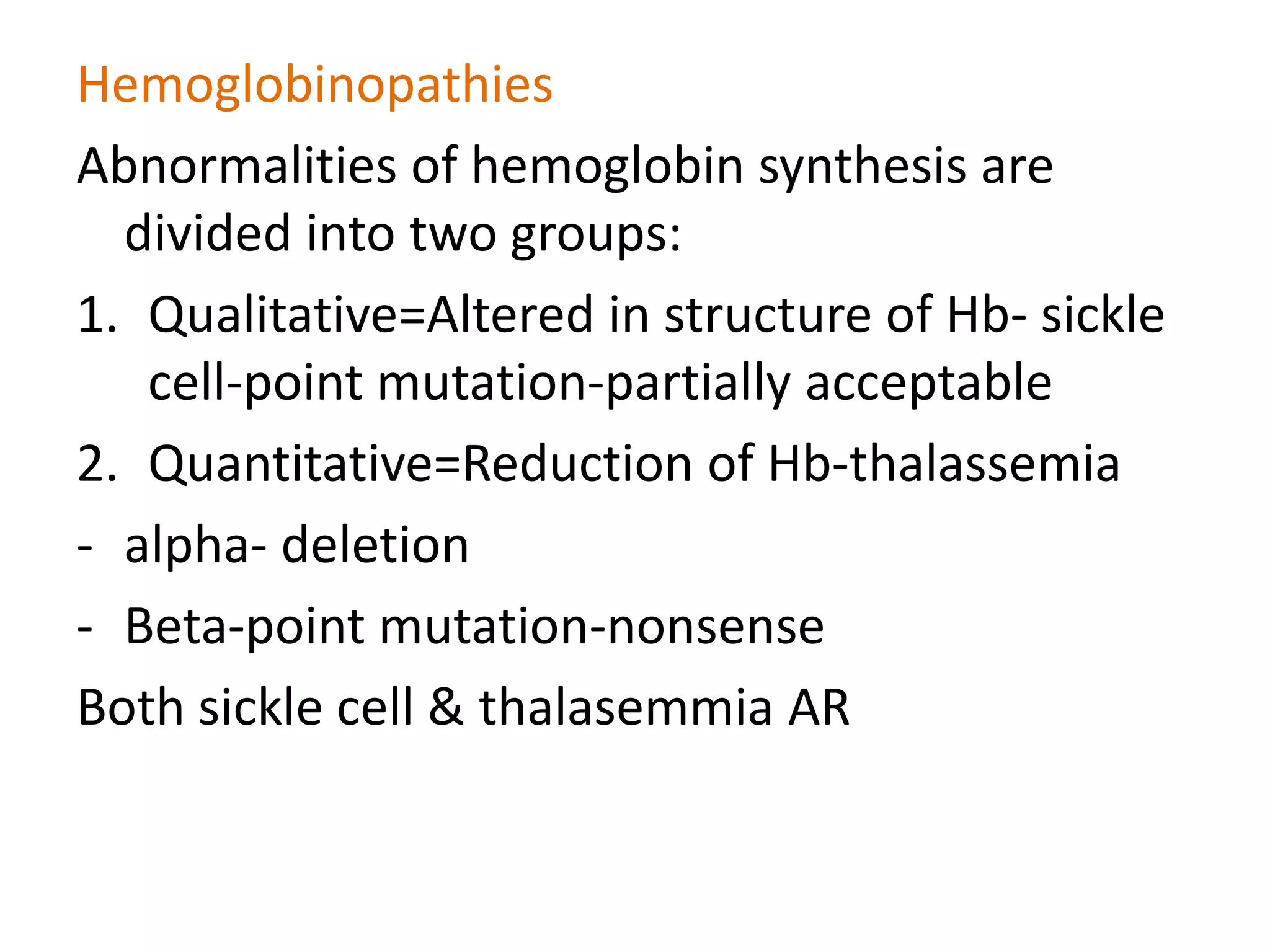
Thalassemia & Sickle cell disease
Dr. Kalpana Arora
HEMOGLOBIN
• Embryonic HB 8 wks intrauterine life
1. Gower I zeta2, epsilon 2
2. Gower II alpha 2 epsilon2
3. Portland I zeta2 gamma2
4. portland II-zeta 2 beta2
• Fetal hemoglobin, HBF- α2ϒ2
• Adult HbA-α2β2
• Normal adult has HbA-96%, HbF<2% 6months
• HbA2-3.5%
HbA HbF HbA2
97% ~1% 2-3.3%
Hemoglobins in normal adults
Hemoglobinopathies
Abnormalities of hemoglobin synthesis are
divided into two groups:
1. Qualitative=Altered in structure of Hb- sickle
cell-point mutation-partially acceptable
2. Quantitative=Reduction of Hb-thalassemia
- alpha- deletion
- Beta-point mutation-nonsense
Both sickle cell & thalasemmia AR
Thalassemia
• AR
• MC hemoglobinopathies both in world & India
• due to decreased synthesis of either the α or beta globin chain of
HbA:
 Alpha thalassemia- chromosome 16,deletion
 Beta thalassemia-chromosome 11, non sense point mutation-mc
mutation
• The hematologic consequences of decreased synthesis of one globin
chain stem not only from low intracellular hemoglobin but also from
relative excess of unpaired chain.
a thalassemia
• a globin chain is made by 4 genes, each gene
contributes to25% of the a-globin chain. The
severity of a thalassemia varies depending on
the number of a globin chains affected.
• Deletion is seen
1) silent carrier -Single alpha globin gene deleted. Asymptomatic
2)a -THALASSEMIA TRAIT – 2 a globin chains deleted. Asymptomatic
with some RBC abnormalities
3) Hemoglobin H Disease – 3 genes of α globin chain are deleted.
There is only 1 functional α gene. Resembles beta thal intermedia.
The synthesis of α chain is markedly reduced & tetramers of excess
β globin called HbH.
HbH has extremely high affinity for oxygen & therefore is not useful for
oxygen exchange, leading to tissue hypoxia.
Patients have moderate to severe anemia & may need blood transfusion
4) Hydrops Fetalis /Barts Hb-No alpha genes in the individual's DNA
• The gamma globins produced by the fetus form tetramers called
hemoglobin Bart. Hemoglobin Bart has very high affinity for
oxygen that it delivers no oxygen to tissues.
• This condition is usually not compatible with postnatal life, and
affected fetuses are either stillborn or die soon after birth .
• Fetus shows severe pallor, edema, hepatosplenomegaly
similar to erythroblastosis fetalis.
Hb H inclusions
ß-Thalassemia
• MC are nonsense point mutations, others are splicing site
mutation, chain termination, chain terminator mutation
• Consequences of thalassemia:
1. Decease HbA synthesis- decreased β chain
2. Increased alpha chains-Excess free α chains aggregate into
insoluble inclusions within RBCs & their precursors.
-They cause membrane damage, which results in apoptosis of
normoblasts in marrow- ineffective erythropoiesis.
- The RBC which escape death in BM are prone to spleenic
sequestration & destruction due to cell membrane damage leading
to extravascular hemolysis- this is MC
,
important
β- Thalassemias
• Thalassemia major/ Cooley’s anemia- homozygous for β
thalassemia genes. Severe requires blood transfusions.
Severe anemia manifests 6-9mths after birth, as hemoglobin
synthesis switches from HbF to HbA. hb is very low. There is
markedly increased HbF. repeated blood transfusion. MC
cause of death is cardiac failure due to iron overload-dilated >
restrictive CMP. Survival 10-20yrs
• Thalassemia minor/β thalassemia trait-has only 1 copy of β
thalassemia gene ie heterozygous. Mildest from with mild or
absent anemia. Pts are asymptomatic & identified with lab
investigations. There is increased HbA2(>3.5%)
• Thalassemia intermediate- b/n major & minor, require multiple
transfusion in adolescent
Thalassemia facies-Frontal bossing, flat nasal bridge
X ray skull- crew cut appearance & hair on end appearance
PERIPHERAL SMEAR
LABORATORY FEATURES
• red cell count - increased
• Reticulocytes-
• Serum ferritin, serum iron-
• TIBC- decreased
• MCV, MCH & MCHC-
• Osmotic fragility - decreased.
• HbA2, HbF - increased
• Hb electrophoresis-HbA is markedly decreased with
proportionate increase in HbA2 & HbF
• HPLC-
NESTROFT
Naked Eye Single Tube Reduced Osmotic Fragility Test
• 20ml of blood + 5 ml 0.36% buffered saline
• Mix and observe for reduced fragility
• This can serve as an initial screening process if
electronic measurement of MCV is not available
• screening test for detection of beta thalassemia
trait
Sickle Cell Anemia
• AR. missense point mutation in  globin chain. Substitution of
glutamic acid by valine ( glutamic acid goes & valine is
welcomed) in the 6th position of  chain
• homozygous for sickle cell mutation, almost all the hemoglobin in
red cells is HbS
• heterozygotes only 40% of hemoglobin is Hbs; it provides
protection against falciparum malaria
Molecular pathology of SCD
The sickle mutation
Sickle Cell Disease: Pharmacologic Treatment
GAG
Glutamic acid
GTG
Valine
The s Mutation
The same mutation found in all
s genes around the world
6th Codon of -Globin Gene
mutation 6th position of  chain-
Red Blood Cells from Sickle Cell Anemia
OXY-STATE DEOXY-STATE
• Deoxygenation of SS erythrocytes leads to
intracellular hemoglobin polymerization, loss of
deformability and changes in cell morphology.
Factors affecting sickling
• Amount of HbS- most important factor for sickling is amount of
HbS . In homozygous individuals, all the hemoglobin in RBC is HbS
& undergo sickling. In heterozygous, only 40% of Hb is HbS &
sickling does not occur as HbA has inhibitor effect on
polymerization of HbS
• HbF also has inhibitory effect on sickling of HbS-newborns do not
develop the disease till they are 5-6months of age
• Hemoglobin concentration- MCHC-higher HbS concentration
increase the probability that aggregation & polymerisation will
occur during any given period of time. Thus the conditions which
decease MCHC reduce disease severity
• Deoxygenation with HbS- polymerization of HbS occurs only in
deoxygenated state. A decrease in pH reduces the oxygen affinity
of Hb, thereby increasing the fraction of deoxygenated HbS at any
given oxygen tension & augmenting the tendency of sickling
• The length of time RBC are exposed to low
oxygen tension- sickling of RBC is confined to
microvascular beds where blood flow is
sluggish. This is the case in spleen & BM which
are predominantly affected by sickling
• Thalassemia & sickle cell-decrease severity
C/F
• Chronic hemolysis- irreversible sickle cells have rigid, non
deformable cell membranes which cause difficulty in negotiating
the spleenic sinusoids, sequestration & rapid phagocytosis. This
leads to extra vascular hemolysis. Some intravascular hemolysis
also occurs due to increased mechanical fragility of damaged cells.
This leads to increased bilirubin & pigment gall stones
• Vaso-occlusive symptoms- MC manifestation. reversible sickle cells
express higher than normal levels of adhesion molecules & are
abnormally sticky that is responsible for occlusion of
microcirculation –thrombosis-esp of bones, lungs, spleen & penis.
These manifest as:
1.Painful bone crisis
2.Hand-foot syndrome-dactylitis of bones of hand/feet
3.Autospleenectomy-predisposes to infection by encapsulated
bacteria eg H influenza & pneumococcus
4.Acute chest syndrome
Hematologic Crises
Aplastic Crises
• Parvovirus B19
• CFU-E
Splenic Sequestration
• Hb↓2g,
• compensatory marrow erythropoiesis,
• acutely enlarging spleen.
Hemolytic Crises (Hyperhemolytic Crises)
• Infection
Megaloblastic crisis
• CNS symptoms- seizure, stroke
• Priapism & infarction of penis
• Leg ulcers
• MC cause of osteomyelitis is salmonella
Peripheral smear
• ESR- decreased
• Hb electrophoresis- two bands in heterozygous state/sickle cell
trait & 1 band in homozygous state. HbS is slower moving than
HbA towards positive electrode
• HPLC- best test for sickle cell
• leukocytosis
Sickling
Test
• Sickling phenomenon may be demonstrated in a thin
wet film of blood .
• If Hb S is present, the red cells lose their smooth,
round shape and become sickled.
• This process may take up to 12 hours in Hb S trait,
whereas changes are apparent in homozygotes and
compound heterozygotes after 1 hour at 37°C.
• These changes can be hastened by the addition of a
reducing agent such as sodium dithionite
Solubility test
• The decreased solubility of deoxy Hb S forms the basis for tests in
which blood is added to a buffered solution of a reducing agent
such as sodium dithionite.
• Hb S is insoluble and precipitates in solution, rendering it turbid,
whereas solutions containing hemoglobins other than Hb S remain
clear.
Treatment
There is no known cure for sickle cell anemia.
The four main treatment options are:
• Drug Treatment
• Blood Transfusions
• Blood and Marrow Stem Cell Transplantation
• Gene Therapy
These main treatment options for the painful crisis involves
heavy reliance on painkilling drugs and oral and intravenous
fluids whose main functions are to reduce pain and prevent
complications.
Prevention
• Screening
– Premarital screening
– Preconception screening
• Genetic counselling
• Prenatal diagnosis
thalassemia and sickle cell disease

thalassemia and sickle cell disease

  • 1.
    Thalassemia & Sicklecell disease Dr. Kalpana Arora
  • 2.
    HEMOGLOBIN • Embryonic HB8 wks intrauterine life 1. Gower I zeta2, epsilon 2 2. Gower II alpha 2 epsilon2 3. Portland I zeta2 gamma2 4. portland II-zeta 2 beta2 • Fetal hemoglobin, HBF- α2ϒ2 • Adult HbA-α2β2 • Normal adult has HbA-96%, HbF<2% 6months • HbA2-3.5%
  • 3.
    HbA HbF HbA2 97%~1% 2-3.3% Hemoglobins in normal adults
  • 4.
    Hemoglobinopathies Abnormalities of hemoglobinsynthesis are divided into two groups: 1. Qualitative=Altered in structure of Hb- sickle cell-point mutation-partially acceptable 2. Quantitative=Reduction of Hb-thalassemia - alpha- deletion - Beta-point mutation-nonsense Both sickle cell & thalasemmia AR
  • 5.
    Thalassemia • AR • MChemoglobinopathies both in world & India • due to decreased synthesis of either the α or beta globin chain of HbA:  Alpha thalassemia- chromosome 16,deletion  Beta thalassemia-chromosome 11, non sense point mutation-mc mutation • The hematologic consequences of decreased synthesis of one globin chain stem not only from low intracellular hemoglobin but also from relative excess of unpaired chain.
  • 6.
    a thalassemia • aglobin chain is made by 4 genes, each gene contributes to25% of the a-globin chain. The severity of a thalassemia varies depending on the number of a globin chains affected. • Deletion is seen
  • 7.
    1) silent carrier-Single alpha globin gene deleted. Asymptomatic 2)a -THALASSEMIA TRAIT – 2 a globin chains deleted. Asymptomatic with some RBC abnormalities 3) Hemoglobin H Disease – 3 genes of α globin chain are deleted. There is only 1 functional α gene. Resembles beta thal intermedia. The synthesis of α chain is markedly reduced & tetramers of excess β globin called HbH. HbH has extremely high affinity for oxygen & therefore is not useful for oxygen exchange, leading to tissue hypoxia. Patients have moderate to severe anemia & may need blood transfusion 4) Hydrops Fetalis /Barts Hb-No alpha genes in the individual's DNA • The gamma globins produced by the fetus form tetramers called hemoglobin Bart. Hemoglobin Bart has very high affinity for oxygen that it delivers no oxygen to tissues. • This condition is usually not compatible with postnatal life, and affected fetuses are either stillborn or die soon after birth . • Fetus shows severe pallor, edema, hepatosplenomegaly similar to erythroblastosis fetalis.
  • 8.
  • 9.
    ß-Thalassemia • MC arenonsense point mutations, others are splicing site mutation, chain termination, chain terminator mutation • Consequences of thalassemia: 1. Decease HbA synthesis- decreased β chain 2. Increased alpha chains-Excess free α chains aggregate into insoluble inclusions within RBCs & their precursors. -They cause membrane damage, which results in apoptosis of normoblasts in marrow- ineffective erythropoiesis. - The RBC which escape death in BM are prone to spleenic sequestration & destruction due to cell membrane damage leading to extravascular hemolysis- this is MC
  • 10.
  • 11.
    β- Thalassemias • Thalassemiamajor/ Cooley’s anemia- homozygous for β thalassemia genes. Severe requires blood transfusions. Severe anemia manifests 6-9mths after birth, as hemoglobin synthesis switches from HbF to HbA. hb is very low. There is markedly increased HbF. repeated blood transfusion. MC cause of death is cardiac failure due to iron overload-dilated > restrictive CMP. Survival 10-20yrs • Thalassemia minor/β thalassemia trait-has only 1 copy of β thalassemia gene ie heterozygous. Mildest from with mild or absent anemia. Pts are asymptomatic & identified with lab investigations. There is increased HbA2(>3.5%) • Thalassemia intermediate- b/n major & minor, require multiple transfusion in adolescent
  • 12.
    Thalassemia facies-Frontal bossing,flat nasal bridge X ray skull- crew cut appearance & hair on end appearance
  • 14.
  • 18.
    LABORATORY FEATURES • redcell count - increased • Reticulocytes- • Serum ferritin, serum iron- • TIBC- decreased • MCV, MCH & MCHC- • Osmotic fragility - decreased. • HbA2, HbF - increased • Hb electrophoresis-HbA is markedly decreased with proportionate increase in HbA2 & HbF • HPLC-
  • 20.
    NESTROFT Naked Eye SingleTube Reduced Osmotic Fragility Test • 20ml of blood + 5 ml 0.36% buffered saline • Mix and observe for reduced fragility • This can serve as an initial screening process if electronic measurement of MCV is not available • screening test for detection of beta thalassemia trait
  • 21.
    Sickle Cell Anemia •AR. missense point mutation in  globin chain. Substitution of glutamic acid by valine ( glutamic acid goes & valine is welcomed) in the 6th position of  chain • homozygous for sickle cell mutation, almost all the hemoglobin in red cells is HbS • heterozygotes only 40% of hemoglobin is Hbs; it provides protection against falciparum malaria
  • 22.
    Molecular pathology ofSCD The sickle mutation Sickle Cell Disease: Pharmacologic Treatment GAG Glutamic acid GTG Valine The s Mutation The same mutation found in all s genes around the world 6th Codon of -Globin Gene
  • 23.
    mutation 6th positionof  chain-
  • 24.
    Red Blood Cellsfrom Sickle Cell Anemia OXY-STATE DEOXY-STATE • Deoxygenation of SS erythrocytes leads to intracellular hemoglobin polymerization, loss of deformability and changes in cell morphology.
  • 25.
    Factors affecting sickling •Amount of HbS- most important factor for sickling is amount of HbS . In homozygous individuals, all the hemoglobin in RBC is HbS & undergo sickling. In heterozygous, only 40% of Hb is HbS & sickling does not occur as HbA has inhibitor effect on polymerization of HbS • HbF also has inhibitory effect on sickling of HbS-newborns do not develop the disease till they are 5-6months of age • Hemoglobin concentration- MCHC-higher HbS concentration increase the probability that aggregation & polymerisation will occur during any given period of time. Thus the conditions which decease MCHC reduce disease severity • Deoxygenation with HbS- polymerization of HbS occurs only in deoxygenated state. A decrease in pH reduces the oxygen affinity of Hb, thereby increasing the fraction of deoxygenated HbS at any given oxygen tension & augmenting the tendency of sickling
  • 26.
    • The lengthof time RBC are exposed to low oxygen tension- sickling of RBC is confined to microvascular beds where blood flow is sluggish. This is the case in spleen & BM which are predominantly affected by sickling • Thalassemia & sickle cell-decrease severity
  • 28.
    C/F • Chronic hemolysis-irreversible sickle cells have rigid, non deformable cell membranes which cause difficulty in negotiating the spleenic sinusoids, sequestration & rapid phagocytosis. This leads to extra vascular hemolysis. Some intravascular hemolysis also occurs due to increased mechanical fragility of damaged cells. This leads to increased bilirubin & pigment gall stones • Vaso-occlusive symptoms- MC manifestation. reversible sickle cells express higher than normal levels of adhesion molecules & are abnormally sticky that is responsible for occlusion of microcirculation –thrombosis-esp of bones, lungs, spleen & penis. These manifest as: 1.Painful bone crisis 2.Hand-foot syndrome-dactylitis of bones of hand/feet 3.Autospleenectomy-predisposes to infection by encapsulated bacteria eg H influenza & pneumococcus 4.Acute chest syndrome
  • 29.
    Hematologic Crises Aplastic Crises •Parvovirus B19 • CFU-E Splenic Sequestration • Hb↓2g, • compensatory marrow erythropoiesis, • acutely enlarging spleen. Hemolytic Crises (Hyperhemolytic Crises) • Infection Megaloblastic crisis
  • 30.
    • CNS symptoms-seizure, stroke • Priapism & infarction of penis • Leg ulcers • MC cause of osteomyelitis is salmonella
  • 31.
  • 32.
    • ESR- decreased •Hb electrophoresis- two bands in heterozygous state/sickle cell trait & 1 band in homozygous state. HbS is slower moving than HbA towards positive electrode • HPLC- best test for sickle cell • leukocytosis
  • 33.
    Sickling Test • Sickling phenomenonmay be demonstrated in a thin wet film of blood . • If Hb S is present, the red cells lose their smooth, round shape and become sickled. • This process may take up to 12 hours in Hb S trait, whereas changes are apparent in homozygotes and compound heterozygotes after 1 hour at 37°C. • These changes can be hastened by the addition of a reducing agent such as sodium dithionite
  • 34.
    Solubility test • Thedecreased solubility of deoxy Hb S forms the basis for tests in which blood is added to a buffered solution of a reducing agent such as sodium dithionite. • Hb S is insoluble and precipitates in solution, rendering it turbid, whereas solutions containing hemoglobins other than Hb S remain clear.
  • 35.
    Treatment There is noknown cure for sickle cell anemia. The four main treatment options are: • Drug Treatment • Blood Transfusions • Blood and Marrow Stem Cell Transplantation • Gene Therapy These main treatment options for the painful crisis involves heavy reliance on painkilling drugs and oral and intravenous fluids whose main functions are to reduce pain and prevent complications.
  • 36.
    Prevention • Screening – Premaritalscreening – Preconception screening • Genetic counselling • Prenatal diagnosis