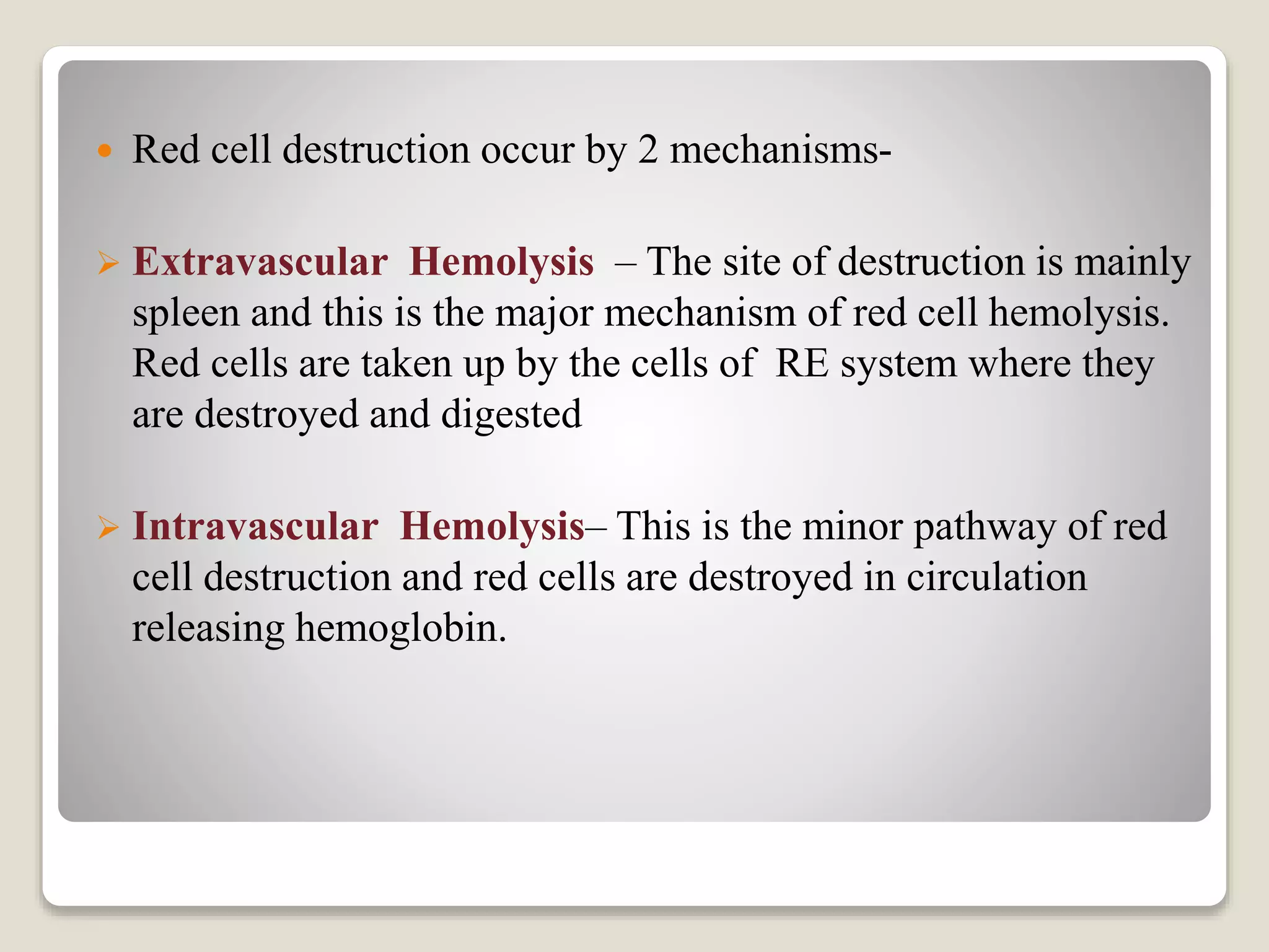
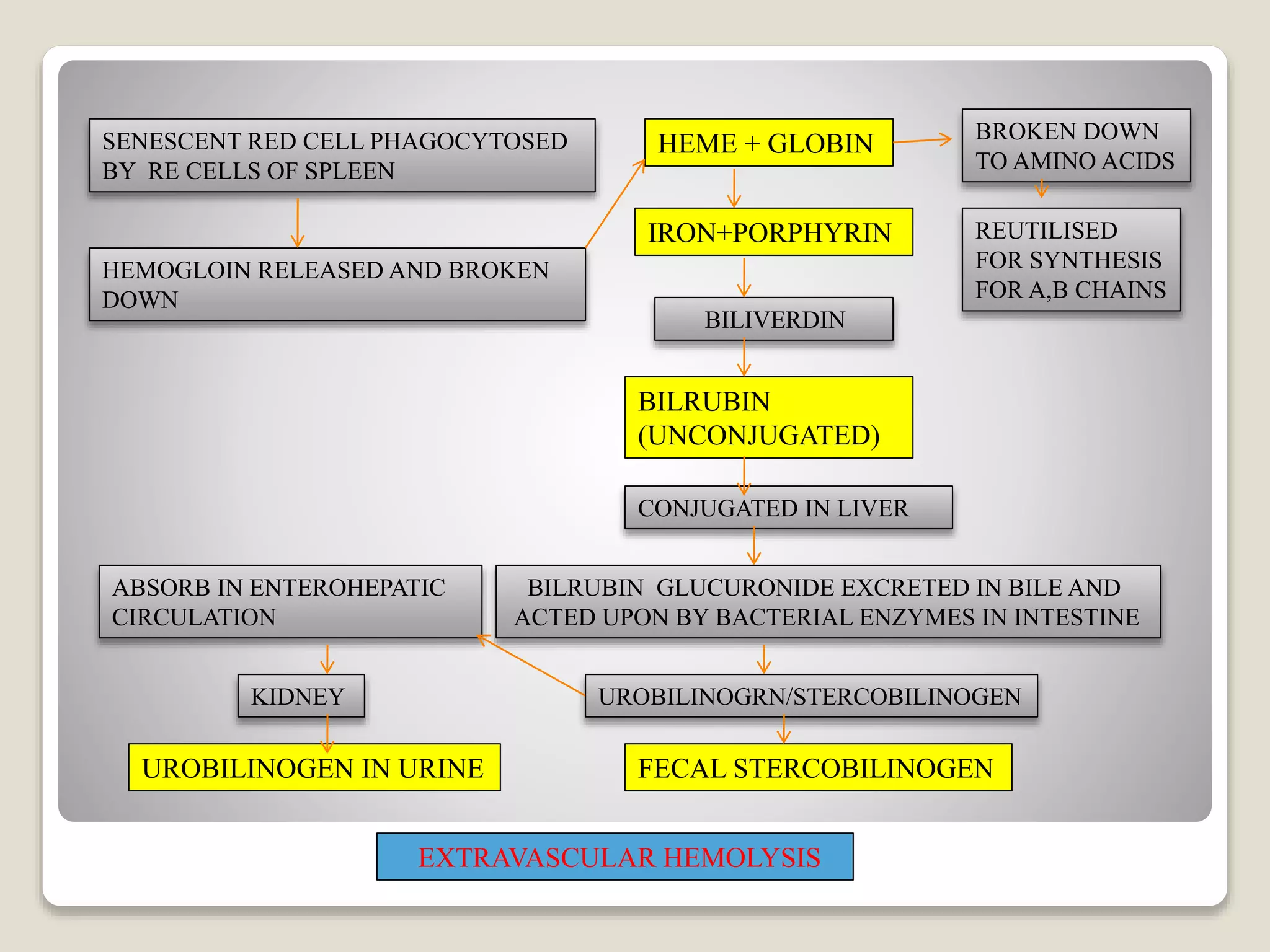
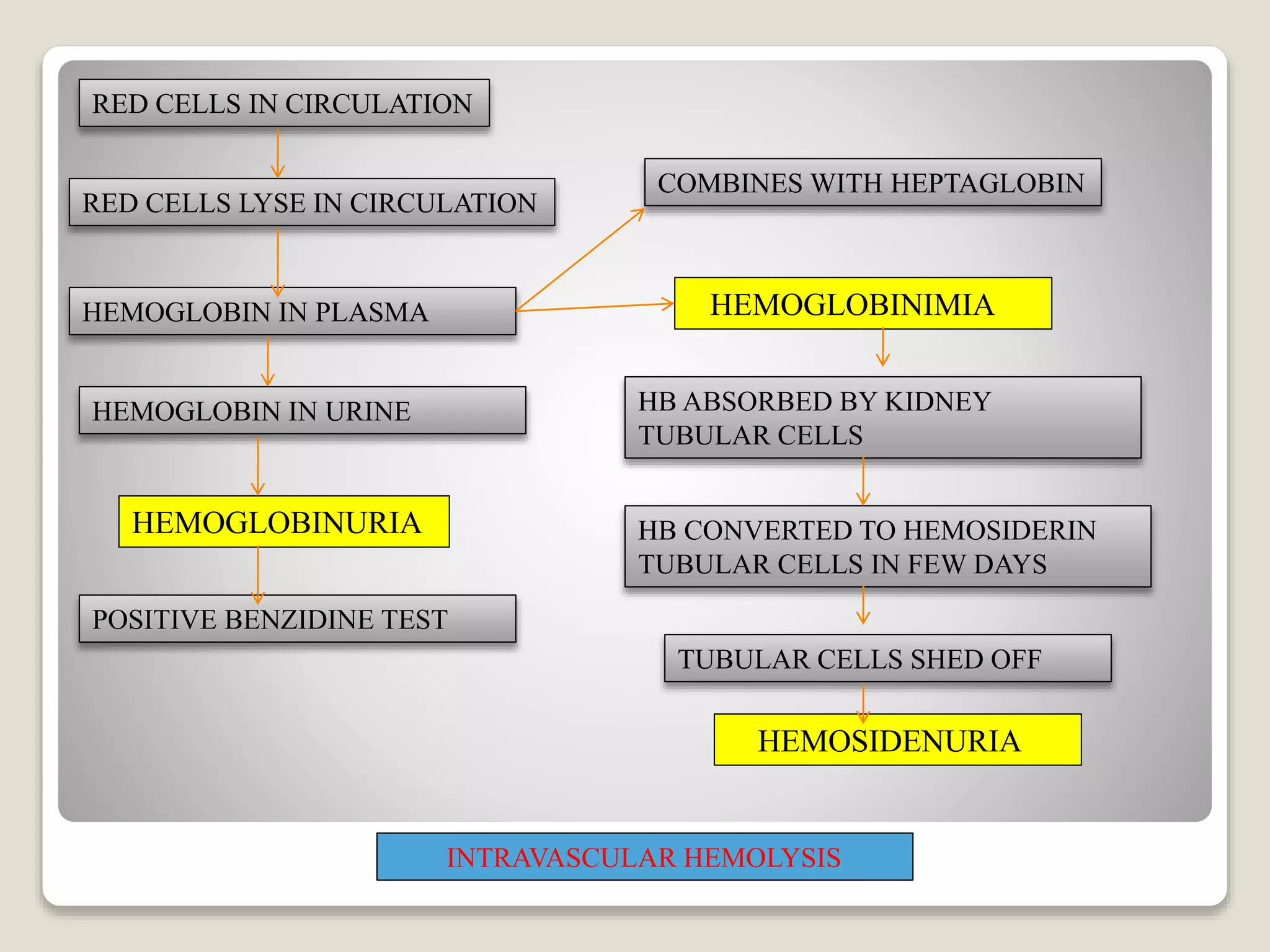
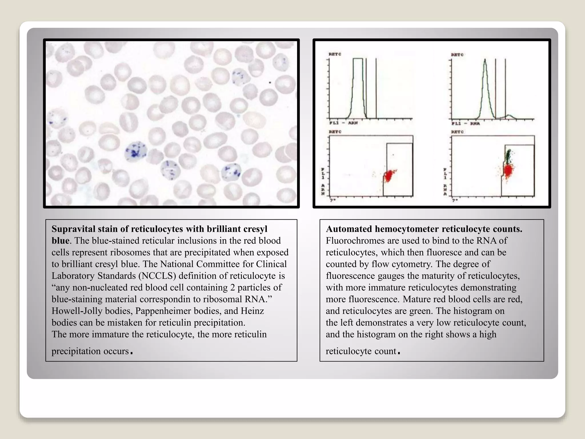
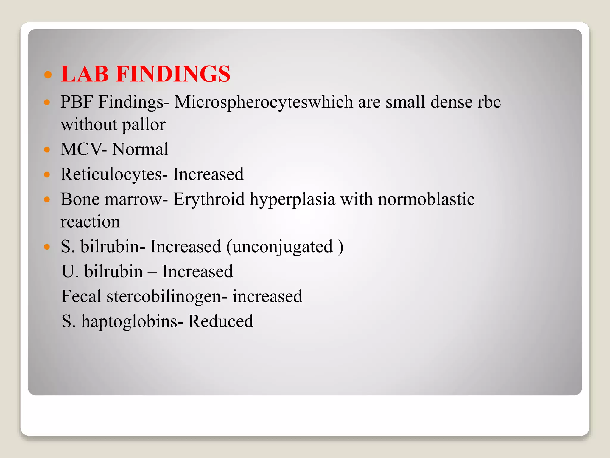
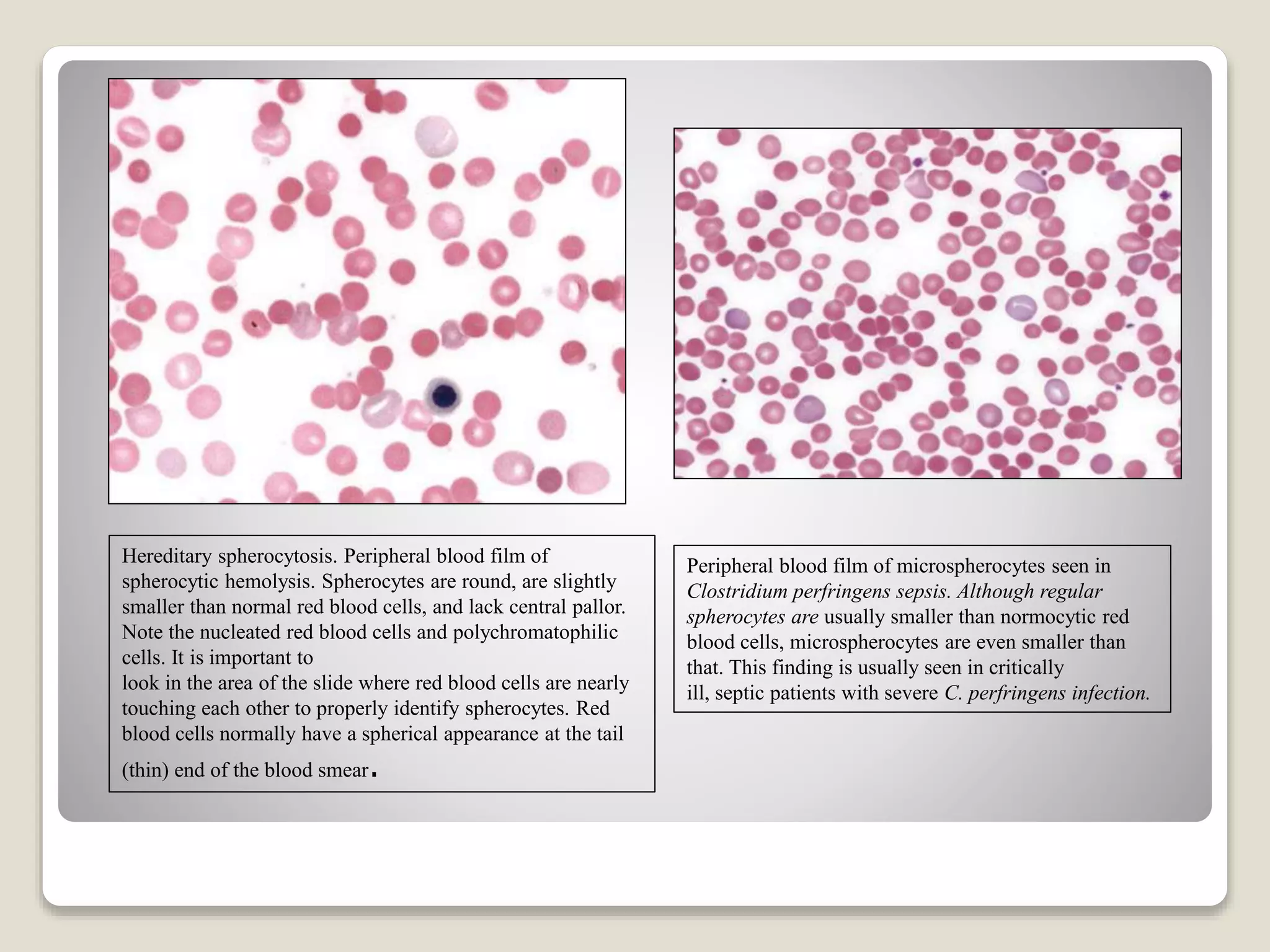
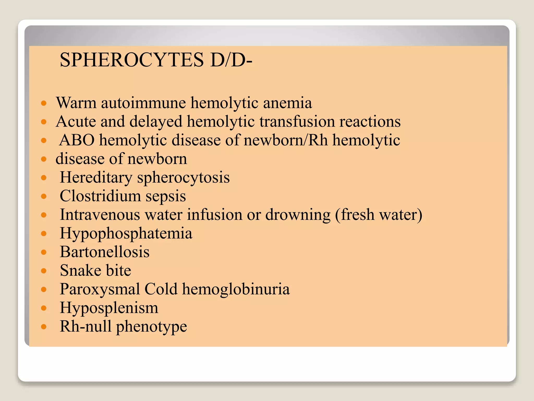
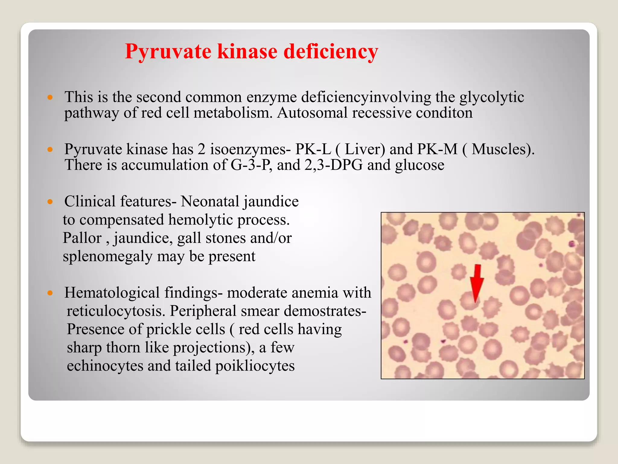
This document provides an overview of hemolytic anemia, including definitions, pathogenesis, classification, clinical features, laboratory findings, and approaches. Hemolytic anemia is characterized by increased red blood cell destruction. It can be hereditary or acquired. Specific hereditary forms discussed include hereditary spherocytosis, elliptocytosis, and pyropoikilocytosis, which are caused by red blood cell membrane defects. Clinical features may include pallor, jaundice, splenomegaly, and gallstones. Laboratory findings aid in diagnosis and include peripheral smear showing abnormal red blood cells, reticulocytosis, and elevated bilirubin. The document also discusses hemolytic anemia evaluation and differential diagnoses.
Hereditary spherocytosis is an inherited condition related to RBC destruction. its diagnosis is require to differentiate immune hemolytic anemia and G-6-P-D deficiency anemia
causes of macrocytic anemia pathopysiology, sign and symptoms and the difference between macrocytic anemia megaloblastIc anemia. causes of hypersegmented neutrophils and its association between them. investigation and medical management plus pictures illustration.
This presentation is about anemia of chronic disease, nowadays also called as anemia of Inflammation. I have dealt with anemia in CKD and malignancy in detail.
Haemolysis indicates that there is shortening of the normal red cell lifespan of 120 days. There are many causes.
To compensate, the bone marrow may increase its output of red cells six- to eightfold by increasing the proportion of red cells produced, expanding the volume of active marrow, and releasing reticulocytes prematurely. Anaemia occurs only if the rate of destruction exceeds this increased production rate.
In this presentation I've tried to summarize classification of hemolytic anemia and in depth review of rbc membrane disorders like hereditary spherocytosis, hereditary elliptocytosis, enzymopathies of hemolytic anemia like g6pd disorder, pyruvate kinase disorders, hemoglobinopathies related to hemolytic anemia like thalassemia, sickle cell anemia and especially pathophysiology and mechanism of hemolysis either extravascular or intravascular. Hope it helps you understand the entity better.
Anaemias, causes, pathophysiology, morphological and aetiological types, Investigations and treatment, including blood transfusion were discussed in this presentation
Hemolytic anemias share the following features:
A shortened red cell life span below the normal 120 days
Elevated erythropoietin levels and a compensatory increase in erythropoiesis
Accumulation of hemoglobin degradation products that are created as part of the process of red cell hemolysis
- Video recording of this lecture in English language: https://youtu.be/kqbnxVAZs-0
- Video recording of this lecture in Arabic language: https://youtu.be/SINlygW1Mpc
- Link to download the book free: https://nephrotube.blogspot.com/p/nephrotube-nephrology-books.html
- Link to NephroTube website: www.NephroTube.com
- Link to NephroTube social media accounts: https://nephrotube.blogspot.com/p/join-nephrotube-on-social-media.html
Title: Sense of Smell
Presenter: Dr. Faiza, Assistant Professor of Physiology
Qualifications:
MBBS (Best Graduate, AIMC Lahore)
FCPS Physiology
ICMT, CHPE, DHPE (STMU)
MPH (GC University, Faisalabad)
MBA (Virtual University of Pakistan)
Learning Objectives:
Describe the primary categories of smells and the concept of odor blindness.
Explain the structure and location of the olfactory membrane and mucosa, including the types and roles of cells involved in olfaction.
Describe the pathway and mechanisms of olfactory signal transmission from the olfactory receptors to the brain.
Illustrate the biochemical cascade triggered by odorant binding to olfactory receptors, including the role of G-proteins and second messengers in generating an action potential.
Identify different types of olfactory disorders such as anosmia, hyposmia, hyperosmia, and dysosmia, including their potential causes.
Key Topics:
Olfactory Genes:
3% of the human genome accounts for olfactory genes.
400 genes for odorant receptors.
Olfactory Membrane:
Located in the superior part of the nasal cavity.
Medially: Folds downward along the superior septum.
Laterally: Folds over the superior turbinate and upper surface of the middle turbinate.
Total surface area: 5-10 square centimeters.
Olfactory Mucosa:
Olfactory Cells: Bipolar nerve cells derived from the CNS (100 million), with 4-25 olfactory cilia per cell.
Sustentacular Cells: Produce mucus and maintain ionic and molecular environment.
Basal Cells: Replace worn-out olfactory cells with an average lifespan of 1-2 months.
Bowman’s Gland: Secretes mucus.
Stimulation of Olfactory Cells:
Odorant dissolves in mucus and attaches to receptors on olfactory cilia.
Involves a cascade effect through G-proteins and second messengers, leading to depolarization and action potential generation in the olfactory nerve.
Quality of a Good Odorant:
Small (3-20 Carbon atoms), volatile, water-soluble, and lipid-soluble.
Facilitated by odorant-binding proteins in mucus.
Membrane Potential and Action Potential:
Resting membrane potential: -55mV.
Action potential frequency in the olfactory nerve increases with odorant strength.
Adaptation Towards the Sense of Smell:
Rapid adaptation within the first second, with further slow adaptation.
Psychological adaptation greater than receptor adaptation, involving feedback inhibition from the central nervous system.
Primary Sensations of Smell:
Camphoraceous, Musky, Floral, Pepperminty, Ethereal, Pungent, Putrid.
Odor Detection Threshold:
Examples: Hydrogen sulfide (0.0005 ppm), Methyl-mercaptan (0.002 ppm).
Some toxic substances are odorless at lethal concentrations.
Characteristics of Smell:
Odor blindness for single substances due to lack of appropriate receptor protein.
Behavioral and emotional influences of smell.
Transmission of Olfactory Signals:
From olfactory cells to glomeruli in the olfactory bulb, involving lateral inhibition.
Primitive, less old, and new olfactory systems with different path
These simplified slides by Dr. Sidra Arshad present an overview of the non-respiratory functions of the respiratory tract.
Learning objectives:
1. Enlist the non-respiratory functions of the respiratory tract
2. Briefly explain how these functions are carried out
3. Discuss the significance of dead space
4. Differentiate between minute ventilation and alveolar ventilation
5. Describe the cough and sneeze reflexes
Study Resources:
1. Chapter 39, Guyton and Hall Textbook of Medical Physiology, 14th edition
2. Chapter 34, Ganong’s Review of Medical Physiology, 26th edition
3. Chapter 17, Human Physiology by Lauralee Sherwood, 9th edition
4. Non-respiratory functions of the lungs https://academic.oup.com/bjaed/article/13/3/98/278874
micro teaching on communication m.sc nursing.pdfAnurag Sharma
Microteaching is a unique model of practice teaching. It is a viable instrument for the. desired change in the teaching behavior or the behavior potential which, in specified types of real. classroom situations, tends to facilitate the achievement of specified types of objectives.
263778731218 Abortion Clinic /Pills In Harare ,sisternakatoto
263778731218 Abortion Clinic /Pills In Harare ,ABORTION WOMEN’S CLINIC +27730423979 IN women clinic we believe that every woman should be able to make choices in her pregnancy. Our job is to provide compassionate care, safety,affordable and confidential services. That’s why we have won the trust from all generations of women all over the world. we use non surgical method(Abortion pills) to terminate…Dr.LISA +27730423979women Clinic is committed to providing the highest quality of obstetrical and gynecological care to women of all ages. Our dedicated staff aim to treat each patient and her health concerns with compassion and respect.Our dedicated group ABORTION WOMEN’S CLINIC +27730423979 IN women clinic we believe that every woman should be able to make choices in her pregnancy. Our job is to provide compassionate care, safety,affordable and confidential services. That’s why we have won the trust from all generations of women all over the world. we use non surgical method(Abortion pills) to terminate…Dr.LISA +27730423979women Clinic is committed to providing the highest quality of obstetrical and gynecological care to women of all ages. Our dedicated staff aim to treat each patient and her health concerns with compassion and respect.Our dedicated group of receptionists, nurses, and physicians have worked together as a teamof receptionists, nurses, and physicians have worked together as a team wwww.lisywomensclinic.co.za/
Basavarajeeyam is an important text for ayurvedic physician belonging to andhra pradehs. It is a popular compendium in various parts of our country as well as in andhra pradesh. The content of the text was presented in sanskrit and telugu language (Bilingual). One of the most famous book in ayurvedic pharmaceutics and therapeutics. This book contains 25 chapters called as prakaranas. Many rasaoushadis were explained, pioneer of dhatu druti, nadi pareeksha, mutra pareeksha etc. Belongs to the period of 15-16 century. New diseases like upadamsha, phiranga rogas are explained.
Tom Selleck Health: A Comprehensive Look at the Iconic Actor’s Wellness Journeygreendigital
Tom Selleck, an enduring figure in Hollywood. has captivated audiences for decades with his rugged charm, iconic moustache. and memorable roles in television and film. From his breakout role as Thomas Magnum in Magnum P.I. to his current portrayal of Frank Reagan in Blue Bloods. Selleck's career has spanned over 50 years. But beyond his professional achievements. fans have often been curious about Tom Selleck Health. especially as he has aged in the public eye.
Follow us on: Pinterest
Introduction
Many have been interested in Tom Selleck health. not only because of his enduring presence on screen but also because of the challenges. and lifestyle choices he has faced and made over the years. This article delves into the various aspects of Tom Selleck health. exploring his fitness regimen, diet, mental health. and the challenges he has encountered as he ages. We'll look at how he maintains his well-being. the health issues he has faced, and his approach to ageing .
Early Life and Career
Childhood and Athletic Beginnings
Tom Selleck was born on January 29, 1945, in Detroit, Michigan, and grew up in Sherman Oaks, California. From an early age, he was involved in sports, particularly basketball. which played a significant role in his physical development. His athletic pursuits continued into college. where he attended the University of Southern California (USC) on a basketball scholarship. This early involvement in sports laid a strong foundation for his physical health and disciplined lifestyle.
Transition to Acting
Selleck's transition from an athlete to an actor came with its physical demands. His first significant role in "Magnum P.I." required him to perform various stunts and maintain a fit appearance. This role, which he played from 1980 to 1988. necessitated a rigorous fitness routine to meet the show's demands. setting the stage for his long-term commitment to health and wellness.
Fitness Regimen
Workout Routine
Tom Selleck health and fitness regimen has evolved. adapting to his changing roles and age. During his "Magnum, P.I." days. Selleck's workouts were intense and focused on building and maintaining muscle mass. His routine included weightlifting, cardiovascular exercises. and specific training for the stunts he performed on the show.
Selleck adjusted his fitness routine as he aged to suit his body's needs. Today, his workouts focus on maintaining flexibility, strength, and cardiovascular health. He incorporates low-impact exercises such as swimming, walking, and light weightlifting. This balanced approach helps him stay fit without putting undue strain on his joints and muscles.
Importance of Flexibility and Mobility
In recent years, Selleck has emphasized the importance of flexibility and mobility in his fitness regimen. Understanding the natural decline in muscle mass and joint flexibility with age. he includes stretching and yoga in his routine. These practices help prevent injuries, improve posture, and maintain mobilit
Flu Vaccine Alert in Bangalore Karnatakaaddon Scans
As flu season approaches, health officials in Bangalore, Karnataka, are urging residents to get their flu vaccinations. The seasonal flu, while common, can lead to severe health complications, particularly for vulnerable populations such as young children, the elderly, and those with underlying health conditions.
Dr. Vidisha Kumari, a leading epidemiologist in Bangalore, emphasizes the importance of getting vaccinated. "The flu vaccine is our best defense against the influenza virus. It not only protects individuals but also helps prevent the spread of the virus in our communities," he says.
This year, the flu season is expected to coincide with a potential increase in other respiratory illnesses. The Karnataka Health Department has launched an awareness campaign highlighting the significance of flu vaccinations. They have set up multiple vaccination centers across Bangalore, making it convenient for residents to receive their shots.
To encourage widespread vaccination, the government is also collaborating with local schools, workplaces, and community centers to facilitate vaccination drives. Special attention is being given to ensuring that the vaccine is accessible to all, including marginalized communities who may have limited access to healthcare.
Residents are reminded that the flu vaccine is safe and effective. Common side effects are mild and may include soreness at the injection site, mild fever, or muscle aches. These side effects are generally short-lived and far less severe than the flu itself.
Healthcare providers are also stressing the importance of continuing COVID-19 precautions. Wearing masks, practicing good hand hygiene, and maintaining social distancing are still crucial, especially in crowded places.
Protect yourself and your loved ones by getting vaccinated. Together, we can help keep Bangalore healthy and safe this flu season. For more information on vaccination centers and schedules, residents can visit the Karnataka Health Department’s official website or follow their social media pages.
Stay informed, stay safe, and get your flu shot today!
2. INTRODUCTION-
I. DEFINATION
II. PATHOGENESIS
III. CLINICAL FEATURES
IV. LAB FINDINGS
CLASSIFICATION
APPROACH TO HEMOLYTIC ANEMIAS
RED CELL MEMBRANE DEFECTS
ENZYMOPATHIES
HEMOGLOBINOPATIES
OTHERS
3. ANEMIA - Anemia is defined as reduction of the total
circulating red cell mass below normal limits.
Anemia reduces the oxygen carrying capacity of blood,
leading to tissue hypoxia.
Anemia is usually diagnosed based on reduction in
hematocrit and Hemoglobin concentration of the blood to
levels that are below the normal range
4.
5. HEMOLYTIC ANEMIA
Hemolytic anemias are characterised by increase red cell
destruction
It shares the following features
1. A shortened red cell life span below the normal 120 days
2. Elevated erythropoietin level and compensatory increase in
erythropoiesis
3. Accumulation of hemoglobin degradation products that are
created as a part of process of red cell hemolysis
6. PATHOGENESIS
RED CELL DESTRUCTION
The physiological destruction of senescent red cells takes
place with in macrophages, which are abundant in spleen,
liver and bone marrow
This process appears to be triggered by age- dependent
changes in red cell surface proteins, which lead to their
recognition and phagocytosis
7. Red cell destruction occur by 2 mechanisms-
Extravascular Hemolysis – The site of destruction is mainly
spleen and this is the major mechanism of red cell hemolysis.
Red cells are taken up by the cells of RE system where they
are destroyed and digested
Intravascular Hemolysis– This is the minor pathway of red
cell destruction and red cells are destroyed in circulation
releasing hemoglobin.
8. SENESCENT RED CELL PHAGOCYTOSED
BY RE CELLS OF SPLEEN
HEMOGLOIN RELEASED AND BROKEN
DOWN
HEME + GLOBIN BROKEN DOWN
TO AMINO ACIDS
REUTILISED
FOR SYNTHESIS
FOR A,B CHAINS
IRON+PORPHYRIN
BILIVERDIN
BILRUBIN
(UNCONJUGATED)
CONJUGATED IN LIVER
BILRUBIN GLUCURONIDE EXCRETED IN BILE AND
ACTED UPON BY BACTERIAL ENZYMES IN INTESTINE
UROBILINOGRN/STERCOBILINOGEN
FECAL STERCOBILINOGEN
ABSORB IN ENTEROHEPATIC
CIRCULATION
KIDNEY
UROBILINOGEN IN URINE
EXTRAVASCULAR HEMOLYSIS
9. RED CELLS IN CIRCULATION
RED CELLS LYSE IN CIRCULATION
HEMOGLOBIN IN PLASMA
HEMOGLOBIN IN URINE
HEMOGLOBINURIA
POSITIVE BENZIDINE TEST
HEMOGLOBINIMIA
HB ABSORBED BY KIDNEY
TUBULAR CELLS
HB CONVERTED TO HEMOSIDERIN
TUBULAR CELLS IN FEW DAYS
TUBULAR CELLS SHED OFF
HEMOSIDENURIA
COMBINES WITH HEPTAGLOBIN
INTRAVASCULAR HEMOLYSIS
10. EXTRAVASCULAR INTRAVSCULAR
S.BILRUBIN UNCONJUGATED++ UNCONJUGATED+
S.HEPTAGLOBIN NORMAL DECREASE
PLASMA
HEMOGLOBIN
ABSENT PRESENT
S.
METHEMALBUMIN
ABSENT PRESENT
LACTATE
DEHYDROGENASE
VARIABLE+ INCREASE++
URINE BILRUBIN PRESENT PRESENT
U. HEMOGLOBIN ABSENT PRESENT
U. HEMOSIDERIN ABSENT PRESENT
LAB EVALUATION OF HEMOLYSIS
11. CLINICAL FEATURES –
Clinical sign and symptoms of hemolytic anemia depend
upon the severity as well as duration of hemolysis. These are
Pallor
Jaundice
Splenomegaly
Gall stones
Skeletal abnormalities in severe hemolysis
Leg ulcers
Dyspnoea
Tachycardia and systolic murmur
12. Lab investigations
1. History of the patient
2. Peripheral blood film
3. Bone marrow findings
4. Biochemical tests
5. Other screening tests
13. PERIPHERAL BLOOD FINDINGS – Peripheral smear
evaluation is the most important investigation in hemolytic
anemias
The following morphological findings alone or in
combination are suggestive of hemolysis :
Polychromatophilia, nucleated red cells, thrombocytosis
and neutrophilia with mild shift to left
Red cell morphologic abnormalities provide a clue to
underlyng disorder. Some are Spherocytes, Sickle cell,
Target cells, Schistocytes (fragmented red cells, helmet
cells, traingular cells) and acanthocytes
14. Peripheral blood film with Romanowsky stain
demonstrating polychromatophilic cells. The
polychromatophilic cells are basophilic because of
increased RNA content. The cells are usually larger than
normocytic red blood cells
Autoimmune hemolytic anemia. Numerous
spherocytes, small round RBCs lacking central
pallor, are shown in this blood smear from a case of
Coombs-positive hemolytic anemia.
15. 2. Bone marrow findings- Compensatory mechanism to
hemolysis
Erythroid hyperplasia of bone marrow- Erythroid hyperplasia with
normoblastic reaction. Reversal of M:E ratio
Reticulocytosis – Increase variabley
- Mild (2-10%)- Hemogobinopathies
- Moderate to marked (10-60%)-
Immune hemolytic anemias,
Hereditary spherocytosis ,
G6PD deficient states
16. Bone marrow findings in hemolytic anemia.
Top panel: Erythroid hyperplasia is present with
a predominance of erythroid precursors. The
normal myeloid to erythroid ratio in a bone
marrow aspirate is 3 to 5:1. In this case, there
occurs a reversal of the myeloid to erythroid ratio
of 1:4.
Bottom panel: Bone marrow biopsy
in a patient with hemolytic anemia. Erythroid
hyperplasia is seen with a predominance of
erythroid precursors
17. Supravital stain of reticulocytes with brilliant cresyl
blue. The blue-stained reticular inclusions in the red blood
cells represent ribosomes that are precipitated when exposed
to brilliant cresyl blue. The National Committee for Clinical
Laboratory Standards (NCCLS) definition of reticulocyte is
“any non-nucleated red blood cell containing 2 particles of
blue-staining material correspondin to ribosomal RNA.”
Howell-Jolly bodies, Pappenheimer bodies, and Heinz
bodies can be mistaken for reticulin precipitation.
The more immature the reticulocyte, the more reticulin
precipitation occurs.
Automated hemocytometer reticulocyte counts.
Fluorochromes are used to bind to the RNA of
reticulocytes, which then fluoresce and can be
counted by flow cytometry. The degree of
fluorescence gauges the maturity of reticulocytes,
with more immature reticulocytes demonstrating
more fluorescence. Mature red blood cells are red,
and reticulocytes are green. The histogram on
the left demonstrates a very low reticulocyte count,
and the histogram on the right shows a high
reticulocyte count.
21. HEREDITARY SPHEROCYTOSIS
Hereditary spheroytosis is an inherited hemolytic anemia
resultimg from red cell mebrane defect leading to
microspherocytosis, splenomegaly and jaundice
ETIOAPTHOGENESIS-
Spectrin deficiency is the most common abnormality
Mutation of b spectrin gene and point mutations affect the
binding of spectrin to protein 4.1
24. CLINICAL FEATURES-
Seen all over the world
Autosomal dominent with variable penetrance
M=F ; present in neonate, childhood or adulthood
Intermittent jaundice is usual presentation
O/E- splenomegaly is a constant feature
Gall stones (pigment type)
Chronic leg ulcers (rare)
25. LAB FINDINGS
PBF Findings- Microspherocyteswhich are small dense rbc
without pallor
MCV- Normal
Reticulocytes- Increased
Bone marrow- Erythroid hyperplasia with normoblastic
reaction
S. bilrubin- Increased (unconjugated )
U. bilrubin – Increased
Fecal stercobilinogen- increased
S. haptoglobins- Reduced
26. Hereditary spherocytosis. Peripheral blood film of
spherocytic hemolysis. Spherocytes are round, are slightly
smaller than normal red blood cells, and lack central pallor.
Note the nucleated red blood cells and polychromatophilic
cells. It is important to
look in the area of the slide where red blood cells are nearly
touching each other to properly identify spherocytes. Red
blood cells normally have a spherical appearance at the tail
(thin) end of the blood smear.
Peripheral blood film of microspherocytes seen in
Clostridium perfringens sepsis. Although regular
spherocytes are usually smaller than normocytic red
blood cells, microspherocytes are even smaller than
that. This finding is usually seen in critically
ill, septic patients with severe C. perfringens infection.
27. Other diagnostic tests-
Osmotic fragility test- shift of curve to right
Incubated osmotic fragiloty test
Glycerol lysis test – Increased (rate of lysis)
Flow cytometry based on EMA (Eosin5-malemide)- lower in
HS ( mean fluooscnt intensity of EMA tagged cells)
28.
29. Osmotic fragility curves of normal and hereditary spherocytosis red blood cells. RBCs are
exposed to decreasing strengths of hypotonic saline solutions, and the degree of hemolysis (%) is measured.
Increased fragility is indicated by a shift of the curve to the left, and is seen in conditions associated with
spherocytosis. In the fresh sample on the left, a tail of HS cells occurs with increased sensitivity. Incubation of the
sample for 24 hours prior (graph on the right) accentuates the osmotic fragility of spherocytes, whereas normal cells
only become more slightly fragile. The osmotic fragility of unincubated blood may be normal in some patients with
HS; therefore, incubated testing should be performed as well.
32. HEREDITARY ELLIPTOCYTOSIS
Group of anemias characterised by the presence of elliptical
or oval RBCs in the peripheral blood. Such cells should be
more than 25%
Autosomal dominent disorder
Membrane protein abnormalities like a b-spectrin defect,
structural defectsor deficiency of protein 4.1 lead to elliptical
shape of rbcs. membrane dysfunction and mild hemolysis
33. Clinically patient are asymptomatic and mild hemolytic anemia is fully
compensated in most cases
Case is diagnosed incidentally when the blood film is examined for other
ailment
Periperal smear demonstrates presence of elliptocytes ( cigar shaped )
which vary from 20-90% of cells. Osmotic fragility normal
Three subtypes of HE are-
1. Common HE- 85% of cases
2. Spherocytic HE- HS-HE hybrid(20% ellipsoid cells); European hybrids
3. Stomatcytic HE
34. Hereditary elliptocytosis. Elliptocytes and
ovalocytes are present in this blood film from a case
of hereditary elliptocytosis. Elliptocytes are
elongated with rounded edges (as opposed to
sharp edges in sickle cells).
•Large numbers of
elliptocytes
• Hereditary elliptocytosis
• Small numbers of
elliptocytes
• Iron deficiency
• Thalassemia trait and
major
• Megaloblastic anemia
• Myelodysplastic
syndrome
• Myelofibrosis
• Southeast Asian
ovalocytosis
35. Hereditary pyropoikliocytosis
Hereditary pyropoikliocytosis is a rare hemolytic anemia
There is a defective spectrin gene transmitted by one parent
and also an elusive thalassemia like defect of spectrin
synthesis inherited from normal parent
This results in a compound inheritance in which a spectrin
abnormality is superimposed upon spectrin deficiency
36. Hereditary pyropoikilocytosis. Peripheral blood film in patient with hereditary
pyropoikilocytosis. Significant variations in size and shape are present: poikliocytes,
teardrops, fragments, microspherocytes, elliptocytes, and small pieces and buds of red blood
cells. The cells are microcytic with low MCV . Incubted osmotic fragility increased
37. Stomatocytosis
Stomatocytes are red cells with a slit like central pallor and
these are uniconcave/bowel shape in wet suspensions
Disorder Stomatocytes %
Normal individual <5%
Hereditary stomatocytosis >30%
Accquired stomatoctosis 5-50%
38. Hereditary stomatocytosis. The red blood cells in this
blood smear demonstrate slit-like central pallor, creating the
appearance of a mouth (stoma in Greek), from which the name
stomatocytes derives. Hereditary stomatocytosis may demonstrate
10% to 50% stomatocytes on the peripheral blood film. Ovalocytes
and macrocytes also may be present.
•STOMATOCYTES
•Artifact
• Alcoholism
• Alcoholic liver
disease
• Obstructive liver
disease
• Hereditary
stomatocytosis
• Hereditary
xerocytosis
• Southeast Asian
ovalocytosis
• Tangier disease
• Rh-null phenotype
• Drugs (hydroxyurea)
40. GLUCOSE 6-PHOSPHATE DEHYDROGENASE
DEFICIENCY
Glucose6-phosphate dehydrogenase is the first enzyme in the hexose
monophosphate shunt pathway (HMP) which protects red cells from
oxidant injury
Deficiency of G6PD may result in episodes of hemolysis following certain
drug intake or chemical exposure or infection
G6PD deficiency is a sex linked disease. Its prevalance is higher in
tropical eastern countries. Prevalance is higher in kurdish jews (60-70%)
and lower in japan (.1%)
41. WHO Classification of G6PD variants
Class/ Variants Severity Activity Hemolysis
Class I
( G6-PD Canton)
SEVERE
DEFICIENCY
CHRONIC
HEMOLYTIC ANEMIA
Class II
(G6-PD
Mediterranean)
SEVERE
DFICIENCY
<10% OF NORMAL INTERMITTENT
HEMOLYSIS
Class III
( G6-PD A-)
MODERATE
DEFICIENCY
10-60% OF
NORMAL
HEMOLYSIS ON
EXPOSURE TO
DRUGS
Class IV
( G6-PD A+)
NO DEFICIENCY 60-100% OF
NORMAL
NO HEMOLYSIS
Class V
( G6PD B*)
- INCREASED
ENZYMATIC
ACTIVITY
NO HEMOLYSIS
42. Clinical and hematological presentation of G6PD deficiency
Acute hemolytic anemia- Occurs following exposure to drugs like
primaquine, infections like pneumonia, typhoid and oxidative chemicals.
CF- appears 1-3 hours after drug adiministration. Sudden development of
pallor, passage of dark urine, jaundice and severe backache
Chronic non-spherocytic anemia- There is moderately severe enyme
deficiency, hemolysis continues throughout life. Seen in neonatal period. CF-
hemolysis is compensated so milder symptoms
Neonatal hyperbilrubinimia- Jaundice in G6PD deficient neonates is
common wit G6PD mediterranean variant (class III). CF- Jaundice,
kernicterus
Favism- Common in children caused by consumption of fava beans.
Glucoside divisine and aglycone isouramil is responsible. Resulting in acute
severe hemolysis within few hours . CF-headache, fever, chills and back
pain.
43. Diagnostic tests-
1. Peripheral blood film evaluation, history and biochemical finding-
Moderate anisopoikliocytosis with polychromatophilia
Microspherocytes and bite cell ( removel of heinz bodies)
Reticulocytosis (20-50%)
Hemogobinuria and increase urobilinogen in urine
2. The commonly employed screening tests for G6PD deficiency are-
Methemaglobin reduction test (MRT)
Ascorbate –cyanide test
Fluooscent spot test
Dye decolourisation test
3. Quantitative G6-PD assay and DNA analysis by PCR
44. Peripheral blood film demonstrating blister cells in a
patient with glucose-6-phosphate dehydrogenase
deficiency. The blister appears as a vacuole in the
erythrocyte’s hemoglobin at the edge of the red blood
cell surface. A thin rim of cytoplasm seems to
enclose this vacuole. This cell is usually a precursor
to a bite cell.
Bite cells. The red blood cells in this peripheral
smear appear bitten. The erythrocyte may retain
or lose central pallor, depending on the size and
numbers of bites. In some cases, the bite cell
may be mistaken for helmet cells, a type of
fragmented erythrocyte.. A double bite cell is
displayed in the center of the figure.
45. Heinz bodies. Peripheral blood stained with crystal violet
supravital stain demonstrating Heinz-body inclusions, which
are not visible with Romanowsky stains alone. Heinz bodies
are purple-blue, large, single or multiple inclusions attached
to the inner surface of the red blood cellmembrane. They
represent precipitated normal or unstable hemoglobins..
Reticulocytes do not stain with crystal violet.
•Heinz bodies
•Oxidative stress
glucose-6-phosphate
dehydrogenase deficiency,
glutathione synthetase deficiency
Drugs
Toxins
• Unstable hemoglobins
46. Pyruvate kinase deficiency
This is the second common enzyme deficiencyinvolving the glycolytic
pathway of red cell metabolism. Autosomal recessive conditon
Pyruvate kinase has 2 isoenzymes- PK-L ( Liver) and PK-M ( Muscles).
There is accumulation of G-3-P, and 2,3-DPG and glucose
Clinical features- Neonatal jaundice
to compensated hemolytic process.
Pallor , jaundice, gall stones and/or
splenomegaly may be present
Hematological findings- moderate anemia with
reticulocytosis. Peripheral smear demostrates-
Presence of prickle cells ( red cells having
sharp thorn like projections), a few
echinocytes and tailed poikliocytes
49. The Thalassemias
Thalassemia syndrome are autosomal recessive disorders
Thalassemia results from defects in the rate of synthesis of a or b chains,
lead to reduced hemoglobin production and accumulation of a or b chains
Thalassemia is considered to be quantitative hemogolobinopathy, since no
structural abnormal hb is synthsised
50. Classification
B thalassemia A thalassemia Misc thalassemia
syndrome
T. Major Hydropes fetais HbS- thal
T. Intermedia Hbh disase HbE- thal
T. Trait A-thalassemia trait HbD-thal
T. Minima A-b-thal
HPHF
Y-Thal,
d-thal
51. B- thalassemia syndromes
Epidemiology- In india , b-thalassemia is commmonly seen in sindhis,
punjabis, bengalis, gujratis, parsis and lohanas
Genetics –
Globin of HbA -2a+2b- chains. Synthesis of a-chains control by 2 gene
cluster on chrosome16 and b chain on chrosome 11
Point mutation of the globin gene cluster- single nucleotide substitution,
lead to supression of b- chain. Divided into-
1. promoter region and chain terminator mutation
2. mutation affecting m- rna processing
52. Thalassemia major (cooleys anemia)
Homozygous form of b0/b0 or b+/b+ or double heterozygous
b0/b+
Pathophysiology –
1. Accumulation of free b chains
2. Extravascular hemolysis
3. Marrrow and bone changes
4. Extrramedullary hematopoisis
5. Synthesis of hbf
6. Iron overload
7. Hepacidin
53. Clinical features- Present within 1st yr of life
Failure to thrive , intermittent infections, palllor
Protuberant abdomen (hepatosplenomegaly)
Frontal bossing (thickening of cranial bones)
Prominent cheek bones (zygomatic bones overgrowth)- mongoloid
facies, thalassemic facies
Mild jaundice
Cholelithiasis
Bone changes- x- ray hair on end appearences
Endocrine changes (due to iron deposition)-
GH defciincy, hypothyroidism, DM
54. -Thalassemia facial bone abnormalities. These
changes include bossing of the skull; hypertrophy
of the maxilla, exposing the upper teeth;
depression of nasal bridge; and periorbital
puffiness.
-Thalassemia major leg ulcer. Leg ulcers can
occur in all types of hereditary hemolytic
anemias, including sickle cell disease and
hereditary spherocytosis.
55. -Thalassemia bone abnormalities. Note the “hair on
end” appearance of the cortical bone caused by expansion of
the bone marrow (arrows). The subperiosteal bone grows in
radiating striations, which appears as “hairs.”
-Thalassemia major.
Note the pallor, short
stature,massive
hepatosplenomegaly,
and wasted limbs in
this undertransfused
case of -thalassemia
major
56. Peripheral blood findings-
RBCs- Microcytic Hypochromic with decreased MCV, MCH, MCHC
Anisooikliocytosi- target cells, basophilic stippling, nucleted RBCs, tear
drop cells, fragmented red cells and occasional howel jolly bodies
Reticulocytes <2%
Bone marrrow- Erythroid hyperplasia, Reversal of M:E ratio
Iron studies- s. ferritin, transferrrin saturaion markedly inreased. S. iron
inc. TIBC reduced
57. -Thalassemia major. Unless they have had
transfusions, patients with this disease usually have
severe anemia. This peripheral blood film
demonstrates many nucleated red blood cells,
microcytosis, and hypochromasia with multiple
morphologic changes: target cells, teardrop cells,
fragments, basophilic stippling, and Pappenheimer
bodies. The nucleated red blood cells may be
dysplastic or show abnormal hemoglobinization.
Neutrophilia and thrombocytosis may occur. This
patient has undergone splenectomy for hypersplenism
and increased transfusion requirements. Howell-Jolly
bodies are present.
58. Thalassemia trait blood film. Peripheral blood films in
-thalassemia trait may demonstrate microcytosis and
possibly hypochromasia. Multiple morphologic changes
including target cells, teardrop cells, and rare fragments
may occur.. Basophilic stippling may help distinguish -
thalassemia trait from iron deficiency, but is not always
present in patients with -thalassemia trait. Red blood cell
indices may help:a normal or slightly decreased
hemoglobin with a low MCV/MCH and a low or mildly
increased RDW suggests thalassemia. Red blood cell
indices may not always distinguish iron deficiency from
thalassemia trait, however. Patients also may have
combined irondeficiency and -thalassemia trait
Basophilic stippling in
thalassemia. Peripheral blood
film demonstrating microcytic
hypochromic RBCs and
basophilic stippling (arrows).
Basophilic stippling occurs in
thalassemia as well as in other
hematologic disorders
Basophilic stippling
•Thalassemia trait and major
• Hemolytic anemia
• Myelodysplastic
syndrome/sideroblastic
anemia
• Megaloblastic anemia
• Pyrimidine 5 nucleotidase
deficiency
• Heavy metal poisoning
(coarse basophilic
stippling)
• Lead, zinc, arsenic, silver,
mercu
59. Bone marrow in thalassemia. Top and bottom panels
show bone marrow aspirate and biopsy,
respectively, from a case of thalassemia trait. The
bone marrow has increased numbers of erythroid
precursors (a low myeloid to erythroid ratio) related
to the increased peripheral RBC destruction in this
disease.
60. Alkaline hemoglobin (Hb) electrophoresis. Top panel: Lane 2: Normal. Lanes 3 and 5: -thalassemia trait. Lane 4:
HbS disease. Bottom panel: Lane 2: Normal. Lane 3: Hb D trait. Lane 4: HbS trait. Lanes 5 and 7: Hb Lepore trait
(faint band around HbS band area). Lane 6: HbC trait. Lane 8: HbH disease (note fast-moving Hb band, arrow).
Hemoglobins that move with HbS on alkaline include D/G/ Lepore, and hemoglobins that move with HbC on
alkaline include E/O/A2
61. Hemoglobinopathies associated with micocytosis
Thalassemia trait (heterozygous)
Thalassemia major (homozygous)
Thalassemia trait
HbH disease
Thalassemia trait and hemoglobin constant spring
HbC heterozygous and homozygous
HbE heterozygous and homozygous
HbD disease
HbO Arab disease
Hb Lepore heterozygous and homozygous
δβ-Thalassemia heterozygous and homozygous
γδβ-Thalassemia heterozygous and homozygous
Hereditary persistence of fetal hemoglobin
homozygous Hereditary persistence of fetal hemoglobin(HPFH)
62. Sickle cell disorders
Sickling syndromes are characterized by the presence of HbS which
imparts sickle shape to red cells in a state of reduced oxygen tension
HbS is prevalant in Africa, Mediterranean countries and India. In India,
seen common in tribals and in ethnic groups of MP, Orissa, AP,
Maharashtra (vidharba region), TN (chetti tribes) and Kerala
There is high prevelance of HbS in areas endemic to malaria falciperum
63. Genetics –
Sickle mutation is caused by substitution of valine in place of glutamic
acid in the 6th position (b6 glu-val) of b-chain
Mutation results in clinical presentation
1. Sickle cell anemia- HbS-HbS, Homozygous state
2. Sickle cell trait - HbA-HbS, heterozygous state
3. Sickle cell disease- Refer to all diseases with HbS in combination with –
normal (HbA), abnormal gene of b-thalassemia, a-thalassemia, HbD,
HbE, HbC,HbQ
64. Pathophysiology of vascular occlusion and
hemolysis
Polymerisation of deoxygenated HbS is the primary event in the
pathogenesis of the disease
Red cell containing HbS pass through microcirculation of spleen –
various cycles of sickling and desickling – Irreversible sickeled RBCs
– Extravascular hemolysis in spleen – Vascular stasis – vascular
occlusion – splenic infarcts – hyposplenism (lead to infection) and
autosplenectomy
65.
66. Clinical features-
Delay in puberty, growth and development
Recurrent leg ulcers
Avascular necrosis of femur head
Dactylitis ( Hand –Foot syndrome )
Pneumonia, meningitis, Osteomylitis
Jaundice and liver enlargement
Pigment gall stones
Acute abdominal pain ( infarcts of abdominal viscera)
Priapism
Acute chest syndrome (fever, chest pain, leucocytosis,
appearance of pulmonary infilterate with sickle anemia)
Sickle retinopathy- Salmon patches- intra retinal
hemmorhages
69. Sickle cell trait
Sickle cell trait usually do not manifest any clinical findings
Hemoglobin varies from 11-13 gm/dl
Red cells are normocytic normochromic and very target cells and
mild degree of anisopoikliocytosis
Clinical and hematological picture is milder in comparison to HbSS
state
Diagnosis is confirmed by Hb electrophoresis, HPLC and sickling
test
70. Hematological findings –
Anemia- moderately severe anemia with Hb 5- 10 gm
PBF demonstrates –
Red cells- Normocytic normochromic to mildly hypochromic moderate to
severe degree of anisopoikliocytosis. Sickle cells, target cells, ovalocytes,
polychromtophila with nucleted RBCs. Howell-jolly bodies alo seen
TLC- Mildly elevated ; Platlets- Increased
Reticulocytosis- 3%-10%
Bone marrow- Erythroid hyperplasia with normoblastic reaction
71. Sickle cell anemia. Top panel:
Peripheral blood film of
hemoglobin SS (HbS disease).
The numerous elongated
erythrocytes with sharp points
are classic sickle cells. Sickle
cells that appear folded over are
called envelope cells. Target cells
are present, in this case because
of hyposplenism from the splenic
infarction that occurs in HbSS
patients. Howell-Jolly bodies
may be seen as well. Middle
panel: Peripheral blood film in
patient with HbSS,demonstrating
sickle cells with Hb concentrated
at one end and absent at the
other, called hemi-lunes(arrows),
a finding seen in HbSS or HbSC.
Bottom panel: Peripheral blood
film in patient with HbSS,
demonstrating short, stubby, and
rhomboid-shaped sickle cells
called oat and boat cells
(arrows).
72. Other diagnostic tests-
1. Sickling tests- Presence of HbS demostrated by using reducing
agent like 2% sodium metabisulphite
2. Sickling solubility test
2. Hb electrophoresis- Hb electrophoresis can be carried out on
cellulose acetate membrane (pH8.9) or starch agarose (pH 8.6). HbS is a
slow moving Hb as compared to HbA and HbF. Howeever, electrophoretic
mobility of HbD/HbQ india is similar to HbS , therefore sickling test is
essential to differentiate.
73. 3. HPLC- On HPLC, HbS has a retention time of 4.40 to 4.50 min, while
HbD punjab is is 4.50-4.15 min. HbSS/HbSA- In HbSS, major abnormal
Hb is HbSconstituting 70-90% of total Hb, HbF is 10-30% but HbA is nil.
This differentiates homozygous state from heterozygous state, since the
latter demonstrates 2 bands of HbS and HbA
HPLC is a sensitive method for confirmation of HbS
74. Sickle cell solubility test. In this test, whole
blood is added to a high phosphate buffer with
saponin and sodium dithionite, which causes the
hemoglobin to become deoxyhemoglobin.
Deoxyhemoglobin S is insoluble. The turbidity
of the sample on the left indicates the presence
of HbS. The clear sample onthe right contains
no HbS.
Sickling test – 2% metabisulphite
prepration show sickled red cells
75. High-performance liquid chromatography (HPLC)
sample demonstrating hemoglobin S trait (HbA 60%,
HbS 40%). HPLC can separate HbS from
HbD/G/Lepore, which are seen in the same band on
alkaline Hb electrophoresis. Lower panel: HPLC
sample demonstrating hemoglobin S disease (HbS
90%). Note the absence of hemoglobin A.
76. Hemoglobin S/-thalassemia. Sickle cells and
target cells are present in this blood film. The red
blood cells are microcytic, demonstrated by a
diameter smaller than the nucleus of the
mature lymphocyte in the bottom central region
of this picture. The morphology may appear the
same as in a patient with hemoglobin SS/-
thalassemia or HBSS with iron deficiency.
Hemoglobin SC disease. Most of the erythrocytes in
this blood smear are target cells. Few sickle cells are present,
and they tend to be short, stubby, and rhomboid-shaped (oat
or boat cells). Irregularly contracted cells also are present.
Rarely, hemoglobin C crystals are visible. The diagnosis of
hemoglobin SC disease can be difficult using peripheral
blood films alone because few sickle cells are present. It
may appear very similar to HbC disease. These patients may
not demonstrate hyposplenic changes and may have fewer
nucleated red cells than do HbSS patients.
77. Hemoglobin C disease. Target cells, irregularly
contracted cells, and hemoglobin C crystals are
present with microcytosis in this blood smear.
Hemoglobin C crystals (arrows) are seen
in cells that are otherwise empty of hemoglobin.
Hemoglobin C crystals are an uncommon
finding. More frequent are target cells,
irregularly contracted erythrocytes, and
microcytosis.
HbSC disease. The condensation of Hb crystals
in this blood film produces dark, blunt
protuberances and other distortions. (From
Diggs LW, Bell A. 1965. Intraerythrocytic
crystals in sickle cell-hemoglobin C disease,
American Society of Hematology, with
permission.)