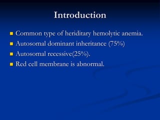
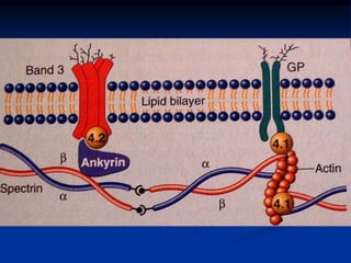
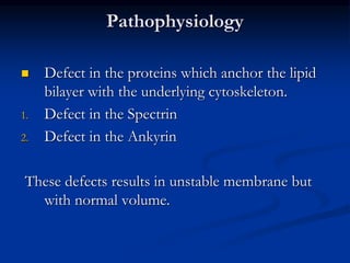
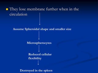
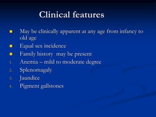
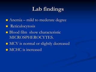
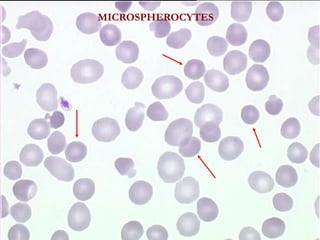
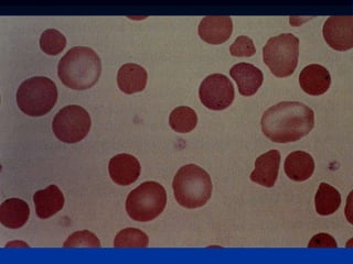
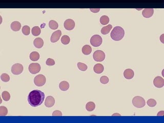
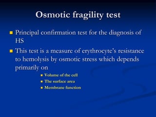
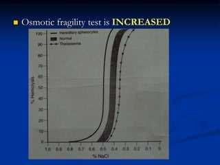
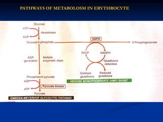
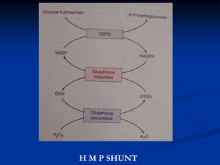
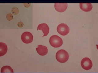
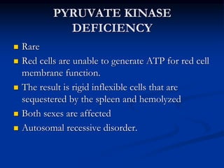
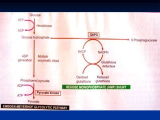
HERIDITARY HEMOLYTIC
ANEMIAS
(INTRACORPUSCULAR)
Dr Vijay Shankar . S
ABNORMALITIES OF
RED CELL
MEMBRANE
Heriditary spherocytosis
Heriditary elliptocytosis
Introduction
 Common type of heriditary hemolytic anemia.
 Autosomal dominant inheritance (75%)
 Autosomal recessive(25%).
 Red cell membrane is abnormal.
Pathophysiology
 Defect in the proteins which anchor the lipid
bilayer with the underlying cytoskeleton.
1. Defect in the Spectrin
2. Defect in the Ankyrin
These defects results in unstable membrane but
with normal volume.
 They lose membrane further when in the
circulation
Assume Spheroidal shape and smaller size
Microspherocytes
Reduced cellular
flexibility
Destroyed in the spleen
Clinical features
 May be clinically apparent at any age from infancy to
old age
 Equal sex incidence
 Family history may be present
1. Anemia – mild to moderate degree
2. Splenomagaly
3. Jaundice
4. Pigment gallstones
Lab findings
 Anemia – mild to mederate degree
 Reticulocytosis
 Blood film show characteristic
MICROSPHEROCYTES.
 MCV is normal or slightly decreased
 MCHC is increased
MICROSPHEROCYTES
Osmotic fragility test
 Principal confirmation test for the diagnosis of
HS
 This test is a measure of erythrocyte’s resistance
to hemolysis by osmotic stress which depends
primarily on
 Volume of the cell
 The surface area
 Membrane function
 Osmotic fragility test is INCREASED
 Direct Coombs test is NEGATIVE
 Cholelithiasis occurs in 40 to 50% of the
affected adults.
 Moderate splenic enlargement is seen.
HEMOLYTIC DEFECTS DUE TO
ENZYME DEFICIENCY
 G6PD Deficiency.
 PK deficiency
PATHWAYS OF METABOLOSM IN ERYTHROCYTE
H M P SHUNT
G6PD DEFICIENCY
 Most common human enzyme deficiency in the
world
 X linked trait affecting males.
 females are carriers.
 Patients with G^PD deficiency develop
hemolytic anemia when they are exposed to
oxidant stress like
1. Viral and bacterial infections
2. Drugs( antimalarials, sulfonamides , aspirin, vit
K)
3. Metabolic acidosis
4. Ingestion of Fava beans ( Favism)
Clinical features
 That of acute hemolytic anemia within hours of
exposure to oxidant stress.
 self limited since it affects the older cells only.
 some patients have darkening of urine due to
hemoglobinuria
 More severe cases present with jaundice.
Lab findings
 During the period of acute hemolysis
1. Fall in hematocrit.
2. Features of intravascular hemolysis
3. Formation of Heinz bodies seen in supravital
stain
e
 Between the crisis
1. red cell survival is shortened.
DIAGNOSIS – by direct enzyme assay
PYRUVATE KINASE
DEFICIENCY
 Rare
 Red cells are unable to generate ATP for red cell
membrane function.
 The result is rigid inflexible cells that are
sequestered by the spleen and hemolyzed
 Both sexes are affected
 Autosomal recessive disorder.
Summary
Thank you

Heriditary spherocytosis

  • 1.
  • 2.
    ABNORMALITIES OF RED CELL MEMBRANE Heriditaryspherocytosis Heriditary elliptocytosis
  • 3.
    Introduction  Common typeof heriditary hemolytic anemia.  Autosomal dominant inheritance (75%)  Autosomal recessive(25%).  Red cell membrane is abnormal.
  • 5.
    Pathophysiology  Defect inthe proteins which anchor the lipid bilayer with the underlying cytoskeleton. 1. Defect in the Spectrin 2. Defect in the Ankyrin These defects results in unstable membrane but with normal volume.
  • 6.
     They losemembrane further when in the circulation Assume Spheroidal shape and smaller size Microspherocytes Reduced cellular flexibility Destroyed in the spleen
  • 8.
    Clinical features  Maybe clinically apparent at any age from infancy to old age  Equal sex incidence  Family history may be present 1. Anemia – mild to moderate degree 2. Splenomagaly 3. Jaundice 4. Pigment gallstones
  • 9.
    Lab findings  Anemia– mild to mederate degree  Reticulocytosis  Blood film show characteristic MICROSPHEROCYTES.  MCV is normal or slightly decreased  MCHC is increased
  • 10.
  • 13.
    Osmotic fragility test Principal confirmation test for the diagnosis of HS  This test is a measure of erythrocyte’s resistance to hemolysis by osmotic stress which depends primarily on  Volume of the cell  The surface area  Membrane function
  • 14.
     Osmotic fragilitytest is INCREASED
  • 15.
     Direct Coombstest is NEGATIVE
  • 16.
     Cholelithiasis occursin 40 to 50% of the affected adults.  Moderate splenic enlargement is seen.
  • 17.
    HEMOLYTIC DEFECTS DUETO ENZYME DEFICIENCY  G6PD Deficiency.  PK deficiency
  • 18.
    PATHWAYS OF METABOLOSMIN ERYTHROCYTE
  • 19.
    H M PSHUNT
  • 20.
    G6PD DEFICIENCY  Mostcommon human enzyme deficiency in the world  X linked trait affecting males.  females are carriers.
  • 21.
     Patients withG^PD deficiency develop hemolytic anemia when they are exposed to oxidant stress like 1. Viral and bacterial infections 2. Drugs( antimalarials, sulfonamides , aspirin, vit K) 3. Metabolic acidosis 4. Ingestion of Fava beans ( Favism)
  • 22.
    Clinical features  Thatof acute hemolytic anemia within hours of exposure to oxidant stress.  self limited since it affects the older cells only.  some patients have darkening of urine due to hemoglobinuria  More severe cases present with jaundice.
  • 23.
    Lab findings  Duringthe period of acute hemolysis 1. Fall in hematocrit. 2. Features of intravascular hemolysis 3. Formation of Heinz bodies seen in supravital stain
  • 25.
    e  Between thecrisis 1. red cell survival is shortened. DIAGNOSIS – by direct enzyme assay
  • 26.
    PYRUVATE KINASE DEFICIENCY  Rare Red cells are unable to generate ATP for red cell membrane function.  The result is rigid inflexible cells that are sequestered by the spleen and hemolyzed  Both sexes are affected  Autosomal recessive disorder.
  • 28.
  • 29.