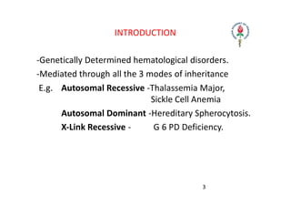
HEMOLYTIC ANEMIA
HEMOLYTIC ANEMIA
Definition:
Those anemias which result from an increase in
RBC destruction coupled with increased
erythropoiesis (i.e.: ability of Bone Marrow to
respond to anemia is unimpaired. )
Classification:
Congenital / Hereditary
Acquired
INTRODUCTION
‐Genetically Determined hematological disorders.
‐Mediated through all the 3 modes of inheritance
E.g. Autosomal Recessive ‐Thalassemia Major,
Sickle Cell Anemia
Autosomal Dominant ‐Hereditary Spherocytosis.
X‐Link Recessive ‐ G 6 PD Deficiency.
3
PATHOGENESIS
RED CELL DESTRUCTION
⚫ The physiological destruction of senescent red cells takes
place with in macrophages, which are abundant in spleen,
liver and bone marrow
⚫ This process appears to be triggered by age- dependent
changes in red cell surface proteins, which lead to their
recognition and phagocytosis
⚫ Red cell destruction occur by 2 mechanisms-
 Extravascular Hemolysis – The site of destruction is mainly
spleen and this is the major mechanism of red cell hemolysis.
Red cells are taken up by the cells of RE system where they
are destroyed and digested
 Intravascular Hemolysis– This is the minor pathway of red
cell destruction and red cells are destroyed in circulation
releasing hemoglobin.
SENESCENT RED CELLPHAGOCYTOSED
BY RE CELLS OF SPLEEN
HEMOGLOIN RELEASEDAND BROKEN
DOWN
HEME + GLOBIN BROKEN DOWN
TOAMINOACIDS
REUTILISED
FOR SYNTHESIS
FORA,B CHAINS
IRON+PORPHYRIN
BILIVERDIN
BILRUBIN
(UNCONJUGA
TED)
CONJUGATED IN LIVER
BILRUBIN GLUCURONIDE EXCRETED IN BILEAND
ACTED UPON BY BACTERIAL ENZYMES IN INTESTINE
UROBILINOGRN/STERCOBILINOGEN
FECAL STERCOBILINOGEN
ABSORB IN ENTEROHEPATIC
CIRCULATION
KIDNEY
UROBILINOGEN IN URINE
EXTRAVASCULAR HEMOLYSIS
RED CELLS IN CIRCULATION
RED CELLS LYSE IN CIRCULATION
HEMOGLOBIN IN PLASMA
HEMOGLOBIN IN URINE
HEMOGLOBINURIA
POSITIVE BENZIDINE TEST
HEMOGLOBINIMIA
HBABSORBED BY KIDNEY
TUBULAR CELLS
HB CONVERTED TO HEMOSIDERIN
TUBULAR CELLS IN FEW DAYS
TUBULAR CELLS SHED OFF
HEMOSIDENURIA
COMBINES WITH HEPTAGLOBIN
INTRAVASCULAR HEMOLYSIS
CLASSIFICATION
Extrinsic Deficit
‐ Isoimmune
‐ Rh incompatibility
‐ Heteroimmune
‐ ABO incompatibility
‐ Autoimmune
‐ Infections & Toxins
Intrinsic Defects
‐ Membrane Defect
Hereditary Spherocytosis
‐ Hb Defects
Thalassemia
Sickle cell anemia
‐ Enzyme Defects
G 6 PD Deficiency
INTRACORPUSCULAR
DEFECTS (INTRINSIC)
EXTRACORPUSCULAR
FACTORS (EXTRINSIC)
Hereditary • Hemoglobinopathies
• Enzymopathies
• Membrane
Cytoskeletal Defects
• Familial Hemolytic
Uremic Syndrome
Acquired • Paroxysmal
Nocturnal
Hemoglobinuria
• Mechanical
Destruction
[Microangiopathic]
• Toxic Agents
• Drugs
• Infectious
• Autoimmune
GENERAL FEATURES OF HEMOLYTIC DISORDERS
General examination ‐ Jaundice, pallor ,bossing of
skull
Systemic examination findings ‐ Enlarged spleen
Hemoglobin
MCV
Reticulocytes
Bilirubin
LDH
‐ From normal to severely reduced
‐ usually increased
‐ increased
‐ increased[mostly unconjugated]
‐ increased
LAB FINDINGS (COND..)
•Serum Haptoglobin &Hemopexin – reduced
•Urine urobilinogen ‐ increased
•Blood film‐polychromasia & Normoblast
•Bone marrow‐ Erythroid hyperplasia
In Intravascular hemolysis
•Serum Haemoglobin‐increased
•Urine Hemosiderin & Haemoglobin ‐positive
POLYCHROMATIC CELLS
TYPES OF HEMOGLOBINS AT VARIOUS STAGES
CONTD..
• Hb A(α2β2) appears at ~1 month of fetal life but
does not become the dominant hemoglobin until
after birth, when Hb F levels start to decline.
• Hb A2 (α2δ2) is a minor hemoglobin that appears
shortly before birth and remains at a low level after
birth.
• The final hemoglobin distribution pattern is not
achieved until at least 6 month of age and
sometimes later.
• The normal hemoglobin pattern is ≥95% Hb A, ≤3.5
Hb A2, and <2.5% Hb F
THALASSEMIA
• Autosomal Recessive Disorder
• Single Gene disorder
• Defect in globin chain synthesis of Hb
• Types
‐ α Thalassemia
• α chain is defective: incompatible with life
‐ β Thalassemia
• β˚ thalassemia if β chain is absent
• βᶧ thalassemia is β is partially produced
CLASSIFICATION
α – Thalassemia β‐ Thalassemia
Silent carrier Silent carrier
α‐ Thalassemia trait Thalassemia trait
Hb H disease Thalassemia intermedia
Hydrops fetalis Thalassemia Major
PATHOPHYSIOLOGY
•Hb. consist of two pairs of amino acid chains α & β.
•Imbalance in the production of these peptide chains of
globin leads to abnormal hemoglobinopathies
•Unpaired peptide chains in Hb. is precipitated on RBC
membrane
•Premature destruction of RBC in bone‐marrow and in
peripheral circulation particularly in R‐E system of
spleen(Ineffective erythropoiesis)
•ɤ chain synthesis persist after fetal life postnatally also
PATHOPHYSIOLOGY
• Increased fetal HB. With its high affinity for oxygen
• leads to tissue hypoxia
• Stimulate erythropoietin secretion
• Leading to both medullary and extra medullary
erythropoiesis
• Resulting in expansion of bone marrow space with
characteristic hemolytic facies, pathological fractures
of long bones , splenomegaly, and its related
complications along with severe degree of Anemia
CLINICAL PROFILE OF THALASSEMIA MAJOR
(HOMOZYGOUS)
CLINICAL FEATURES
• Age of onset – From 6months onwards.
• Thalassemic Facies (Maxillary hyperplasia, flat nasal
bridge, frontal bossing)
• Hepatosplenomegaly
• Greenish Brown Complexion (Due to pallor,
hemosiderosis & Jaundice)
• Severe degree of persistent and progressive Anemia
needs repeated blood transfusion
• Iron overload ‐ endocrine dysfunction, cardiac &
liver dysfunction
• Growth, Development & SMR delayed
PERIPHERAL BLOOD SMEAR OF THALASSEMIA
MAJOR
CONT..
• Microcytic hypochromic anemia showing severe
degree of anisocytosis and poikilocytosis.
• Target cells, Pencil cells, stomatocytes, helmet cells.
• Immature RBC’s like normoblasts .
• Hb & HCT is extremely low with Reticulocytosis.
• Hb electrophoresis demonstrating Fetal Hb. (α2 ϒ2 )
is confirmatory of diagnosis.‐ HbF ‐ 90‐96 % , HbA2 ‐
3.5% ‐ 5.5% & HbA – 0%.
• Mutation in beta‐globin chain expression.
THALASSAEMIA MAJOR : X – RAY SKULL
Showing Hair on end appearance due to increased diploic
space in the skull bones
OTHER TYPES OF THALASSEMIA
Beta thalassemia
intermedia (double
heterozygous)
• Later age of onset –
> 2yrs.Mod degree
of Anemia
•Hepatosplenomegaly
Hb electro:‐HbF‐20‐
100%, HbA2 ‐3.5‐5.5% &
HbA ‐0‐30%
OTHER TYPES OF THALASSEMIA
MANAGEMENT OF THALASSEMIA MAJOR
• Anemia correction‐Packed red cell transfusions
• Excess iron removal‐ chelation therapy
• Management of transfusion transmitted infections
• Management of cardiac and endocrine
complications‐pituitary, thyroid ,parathyroid and
gonads
• Treatment of hypersplenism‐ splenectomy
• Treatment of Osteopenia, leg ulcers & gall stones
Curative treatment – stem cell transplantation
Prevention ‐ Prenatal diagnosis & Genetic counseling
A
TRANSFUSION REGIMENS IN THALASSEMI
• PALLIATIVE TRANSFUSION REGIMEN – Increase Hb
to 8.5 gm.%
• MODERATE TRANSFUSION – Increase Hb to 9‐10.5
gm.%
• HYPERTRANSFUSION REGIMEN ‐ Increase Hb to 10
gm.%
• SUPERTRANSFUSION ‐ Increase Hb to 12gm%
• NEOCYTE TRANSFUSION ‐ To make requirement of
transfusion interval longer.
CHELATION THERAPY
Iron overload – Excess GI absorption, Lack of
mechanism for excreation of Iron from body & C/C red
cell transfusion
Chelation to be started when Sr. Ferritin > 1000 ng/ml
or liver iron >2500 micrograms/gm of dry weight.
Drugs used‐
•Desferrioxamine –(parenteral) remove extracellular
iron
•Deferiprone –( oral) both intra and extra cellular iron
•Deferasirox – (oral) both intra and extra cellular iron
SICKLE CELL DISEASE
Mutation of beta globin‐6 Glutamate
Deoxy HbS (polymerised)
Val.
Ca2+ influx, K+ leakage
Stiff, viscous sickle cell
venocclusion decreased RBC survival
microinfarctions, ischemic pains
autoinfarction of spleen
anemia, jaundice,
gallstones, leg ulcers
CONT..
‐ The disease can manifest only if Homozygous.
‐ Heterozygous individuals ‐ Trait or Carrier state.
‐ Generally Asymptomatic .Only under stress situation
manifest clinically in the form of crises. E.g.
Hypoxia/Sepsis
• Hemoglobin electrophoresis
‐HbS >80%, HbF ‐1‐20%,
HbA2 ‐2‐ 4.5%
• Sickling test ‐ Positive
CRISIS
• Aplastic crisis ‐ Severe Anemia of BM suppression due
to Parvo virus B19
• Hyperhemolytic crisis ‐ Severe hemolysis in peripheral
circulation and R‐E system
• Vaso‐occlusive crisis ‐ Hand‐Foot disease ,avascular
necrosis of bones, acute chest syndrome, stroke,
priapism, painful abdominal crisis
• Sequestration crisis ‐ Shock because of the pooling of
the blood in R‐E system : Spleen and Liver
HEREDITARY SPHEROCYTOSIS PATHOPHYSIOLOGY
•Sodium pump failure because of the membrane defect
due to spectrin & ankyrin deficiency resulting
in
Influx of sodium and water
Loss of biconcave shape
Spherical shape (Microspherocytosis)
• Clinical Triad ‐ Anaemia ,
Jaundice, Splenomegaly
• Complications ‐ Hemolytic &
aplastic crisis, gall
stones
• PS ‐ Spherocytosis
• Osmotic fragility increased
• Treatment‐Splenectomy
• Screen family members
G‐6 P D DEFICIENCY
Generally asymptomatic
Manifests only when
exposed to Oxidant Drugs
Intra‐vascular Hemolysis
•Hemoglobinuria
•Heinz Bodies&
blister cells in
Peripheral smear
•G6PD estimation
diagnostic
KNOWN OXIDANT INSULTS
RIA
PAROXYSMAL NOCTURNAL HEMOGLOBINU
• Acquired chronic H.A
• Persistent intra vascular hemolysis
• Pancytopenia
• Lab : Hemoglobinuria, hemosiderinuria, increased
LDH, bilirubin
• Risk of venous thrombosis
• C/F – Hemoglobinuria during night
• P.S – Polychromatophilia, normoblasts
• B.M – Normoblastic hyperplasia
• Differential diagnosis‐ flow cytometry CD59‐, CD55‐
RBC, WBC
‐ Hams’ acidified serum test
AUTOIMMUNE HEMOLYTIC ANEMIA
• Result from RBC destruction due to RBC
autoantibodies
AUTOIMMUNE HEMOLYTIC ANEMIA‐ TYPES
Primary AIHA
•Warm AI hemolysis: Antibody binds at 37degree C(IgG)
• Cold AI Hemolysis: Antibody binds at 4 degree C(IgM
& complement)
•Paroxysmal cold Hemoglobinuria (IgG & compliment)
Secondary AIHA
•Drugs, infections, primary immunodeficiency,
malignancy, autoimmune disorders
TO CONCLUDE
Hemolytic anemia recognised by clinical picture ‐
history & physical exam, lab test to confirm hemolysis
and peripheral smear to guide further tests.
Spherocytes‐ AIHA, hereditary spherocytosis
Schistocytes‐ HUS, TTP or DIC & heart valve hemolysis
• Blister Cells‐ oxidative damage‐ G6PD
•Sickle cells ‐ sickle cell anemia
•Heinz bodies ‐ G6PD deficiency
THANK YOU

Hemolytic-Anemia.pptx

  • 1.
  • 2.
    HEMOLYTIC ANEMIA Definition: Those anemiaswhich result from an increase in RBC destruction coupled with increased erythropoiesis (i.e.: ability of Bone Marrow to respond to anemia is unimpaired. ) Classification: Congenital / Hereditary Acquired
  • 3.
    INTRODUCTION ‐Genetically Determined hematologicaldisorders. ‐Mediated through all the 3 modes of inheritance E.g. Autosomal Recessive ‐Thalassemia Major, Sickle Cell Anemia Autosomal Dominant ‐Hereditary Spherocytosis. X‐Link Recessive ‐ G 6 PD Deficiency. 3
  • 4.
    PATHOGENESIS RED CELL DESTRUCTION ⚫The physiological destruction of senescent red cells takes place with in macrophages, which are abundant in spleen, liver and bone marrow ⚫ This process appears to be triggered by age- dependent changes in red cell surface proteins, which lead to their recognition and phagocytosis
  • 5.
    ⚫ Red celldestruction occur by 2 mechanisms-  Extravascular Hemolysis – The site of destruction is mainly spleen and this is the major mechanism of red cell hemolysis. Red cells are taken up by the cells of RE system where they are destroyed and digested  Intravascular Hemolysis– This is the minor pathway of red cell destruction and red cells are destroyed in circulation releasing hemoglobin.
  • 6.
    SENESCENT RED CELLPHAGOCYTOSED BYRE CELLS OF SPLEEN HEMOGLOIN RELEASEDAND BROKEN DOWN HEME + GLOBIN BROKEN DOWN TOAMINOACIDS REUTILISED FOR SYNTHESIS FORA,B CHAINS IRON+PORPHYRIN BILIVERDIN BILRUBIN (UNCONJUGA TED) CONJUGATED IN LIVER BILRUBIN GLUCURONIDE EXCRETED IN BILEAND ACTED UPON BY BACTERIAL ENZYMES IN INTESTINE UROBILINOGRN/STERCOBILINOGEN FECAL STERCOBILINOGEN ABSORB IN ENTEROHEPATIC CIRCULATION KIDNEY UROBILINOGEN IN URINE EXTRAVASCULAR HEMOLYSIS
  • 7.
    RED CELLS INCIRCULATION RED CELLS LYSE IN CIRCULATION HEMOGLOBIN IN PLASMA HEMOGLOBIN IN URINE HEMOGLOBINURIA POSITIVE BENZIDINE TEST HEMOGLOBINIMIA HBABSORBED BY KIDNEY TUBULAR CELLS HB CONVERTED TO HEMOSIDERIN TUBULAR CELLS IN FEW DAYS TUBULAR CELLS SHED OFF HEMOSIDENURIA COMBINES WITH HEPTAGLOBIN INTRAVASCULAR HEMOLYSIS
  • 8.
    CLASSIFICATION Extrinsic Deficit ‐ Isoimmune ‐Rh incompatibility ‐ Heteroimmune ‐ ABO incompatibility ‐ Autoimmune ‐ Infections & Toxins
  • 9.
    Intrinsic Defects ‐ MembraneDefect Hereditary Spherocytosis ‐ Hb Defects Thalassemia Sickle cell anemia ‐ Enzyme Defects G 6 PD Deficiency
  • 10.
    INTRACORPUSCULAR DEFECTS (INTRINSIC) EXTRACORPUSCULAR FACTORS (EXTRINSIC) Hereditary• Hemoglobinopathies • Enzymopathies • Membrane Cytoskeletal Defects • Familial Hemolytic Uremic Syndrome Acquired • Paroxysmal Nocturnal Hemoglobinuria • Mechanical Destruction [Microangiopathic] • Toxic Agents • Drugs • Infectious • Autoimmune
  • 11.
    GENERAL FEATURES OFHEMOLYTIC DISORDERS General examination ‐ Jaundice, pallor ,bossing of skull Systemic examination findings ‐ Enlarged spleen Hemoglobin MCV Reticulocytes Bilirubin LDH ‐ From normal to severely reduced ‐ usually increased ‐ increased ‐ increased[mostly unconjugated] ‐ increased
  • 12.
    LAB FINDINGS (COND..) •SerumHaptoglobin &Hemopexin – reduced •Urine urobilinogen ‐ increased •Blood film‐polychromasia & Normoblast •Bone marrow‐ Erythroid hyperplasia In Intravascular hemolysis •Serum Haemoglobin‐increased •Urine Hemosiderin & Haemoglobin ‐positive
  • 13.
  • 14.
    TYPES OF HEMOGLOBINSAT VARIOUS STAGES
  • 15.
    CONTD.. • Hb A(α2β2)appears at ~1 month of fetal life but does not become the dominant hemoglobin until after birth, when Hb F levels start to decline. • Hb A2 (α2δ2) is a minor hemoglobin that appears shortly before birth and remains at a low level after birth. • The final hemoglobin distribution pattern is not achieved until at least 6 month of age and sometimes later. • The normal hemoglobin pattern is ≥95% Hb A, ≤3.5 Hb A2, and <2.5% Hb F
  • 16.
    THALASSEMIA • Autosomal RecessiveDisorder • Single Gene disorder • Defect in globin chain synthesis of Hb • Types ‐ α Thalassemia • α chain is defective: incompatible with life ‐ β Thalassemia • β˚ thalassemia if β chain is absent • βᶧ thalassemia is β is partially produced
  • 17.
    CLASSIFICATION α – Thalassemiaβ‐ Thalassemia Silent carrier Silent carrier α‐ Thalassemia trait Thalassemia trait Hb H disease Thalassemia intermedia Hydrops fetalis Thalassemia Major
  • 18.
    PATHOPHYSIOLOGY •Hb. consist oftwo pairs of amino acid chains α & β. •Imbalance in the production of these peptide chains of globin leads to abnormal hemoglobinopathies •Unpaired peptide chains in Hb. is precipitated on RBC membrane •Premature destruction of RBC in bone‐marrow and in peripheral circulation particularly in R‐E system of spleen(Ineffective erythropoiesis) •ɤ chain synthesis persist after fetal life postnatally also
  • 19.
    PATHOPHYSIOLOGY • Increased fetalHB. With its high affinity for oxygen • leads to tissue hypoxia • Stimulate erythropoietin secretion • Leading to both medullary and extra medullary erythropoiesis • Resulting in expansion of bone marrow space with characteristic hemolytic facies, pathological fractures of long bones , splenomegaly, and its related complications along with severe degree of Anemia
  • 20.
    CLINICAL PROFILE OFTHALASSEMIA MAJOR (HOMOZYGOUS)
  • 21.
    CLINICAL FEATURES • Ageof onset – From 6months onwards. • Thalassemic Facies (Maxillary hyperplasia, flat nasal bridge, frontal bossing) • Hepatosplenomegaly • Greenish Brown Complexion (Due to pallor, hemosiderosis & Jaundice) • Severe degree of persistent and progressive Anemia needs repeated blood transfusion • Iron overload ‐ endocrine dysfunction, cardiac & liver dysfunction • Growth, Development & SMR delayed
  • 22.
    PERIPHERAL BLOOD SMEAROF THALASSEMIA MAJOR
  • 23.
    CONT.. • Microcytic hypochromicanemia showing severe degree of anisocytosis and poikilocytosis. • Target cells, Pencil cells, stomatocytes, helmet cells. • Immature RBC’s like normoblasts . • Hb & HCT is extremely low with Reticulocytosis. • Hb electrophoresis demonstrating Fetal Hb. (α2 ϒ2 ) is confirmatory of diagnosis.‐ HbF ‐ 90‐96 % , HbA2 ‐ 3.5% ‐ 5.5% & HbA – 0%. • Mutation in beta‐globin chain expression.
  • 24.
    THALASSAEMIA MAJOR :X – RAY SKULL Showing Hair on end appearance due to increased diploic space in the skull bones
  • 25.
    OTHER TYPES OFTHALASSEMIA Beta thalassemia intermedia (double heterozygous) • Later age of onset – > 2yrs.Mod degree of Anemia •Hepatosplenomegaly Hb electro:‐HbF‐20‐ 100%, HbA2 ‐3.5‐5.5% & HbA ‐0‐30%
  • 26.
    OTHER TYPES OFTHALASSEMIA
  • 27.
    MANAGEMENT OF THALASSEMIAMAJOR • Anemia correction‐Packed red cell transfusions • Excess iron removal‐ chelation therapy • Management of transfusion transmitted infections • Management of cardiac and endocrine complications‐pituitary, thyroid ,parathyroid and gonads • Treatment of hypersplenism‐ splenectomy • Treatment of Osteopenia, leg ulcers & gall stones Curative treatment – stem cell transplantation Prevention ‐ Prenatal diagnosis & Genetic counseling
  • 28.
    A TRANSFUSION REGIMENS INTHALASSEMI • PALLIATIVE TRANSFUSION REGIMEN – Increase Hb to 8.5 gm.% • MODERATE TRANSFUSION – Increase Hb to 9‐10.5 gm.% • HYPERTRANSFUSION REGIMEN ‐ Increase Hb to 10 gm.% • SUPERTRANSFUSION ‐ Increase Hb to 12gm% • NEOCYTE TRANSFUSION ‐ To make requirement of transfusion interval longer.
  • 29.
    CHELATION THERAPY Iron overload– Excess GI absorption, Lack of mechanism for excreation of Iron from body & C/C red cell transfusion Chelation to be started when Sr. Ferritin > 1000 ng/ml or liver iron >2500 micrograms/gm of dry weight. Drugs used‐ •Desferrioxamine –(parenteral) remove extracellular iron •Deferiprone –( oral) both intra and extra cellular iron •Deferasirox – (oral) both intra and extra cellular iron
  • 30.
    SICKLE CELL DISEASE Mutationof beta globin‐6 Glutamate Deoxy HbS (polymerised) Val. Ca2+ influx, K+ leakage Stiff, viscous sickle cell venocclusion decreased RBC survival microinfarctions, ischemic pains autoinfarction of spleen anemia, jaundice, gallstones, leg ulcers
  • 31.
    CONT.. ‐ The diseasecan manifest only if Homozygous. ‐ Heterozygous individuals ‐ Trait or Carrier state. ‐ Generally Asymptomatic .Only under stress situation manifest clinically in the form of crises. E.g. Hypoxia/Sepsis • Hemoglobin electrophoresis ‐HbS >80%, HbF ‐1‐20%, HbA2 ‐2‐ 4.5% • Sickling test ‐ Positive
  • 32.
    CRISIS • Aplastic crisis‐ Severe Anemia of BM suppression due to Parvo virus B19 • Hyperhemolytic crisis ‐ Severe hemolysis in peripheral circulation and R‐E system • Vaso‐occlusive crisis ‐ Hand‐Foot disease ,avascular necrosis of bones, acute chest syndrome, stroke, priapism, painful abdominal crisis • Sequestration crisis ‐ Shock because of the pooling of the blood in R‐E system : Spleen and Liver
  • 33.
    HEREDITARY SPHEROCYTOSIS PATHOPHYSIOLOGY •Sodiumpump failure because of the membrane defect due to spectrin & ankyrin deficiency resulting in Influx of sodium and water Loss of biconcave shape Spherical shape (Microspherocytosis)
  • 34.
    • Clinical Triad‐ Anaemia , Jaundice, Splenomegaly • Complications ‐ Hemolytic & aplastic crisis, gall stones • PS ‐ Spherocytosis • Osmotic fragility increased • Treatment‐Splenectomy • Screen family members
  • 35.
    G‐6 P DDEFICIENCY Generally asymptomatic Manifests only when exposed to Oxidant Drugs Intra‐vascular Hemolysis •Hemoglobinuria •Heinz Bodies& blister cells in Peripheral smear •G6PD estimation diagnostic
  • 36.
  • 37.
    RIA PAROXYSMAL NOCTURNAL HEMOGLOBINU •Acquired chronic H.A • Persistent intra vascular hemolysis • Pancytopenia • Lab : Hemoglobinuria, hemosiderinuria, increased LDH, bilirubin • Risk of venous thrombosis • C/F – Hemoglobinuria during night • P.S – Polychromatophilia, normoblasts • B.M – Normoblastic hyperplasia • Differential diagnosis‐ flow cytometry CD59‐, CD55‐ RBC, WBC ‐ Hams’ acidified serum test
  • 38.
    AUTOIMMUNE HEMOLYTIC ANEMIA •Result from RBC destruction due to RBC autoantibodies
  • 39.
    AUTOIMMUNE HEMOLYTIC ANEMIA‐TYPES Primary AIHA •Warm AI hemolysis: Antibody binds at 37degree C(IgG) • Cold AI Hemolysis: Antibody binds at 4 degree C(IgM & complement) •Paroxysmal cold Hemoglobinuria (IgG & compliment) Secondary AIHA •Drugs, infections, primary immunodeficiency, malignancy, autoimmune disorders
  • 40.
    TO CONCLUDE Hemolytic anemiarecognised by clinical picture ‐ history & physical exam, lab test to confirm hemolysis and peripheral smear to guide further tests. Spherocytes‐ AIHA, hereditary spherocytosis Schistocytes‐ HUS, TTP or DIC & heart valve hemolysis • Blister Cells‐ oxidative damage‐ G6PD •Sickle cells ‐ sickle cell anemia •Heinz bodies ‐ G6PD deficiency
  • 41.