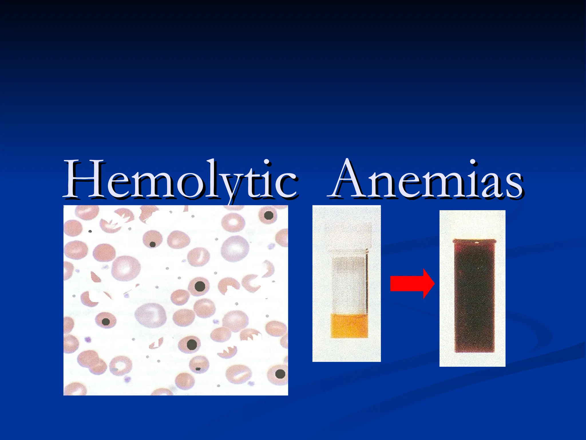
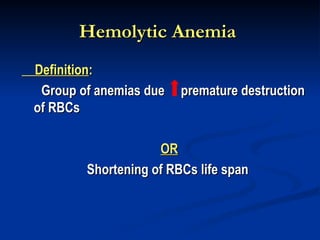
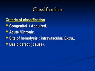
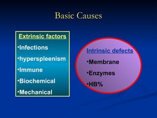
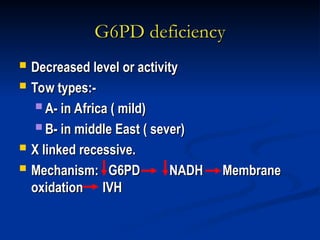
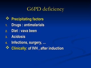
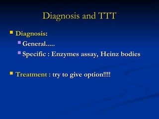
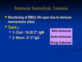
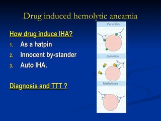
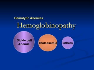
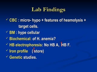
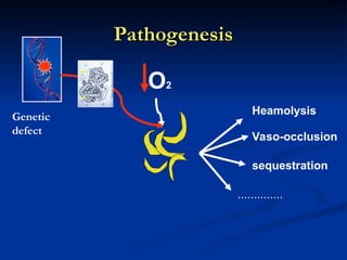
Hemolytic Anemias
Hemolytic Anemias
Hemolytic
Hemolytic Anemia
Anemia
Definition
Definition:
:
Group of anemias due premature destruction
of RBCs
of RBCs
OR
OR
Shortening of RBCs life span
Classification
Classification
Criteria of classification
 Congenital / Acquired.
Congenital / Acquired.
 Acute /Chronic.
Acute /Chronic.
 Site of hemolysis : intravascular/ Extra..
Site of hemolysis : intravascular/ Extra..
 Basic defect ( cause).
Basic defect ( cause).
Basic Causes
Basic Causes
Intrinsic defects
•Membrane
•Enzymes
•HB%
Extrinsic factors
•Infections
•hyperspleenism
•Immune
•Biochemical
•Mechanical
General C/F
General C/F
 ...of anemia…
...of anemia…
 Marked Jaundice , stones.
Marked Jaundice , stones.
 Black urine.
Black urine.
 Splenomegally.
Splenomegally.
 Skeletal deformities.
Skeletal deformities.
 Leg ulcrs.
Leg ulcrs.
Lab Features of Hemolysis
 CBC:
CBC: normo-normo, Retic count, nucleated
normo-normo, Retic count, nucleated
RBCs, polychromasia, Poikelocytosis,
fragmented cells...
fragmented cells...
 BM
BM:
: marked
marked cellular
cellular
 Biochemical
Biochemical : bilirubin,free HB, HB urea, LDH,
: bilirubin,free HB, HB urea, LDH,
ALP.
ALP.
 RBCs studies
RBCs studies: fragility, life span
: fragility, life span
Membrane Defects
Membrane Defects
 Hereditary spherocytosis.
Hereditary spherocytosis.
 Hereditary eleptocytosis
Hereditary eleptocytosis
 Pyropoikelocytosis
Pyropoikelocytosis
 PNH.
PNH.
Hereditary Spherocytosis
 Common ( AD).
Common ( AD).
 Due to defect in cell membrane proteins;
Due to defect in cell membrane proteins;
spectren, anchyren, B and 4 band.
 Deformidibility exra-vascular H.
Deformidibility exra-vascular H.
 Clinically
Clinically: early jaundice, large spleen
: early jaundice, large spleen
 Complications
Complications : leg ulcer, bile stones, Aplastic
: leg ulcer, bile stones, Aplastic
anemia
anemia
 Management?
Management?
Hereditary Spherocytosis
Diagnosis
Diagnosis
 .. Of H. anemia
.. Of H. anemia
 Specific tests:
Specific tests:
 Osmotic fragility test
Osmotic fragility test
 Auto- heamolysis test
Auto- heamolysis test
 Membrane protein studies
Membrane protein studies
 Genetic studied
Genetic studied
OFT
OFT
Normal
High fragility
eg: HS
Leg ulcer
Paroxysmal nocturnal Hurea
 Acquired stem cell disorder resulting in abnormal
Acquired stem cell disorder resulting in abnormal
cell membrane defect.
 DAF complements activity heamolysis
DAF complements activity heamolysis
 Clinically
Clinically: of IVH, morning black urine, thrombosis,
: of IVH, morning black urine, thrombosis,
iron deficiency. AA
iron deficiency. AA
 Specific tests
Specific tests: Ham test, stem cell study
: Ham test, stem cell study
Enzymes Deficiency
Enzymes Deficiency
1.
1. G6PD
G6PD
Function: Antioxdation
1.
1. Pyrophate kinas
Pyrophate kinas
G6PD deficiency
G6PD deficiency
 Decreased level or activity
Decreased level or activity
 Tow types:-
Tow types:-
 A- in Africa ( mild)
A- in Africa ( mild)
 B- in middle East ( sever)
B- in middle East ( sever)
 X linked recessive.
X linked recessive.
 Mechanism: G6PD NADH Membrane
Mechanism: G6PD NADH Membrane
oxidation IVH
oxidation IVH
G6PD deficiency
G6PD deficiency
 Precipitating factors
Precipitating factors
1.
1. Drugs : antimalarials
Drugs : antimalarials
2.
2. Diet : vava been
Diet : vava been
3.
3. Acidosis
Acidosis
4.
4. Infections, surgery, ...
Infections, surgery, ...
 Clinically:
Clinically: of IVH , after induction
of IVH , after induction
Diagnosis and TTT
Diagnosis and TTT
 Diagnosis
Diagnosis:
:
 General.....
General.....
 Specific : Enzymes assay, Heinz bodies
Specific : Enzymes assay, Heinz bodies
 Treatment :
Treatment : try to give option!!!!
try to give option!!!!
Immune hemolytic Anemia
 Shortening of RBCs life span due to immune
Shortening of RBCs life span due to immune
mechanisms (Abs)
mechanisms (Abs)
 Types
Types :-
:-
 1- Clod : 15-20 Cº: IgM
1- Clod : 15-20 Cº: IgM
 2- Worm: 37 Cº IgG
2- Worm: 37 Cº IgG
Allo-immune
Auto-immune
Drug induced
Worm IHA
Worm IHA
 37 Cº, IgG
37 Cº, IgG
 Causes
Causes
 Idiopathic
Idiopathic
 Drugs ( methyledopa)
Drugs ( methyledopa)
 Lymphoma / leukemia
Lymphoma / leukemia
 CT. diseases : Rh artheritis, SLE,..
CT. diseases : Rh artheritis, SLE,..
Mechanism of worm IHA
B Abs
IgG
Complement
activation
Removal by
spleen
IVH
EVH
Opsonization
Diagnosis and treatment
 ..General
..General!
! Autoagglutination and spherocytosis in
Autoagglutination and spherocytosis in
Blood film.
Blood film.
 Specific (serology)
Specific (serology)
 DCT (direct Coomb test)(100%)
DCT (direct Coomb test)(100%)
 IDCT (75%)
IDCT (75%)
 TTT:
TTT:
 Of the cause
Of the cause
 Supportive
Supportive
 Immunosuppressive drugs
Immunosuppressive drugs
Autoaglutination Spherocytosis
Cold IHA
Cold IHA
 15 C
15 Cº
º. IgM
. IgM
 Caused mainly by infection( Mycoplasma).
Caused mainly by infection( Mycoplasma).
 Agg > hemolysis
Agg > hemolysis
 Clinically : peripheral cyanosis( Rhenould)
Clinically : peripheral cyanosis( Rhenould)
 Diagnosed by DCT only ( at 4 C
Diagnosed by DCT only ( at 4 Cº
º)
)
Drug induced hemolytic aneamia
How drug induce IHA?
1.
1. As a hatpin
As a hatpin
2.
2. Innocent by-stander
Innocent by-stander
3.
3. Auto IHA.
Auto IHA.
Diagnosis and TTT ?
Diagnosis and TTT ?
Hemoglobinopathy
Hemoglobinopathy
Hemolytic Anemias
Sickle cell
Anemia Thalassemia Others
Normal HB.
Normal HB.
 Hem + 4 chains of globing
Hem + 4 chains of globing
 Deferent types of chains:-
Deferent types of chains:-
HB Abnormalites
HB Abnormalites
1.
1. Reduced chains production (thalassemia)
Reduced chains production (thalassemia)
2.
2. Abn HB. eg (HBS, C,D, E,...)
Abn HB. eg (HBS, C,D, E,...)
3.
3. Altered O
Altered O2
2 affinity
affinity
4.
4. Failure of reduction (Met HB)
Failure of reduction (Met HB)
Thalassemia
Thalassemia
Definition
Definition
Group of genetic disorders due to reduced rate
of chain production.
Classification
Classification
_
_ alfa thal.
alfa thal.
- B thal
- B thal
Clinical classification
1. Major
2. Intermedia
3. Minor
Genetic Base
Genetic Base
B thalassemia
B thalassemia
 Controlled by tow genes
Controlled by tow genes
 (Point) mutation either complete loss B º
(Point) mutation either complete loss B º
Or partial loss B
Or partial loss B
 So there are many combinations of B, Bº and B .
So there are many combinations of B, Bº and B .
 Clinical severity depends on this combinations
Clinical severity depends on this combinations
+
+
Clinical features
Clinical features
 Clinical severity depends on the genetic
Clinical severity depends on the genetic
defect : Major, intermedius, minor.
Generally:
Generally:
 .. Transfusion dependent H. anemia
.. Transfusion dependent H. anemia
 Hepatosplenomegaly
Hepatosplenomegaly
 Skeletal deformities (Hair on the end, short, ...)
Skeletal deformities (Hair on the end, short, ...)
 Infections
Infections
 Iron over load ( heart, liver , glands,...)
Iron over load ( heart, liver , glands,...)
Hair on the end
Thal. Faces
Lab Findings
Lab Findings
 CBC
CBC : micro- hypo + features of heamolysis +
: micro- hypo + features of heamolysis +
target cells.
target cells.
 BM
BM : hype cellular
: hype cellular
 Biochemical:
Biochemical: of H. anemia?
of H. anemia?
 HB electrophoresis
HB electrophoresis: No HB A, HB F.
: No HB A, HB F.
 Iron profile
Iron profile ( store)
( store)
 Genetic
Genetic studies.
studies.
Electrophoresis
Normal
B thal.Major
B thal trait
S/ B thal
HB H
SS
Treatment
Treatment
 Recurrent transfusion ( every 4-6 weeks)
Recurrent transfusion ( every 4-6 weeks)
 Folic acid.
Folic acid.
 Iron chelating (Dysferexamine+ Vit C).
Iron chelating (Dysferexamine+ Vit C).
 Treat complications:
Treat complications:
 Infections
Infections
 Endocrinal disorders
Endocrinal disorders
 skeletal deformities
skeletal deformities
 Spleenectomy , gene therapy.
Spleenectomy , gene therapy.
Sickle Cell Disorders
 Group of genetic disorders in which HB S is
Group of genetic disorders in which HB S is
inherited.
inherited.
Types :
Types :
 Sickle cell aneamia: HB SS
Sickle cell aneamia: HB SS
 Sickle cell trait :HB AS
Sickle cell trait :HB AS
 HB SC disease
HB SC disease
 Sickle cell thalassemia : HB SB thl.
Sickle cell thalassemia : HB SB thl.
 ......
......
Pathogenesis
Pathogenesis
O2
Heamolysis
Vaso-occlusion
sequestration
..............
Genetic
defect
Sickle cell anemia C/F
 H. anemia, mild in relation to low HB% ( 6 g/dl),
H. anemia, mild in relation to low HB% ( 6 g/dl),
start at 6
start at 6th
th
month.
month.
 Acute complications (crisis)
Acute complications (crisis)
Hemolytic crisis
Hemolytic crisis
Vasso- occlusive
Vasso- occlusive
Visceral sequestration
Visceral sequestration
A plastic crisis
A plastic crisis
Sickle cell anemia C/F
 Spleen first , then autosplenectomy (6
Spleen first , then autosplenectomy (6th
th
year)
year)
 Infections ( pneumonia, osteomyelitis,..)
Infections ( pneumonia, osteomyelitis,..)
 Leg ulceration
Leg ulceration
 Bile stones
Bile stones
 Skeletal deformities
Skeletal deformities
Hemolytic__Anemia presentation lecture.ppt

Hemolytic__Anemia presentation lecture.ppt

  • 1.
  • 2.
    Hemolytic Hemolytic Anemia Anemia Definition Definition: : Group ofanemias due premature destruction Group of anemias due premature destruction of RBCs of RBCs OR OR Shortening of RBCs life span Shortening of RBCs life span
  • 3.
    Classification Classification Criteria of classification Criteriaof classification  Congenital / Acquired. Congenital / Acquired.  Acute /Chronic. Acute /Chronic.  Site of hemolysis : intravascular/ Extra.. Site of hemolysis : intravascular/ Extra..  Basic defect ( cause). Basic defect ( cause).
  • 4.
    Basic Causes Basic Causes Intrinsicdefects •Membrane •Enzymes •HB% Extrinsic factors •Infections •hyperspleenism •Immune •Biochemical •Mechanical
  • 5.
    General C/F General C/F ...of anemia… ...of anemia…  Marked Jaundice , stones. Marked Jaundice , stones.  Black urine. Black urine.  Splenomegally. Splenomegally.  Skeletal deformities. Skeletal deformities.  Leg ulcrs. Leg ulcrs.
  • 6.
    Lab Features ofHemolysis Lab Features of Hemolysis  CBC: CBC: normo-normo, Retic count, nucleated normo-normo, Retic count, nucleated RBCs, polychromasia, Poikelocytosis, RBCs, polychromasia, Poikelocytosis, fragmented cells... fragmented cells...  BM BM: : marked marked cellular cellular  Biochemical Biochemical : bilirubin,free HB, HB urea, LDH, : bilirubin,free HB, HB urea, LDH, ALP. ALP.  RBCs studies RBCs studies: fragility, life span : fragility, life span
  • 8.
    Membrane Defects Membrane Defects Hereditary spherocytosis. Hereditary spherocytosis.  Hereditary eleptocytosis Hereditary eleptocytosis  Pyropoikelocytosis Pyropoikelocytosis  PNH. PNH.
  • 9.
    Hereditary Spherocytosis Hereditary Spherocytosis Common ( AD). Common ( AD).  Due to defect in cell membrane proteins; Due to defect in cell membrane proteins; spectren, anchyren, B and 4 band. spectren, anchyren, B and 4 band.  Deformidibility exra-vascular H. Deformidibility exra-vascular H.  Clinically Clinically: early jaundice, large spleen : early jaundice, large spleen  Complications Complications : leg ulcer, bile stones, Aplastic : leg ulcer, bile stones, Aplastic anemia anemia  Management? Management?
  • 10.
    Hereditary Spherocytosis Hereditary Spherocytosis Diagnosis Diagnosis .. Of H. anemia .. Of H. anemia  Specific tests: Specific tests:  Osmotic fragility test Osmotic fragility test  Auto- heamolysis test Auto- heamolysis test  Membrane protein studies Membrane protein studies  Genetic studied Genetic studied
  • 11.
  • 12.
    Paroxysmal nocturnal Hurea Paroxysmalnocturnal Hurea  Acquired stem cell disorder resulting in abnormal Acquired stem cell disorder resulting in abnormal cell membrane defect. cell membrane defect.  DAF complements activity heamolysis DAF complements activity heamolysis  Clinically Clinically: of IVH, morning black urine, thrombosis, : of IVH, morning black urine, thrombosis, iron deficiency. AA iron deficiency. AA  Specific tests Specific tests: Ham test, stem cell study : Ham test, stem cell study
  • 13.
    Enzymes Deficiency Enzymes Deficiency 1. 1.G6PD G6PD Function: Antioxdation Function: Antioxdation 1. 1. Pyrophate kinas Pyrophate kinas
  • 14.
    G6PD deficiency G6PD deficiency Decreased level or activity Decreased level or activity  Tow types:- Tow types:-  A- in Africa ( mild) A- in Africa ( mild)  B- in middle East ( sever) B- in middle East ( sever)  X linked recessive. X linked recessive.  Mechanism: G6PD NADH Membrane Mechanism: G6PD NADH Membrane oxidation IVH oxidation IVH
  • 15.
    G6PD deficiency G6PD deficiency Precipitating factors Precipitating factors 1. 1. Drugs : antimalarials Drugs : antimalarials 2. 2. Diet : vava been Diet : vava been 3. 3. Acidosis Acidosis 4. 4. Infections, surgery, ... Infections, surgery, ...  Clinically: Clinically: of IVH , after induction of IVH , after induction
  • 16.
    Diagnosis and TTT Diagnosisand TTT  Diagnosis Diagnosis: :  General..... General.....  Specific : Enzymes assay, Heinz bodies Specific : Enzymes assay, Heinz bodies  Treatment : Treatment : try to give option!!!! try to give option!!!!
  • 17.
    Immune hemolytic Anemia Immunehemolytic Anemia  Shortening of RBCs life span due to immune Shortening of RBCs life span due to immune mechanisms (Abs) mechanisms (Abs)  Types Types :- :-  1- Clod : 15-20 Cº: IgM 1- Clod : 15-20 Cº: IgM  2- Worm: 37 Cº IgG 2- Worm: 37 Cº IgG Allo-immune Auto-immune Drug induced
  • 18.
    Worm IHA Worm IHA 37 Cº, IgG 37 Cº, IgG  Causes Causes  Idiopathic Idiopathic  Drugs ( methyledopa) Drugs ( methyledopa)  Lymphoma / leukemia Lymphoma / leukemia  CT. diseases : Rh artheritis, SLE,.. CT. diseases : Rh artheritis, SLE,..
  • 19.
    Mechanism of wormIHA Mechanism of worm IHA B Abs IgG Complement activation Removal by spleen IVH EVH Opsonization
  • 20.
    Diagnosis and treatment Diagnosisand treatment  ..General ..General! ! Autoagglutination and spherocytosis in Autoagglutination and spherocytosis in Blood film. Blood film.  Specific (serology) Specific (serology)  DCT (direct Coomb test)(100%) DCT (direct Coomb test)(100%)  IDCT (75%) IDCT (75%)  TTT: TTT:  Of the cause Of the cause  Supportive Supportive  Immunosuppressive drugs Immunosuppressive drugs
  • 21.
  • 22.
    Cold IHA Cold IHA 15 C 15 Cº º. IgM . IgM  Caused mainly by infection( Mycoplasma). Caused mainly by infection( Mycoplasma).  Agg > hemolysis Agg > hemolysis  Clinically : peripheral cyanosis( Rhenould) Clinically : peripheral cyanosis( Rhenould)  Diagnosed by DCT only ( at 4 C Diagnosed by DCT only ( at 4 Cº º) )
  • 23.
    Drug induced hemolyticaneamia Drug induced hemolytic aneamia How drug induce IHA? How drug induce IHA? 1. 1. As a hatpin As a hatpin 2. 2. Innocent by-stander Innocent by-stander 3. 3. Auto IHA. Auto IHA. Diagnosis and TTT ? Diagnosis and TTT ?
  • 24.
  • 25.
    Normal HB. Normal HB. Hem + 4 chains of globing Hem + 4 chains of globing  Deferent types of chains:- Deferent types of chains:-
  • 26.
    HB Abnormalites HB Abnormalites 1. 1.Reduced chains production (thalassemia) Reduced chains production (thalassemia) 2. 2. Abn HB. eg (HBS, C,D, E,...) Abn HB. eg (HBS, C,D, E,...) 3. 3. Altered O Altered O2 2 affinity affinity 4. 4. Failure of reduction (Met HB) Failure of reduction (Met HB)
  • 27.
    Thalassemia Thalassemia Definition Definition Group of geneticdisorders due to reduced rate Group of genetic disorders due to reduced rate of chain production. of chain production. Classification Classification _ _ alfa thal. alfa thal. - B thal - B thal Clinical classification 1. Major 2. Intermedia 3. Minor
  • 28.
    Genetic Base Genetic Base Bthalassemia B thalassemia  Controlled by tow genes Controlled by tow genes  (Point) mutation either complete loss B º (Point) mutation either complete loss B º Or partial loss B Or partial loss B  So there are many combinations of B, Bº and B . So there are many combinations of B, Bº and B .  Clinical severity depends on this combinations Clinical severity depends on this combinations + +
  • 29.
    Clinical features Clinical features Clinical severity depends on the genetic Clinical severity depends on the genetic defect : Major, intermedius, minor. defect : Major, intermedius, minor. Generally: Generally:  .. Transfusion dependent H. anemia .. Transfusion dependent H. anemia  Hepatosplenomegaly Hepatosplenomegaly  Skeletal deformities (Hair on the end, short, ...) Skeletal deformities (Hair on the end, short, ...)  Infections Infections  Iron over load ( heart, liver , glands,...) Iron over load ( heart, liver , glands,...)
  • 30.
    Hair on theend Thal. Faces
  • 31.
    Lab Findings Lab Findings CBC CBC : micro- hypo + features of heamolysis + : micro- hypo + features of heamolysis + target cells. target cells.  BM BM : hype cellular : hype cellular  Biochemical: Biochemical: of H. anemia? of H. anemia?  HB electrophoresis HB electrophoresis: No HB A, HB F. : No HB A, HB F.  Iron profile Iron profile ( store) ( store)  Genetic Genetic studies. studies.
  • 32.
  • 33.
    Treatment Treatment  Recurrent transfusion( every 4-6 weeks) Recurrent transfusion ( every 4-6 weeks)  Folic acid. Folic acid.  Iron chelating (Dysferexamine+ Vit C). Iron chelating (Dysferexamine+ Vit C).  Treat complications: Treat complications:  Infections Infections  Endocrinal disorders Endocrinal disorders  skeletal deformities skeletal deformities  Spleenectomy , gene therapy. Spleenectomy , gene therapy.
  • 34.
    Sickle Cell Disorders SickleCell Disorders  Group of genetic disorders in which HB S is Group of genetic disorders in which HB S is inherited. inherited. Types : Types :  Sickle cell aneamia: HB SS Sickle cell aneamia: HB SS  Sickle cell trait :HB AS Sickle cell trait :HB AS  HB SC disease HB SC disease  Sickle cell thalassemia : HB SB thl. Sickle cell thalassemia : HB SB thl.  ...... ......
  • 35.
  • 36.
    Sickle cell anemiaC/F Sickle cell anemia C/F  H. anemia, mild in relation to low HB% ( 6 g/dl), H. anemia, mild in relation to low HB% ( 6 g/dl), start at 6 start at 6th th month. month.  Acute complications (crisis) Acute complications (crisis) Hemolytic crisis Hemolytic crisis Vasso- occlusive Vasso- occlusive Visceral sequestration Visceral sequestration A plastic crisis A plastic crisis
  • 37.
    Sickle cell anemiaC/F Sickle cell anemia C/F  Spleen first , then autosplenectomy (6 Spleen first , then autosplenectomy (6th th year) year)  Infections ( pneumonia, osteomyelitis,..) Infections ( pneumonia, osteomyelitis,..)  Leg ulceration Leg ulceration  Bile stones Bile stones  Skeletal deformities Skeletal deformities