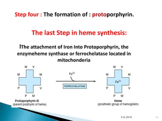
5/6/2018 1
HEMOGLOBIN METABOLISM
Are biconcave discs , with a diameter of
about 7 microns .
 RBCs live for about 120 days in peripheral
circulation, during which time they traverse
about 160 km .
 In a 70 kg person , there will be about
25*1012 RBCs & 750 g Hb.
Mature RBC is non-nucleated,
have no mitochonderia and no
TCA cycle enzymes.
The glycolytic pathway is active which
provides energy and 2,3-
bisphosphoglycerate (2,3-BPG) .
The HMP shunt pathway provides the NADPH
RBC formation in
the bone marrow
requires : amino
acids , iron,
copper, folic acid
, vit B12 , vit C ,
pyridoxal
phosphate , &
pantothenic acid.
Heme is a derivative
of the porphyrin.
Porphyrines are
cyclic compounds
formed by fusion of
4 pyrrol rings
linked by methylene
bridges ( ==CH—)
Hemoglobin is a
conjugated protein
having heme as the
prosthetic group and
the protein, the
globin.
It is a tetrameric
protein with 4
subunits, each
subunit having a
prosthetic heme
group and the globin
polypeptide.
The polypeptide chains are usually 2 alpha &
2 beta chains.
Hb has a MWt of about 67,000 D.
Each gram of Hb contains 3,4 mg of Iron.
Heme present in :
Hemoglobin, myoglobin, cytochromes,
peroxidase, catalase, nitric oxide synthase.
Heme is produced by the combination of iron
with a porphyrin ring .
* Normal level of Hb in blood in males 14—16 g/dl
&in females 13—15 g/dl .
* Hb is globular in shape .
* The adult Hb ( HbA ) has 2 alpha & 2 beta chains
* The fetal Hb ( HbF ) is made up of 2 alpha & 2
gamma chains
* HbA2 is made of 2 alpha & 2 delta chains ..
* Normal adult blood contains 97% HbA , 2% HbA2
& about 1% HbF
* Alpha chain is on chromosome 16 while the
beta, gamma and delta chains are on
chromosome 11 .
* Each alpha chain has 141 amino acids .
* the beta, gamma & delta have 146 amino
acids .
Step one:
The formation of : α-aminolevulinate (ALA) :
From : SUCCINYL-COA + GLYCINE
succinyl-CoA, is derived from citric acid cycle.
This step occurs in the mitochondria.
Step two:
two molecules of ALA are condensed to form:
PORPHO-BILINOGEN (PBG).
This step occurs in the In the cytosol.
HEME SYNTHESIS
5/6/2018 11
Step three:
THE FORMATION OF :
the tetrapyrrole: uro-
porphyrinogen
By condensation of four molecules
of PBG
This step occurs in the In the
cytosol.
HEME SYNTHESIS
5/6/2018 12
lThe attachment of Iron Into Protoporphyrin, the
enzymeheme synthase or ferrochelatase located in
mitochonderia
The last Step in heme synthesis:
5/6/2018 13
Step four : The formation of : protoporphyrin.
1- ALA synthase ( mitochonderial & rate
limiting )
2-ALA dehydratase ( cytoplasmic , contain Zn &
inhibited by lead)
3-PBG-deaminase & UPG-III co-synthase
4-Uroporphyrinogen decarboxylase
mitochonderial))5-Copro porphyrinogen
oxidase
mitochonderial ))6-Protoporphyrinogen
oxidase
7-Heme synthase or Ferrochelatase
*When the ferrous iron ( Fe+2) in heme gets
oxidized to ferric ( Fe+3) form , Hematin is
formed, which loses the property of carrying
the oxygen.
Heme is red in color, but hematin is dark
brown.
** ALA synthase is regulated by repression
mechanism. Heme inhibits the synthesis of
ALA synthase by acting as a co-repressor.
**ALA synthase is also allosterically inhibited by
hematin .
When there is excess of free heme, the Fe+2 is
oxidized to Fe+3 ( ferric) , thus forming
Hematin.
** Drugs like barbiturates induce heme synthesis.
** high cellular concentration of glucose prevents
induction of ALA synthase. This is the basis of
adminstration of glucose to relieve the acute
attach of porphyrias.

Formation of Bilirubin
1-The end product of heme catabolism are bile
pigments. Bilirubin has no function in the body
and is excreated through bile.
2- From hemoglobin, the globin chains are
separated , they are hydrolyzed and the amino
acids are channelled into the body amino acid
pool. The iron librated from the heme is
reutilized .
3-The porphyrin ring is broken down in
reticuloendothelial ( RE) cells of liver, spleen &
bone marrow to bile pigments , mainly bilirubin
4- Heme is degraded primarily by microsomal
enzyme ; heme oxygenase .
5-The ferrous ( Fe+2) librated is oxidized to
Ferric ( Fe+3) and taken up by transferrin .
6- The linear tetrapyrrol formed is bilivedrin ,
which is green in color. In mammals it is
further reduced to bilirubin , a red-yellow
pigment.
Hemoglobin metabolism

Hemoglobin metabolism

  • 1.
  • 2.
    Are biconcave discs, with a diameter of about 7 microns .  RBCs live for about 120 days in peripheral circulation, during which time they traverse about 160 km .  In a 70 kg person , there will be about 25*1012 RBCs & 750 g Hb.
  • 3.
    Mature RBC isnon-nucleated, have no mitochonderia and no TCA cycle enzymes. The glycolytic pathway is active which provides energy and 2,3- bisphosphoglycerate (2,3-BPG) . The HMP shunt pathway provides the NADPH
  • 4.
    RBC formation in thebone marrow requires : amino acids , iron, copper, folic acid , vit B12 , vit C , pyridoxal phosphate , & pantothenic acid.
  • 5.
    Heme is aderivative of the porphyrin. Porphyrines are cyclic compounds formed by fusion of 4 pyrrol rings linked by methylene bridges ( ==CH—)
  • 6.
    Hemoglobin is a conjugatedprotein having heme as the prosthetic group and the protein, the globin. It is a tetrameric protein with 4 subunits, each subunit having a prosthetic heme group and the globin polypeptide.
  • 7.
    The polypeptide chainsare usually 2 alpha & 2 beta chains. Hb has a MWt of about 67,000 D. Each gram of Hb contains 3,4 mg of Iron. Heme present in : Hemoglobin, myoglobin, cytochromes, peroxidase, catalase, nitric oxide synthase. Heme is produced by the combination of iron with a porphyrin ring .
  • 9.
    * Normal levelof Hb in blood in males 14—16 g/dl &in females 13—15 g/dl . * Hb is globular in shape . * The adult Hb ( HbA ) has 2 alpha & 2 beta chains * The fetal Hb ( HbF ) is made up of 2 alpha & 2 gamma chains * HbA2 is made of 2 alpha & 2 delta chains .. * Normal adult blood contains 97% HbA , 2% HbA2 & about 1% HbF
  • 10.
    * Alpha chainis on chromosome 16 while the beta, gamma and delta chains are on chromosome 11 . * Each alpha chain has 141 amino acids . * the beta, gamma & delta have 146 amino acids .
  • 11.
    Step one: The formationof : α-aminolevulinate (ALA) : From : SUCCINYL-COA + GLYCINE succinyl-CoA, is derived from citric acid cycle. This step occurs in the mitochondria. Step two: two molecules of ALA are condensed to form: PORPHO-BILINOGEN (PBG). This step occurs in the In the cytosol. HEME SYNTHESIS 5/6/2018 11
  • 12.
    Step three: THE FORMATIONOF : the tetrapyrrole: uro- porphyrinogen By condensation of four molecules of PBG This step occurs in the In the cytosol. HEME SYNTHESIS 5/6/2018 12
  • 13.
    lThe attachment ofIron Into Protoporphyrin, the enzymeheme synthase or ferrochelatase located in mitochonderia The last Step in heme synthesis: 5/6/2018 13 Step four : The formation of : protoporphyrin.
  • 14.
    1- ALA synthase( mitochonderial & rate limiting ) 2-ALA dehydratase ( cytoplasmic , contain Zn & inhibited by lead) 3-PBG-deaminase & UPG-III co-synthase 4-Uroporphyrinogen decarboxylase mitochonderial))5-Copro porphyrinogen oxidase mitochonderial ))6-Protoporphyrinogen oxidase 7-Heme synthase or Ferrochelatase
  • 17.
    *When the ferrousiron ( Fe+2) in heme gets oxidized to ferric ( Fe+3) form , Hematin is formed, which loses the property of carrying the oxygen. Heme is red in color, but hematin is dark brown. ** ALA synthase is regulated by repression mechanism. Heme inhibits the synthesis of ALA synthase by acting as a co-repressor.
  • 18.
    **ALA synthase isalso allosterically inhibited by hematin . When there is excess of free heme, the Fe+2 is oxidized to Fe+3 ( ferric) , thus forming Hematin. ** Drugs like barbiturates induce heme synthesis. ** high cellular concentration of glucose prevents induction of ALA synthase. This is the basis of adminstration of glucose to relieve the acute attach of porphyrias. 
  • 19.
    Formation of Bilirubin 1-Theend product of heme catabolism are bile pigments. Bilirubin has no function in the body and is excreated through bile. 2- From hemoglobin, the globin chains are separated , they are hydrolyzed and the amino acids are channelled into the body amino acid pool. The iron librated from the heme is reutilized . 3-The porphyrin ring is broken down in reticuloendothelial ( RE) cells of liver, spleen & bone marrow to bile pigments , mainly bilirubin
  • 20.
    4- Heme isdegraded primarily by microsomal enzyme ; heme oxygenase . 5-The ferrous ( Fe+2) librated is oxidized to Ferric ( Fe+3) and taken up by transferrin . 6- The linear tetrapyrrol formed is bilivedrin , which is green in color. In mammals it is further reduced to bilirubin , a red-yellow pigment.