Downloaded 271 times


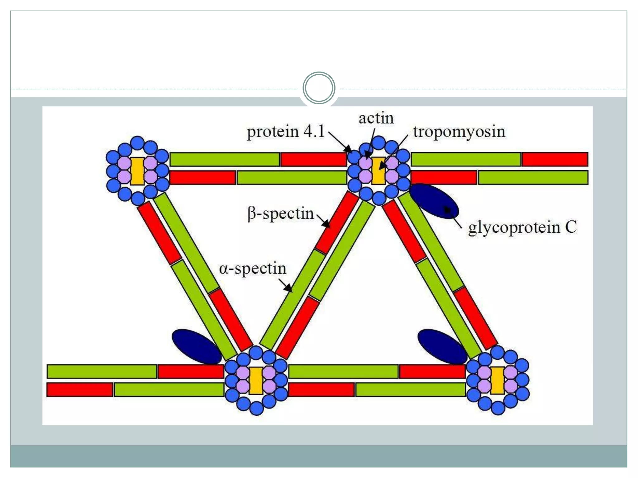


The document discusses the structure and metabolism of red blood cells (RBCs), highlighting their lifespan, membrane composition, and key proteins necessary for functionality and stability. It details the red cell's metabolic pathways, primarily focusing on anaerobic glycolysis as the main source of ATP, along with ancillary pathways like the pentose phosphate pathway and the Leubering-Rapoport shunt. RBCs rely on a complex cytoskeletal structure and membrane proteins to maintain shape, deformability, and functionality, ensuring effective oxygen transport.


