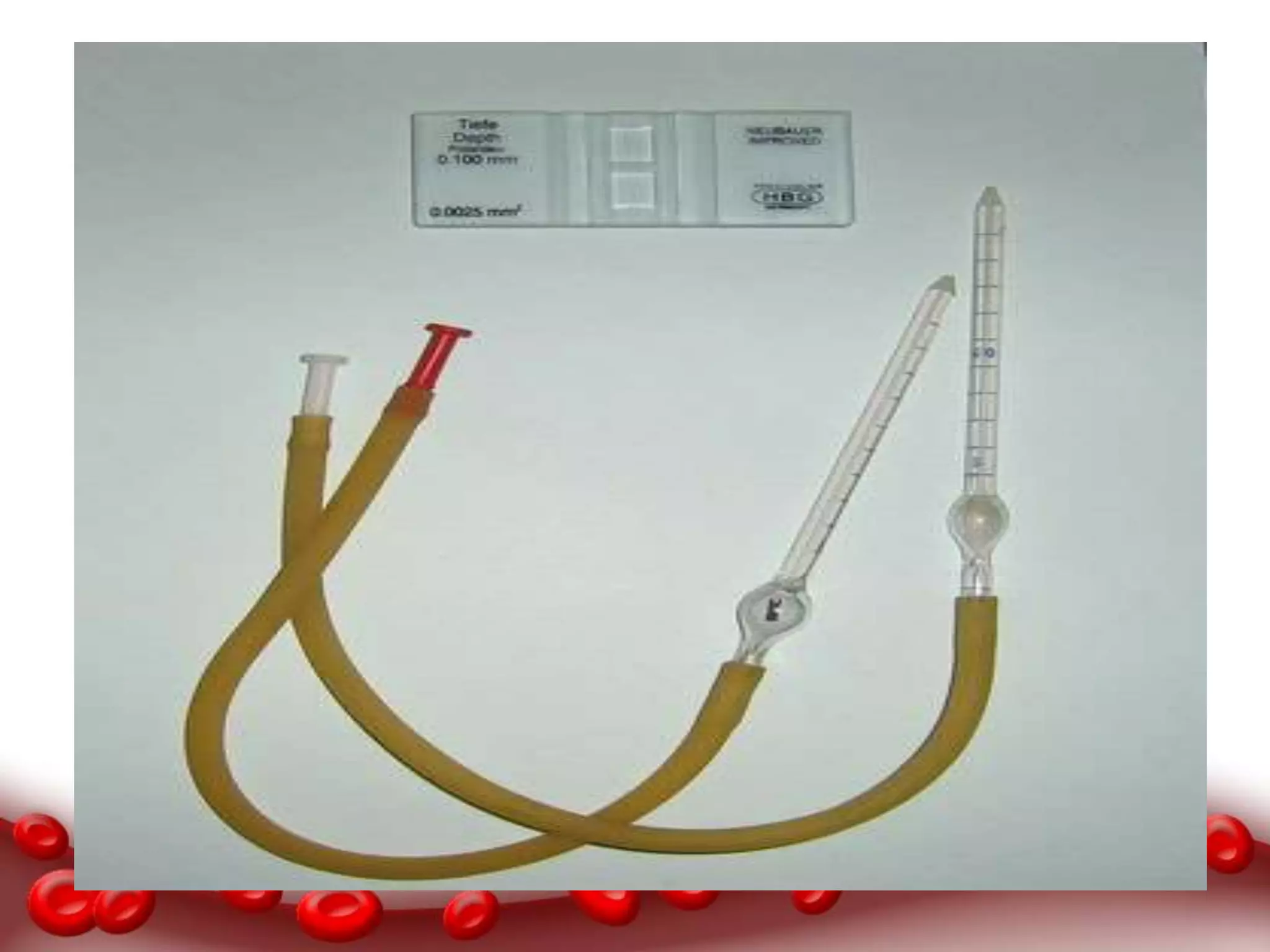
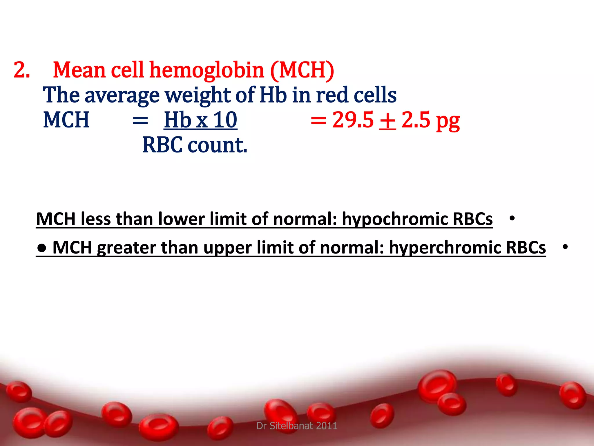
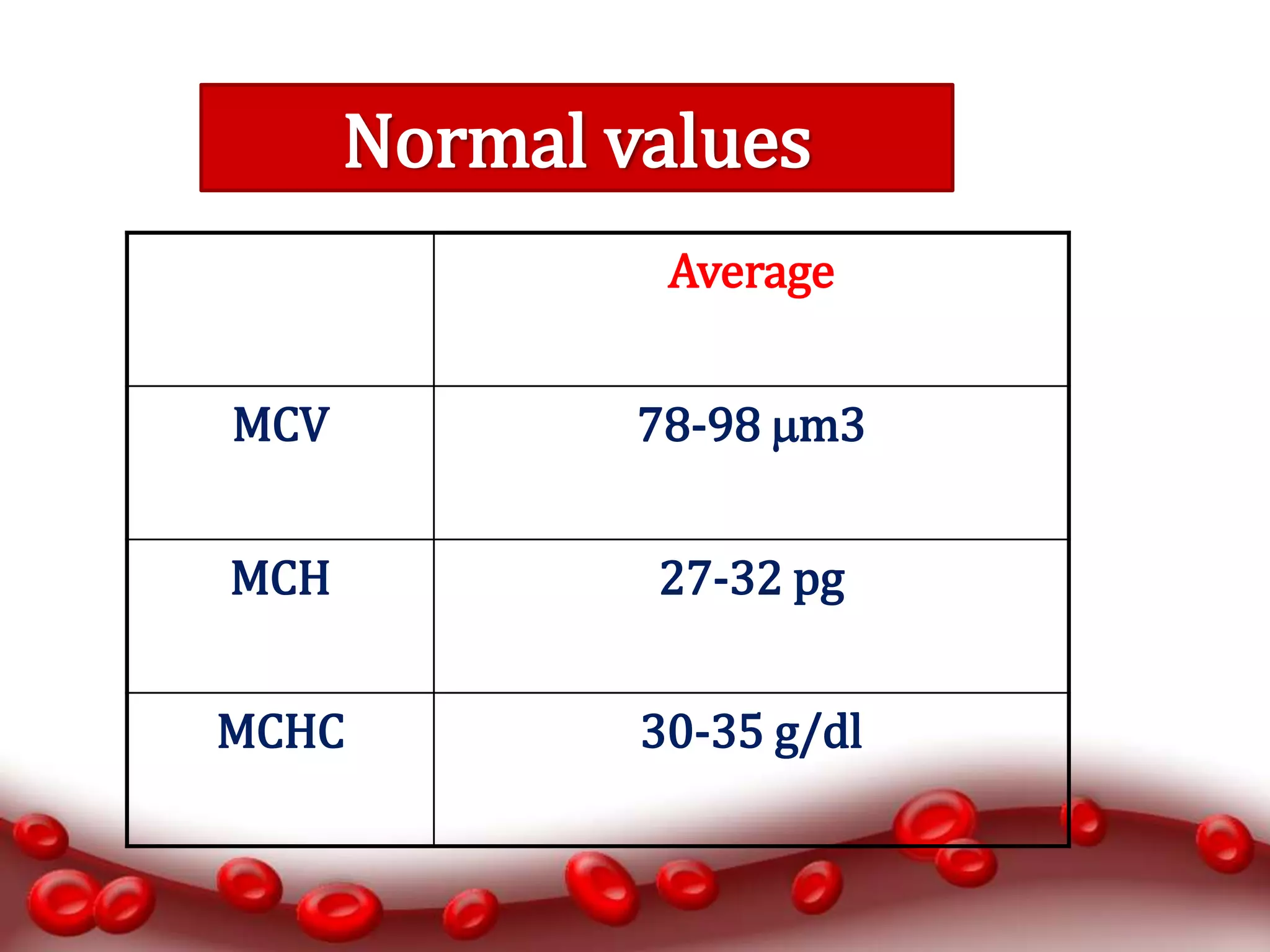
BLOOD PRACTICAL
CBC
Objectives (CBC)
At the end of this lesson the students should be able
to:
1. Recognize the method used to measure the
different hematological values, and compare it
with the normal values.
2. Do the calculation of indices, their normal values
and their importance in diagnosis of different
types of anemia.
Aims of the Practical
1. Counting Red blood cells.
2. Counting White blood cells.
3. Determination of hemoglobin concentration.
4. Determination of packed cell volume (PCV)
hematocrit.
5. Calculation of red blood cell indices.
Complete Blood Count (CBC)
It is a test panel requested by a doctor or other medical
professional that gives information about the cells in a
patient's blood.
Complete Blood Count (cbc)
RBC and WBC count
Manual
•Hemocytometer
Automated
•Coulter Counter
Hemocytometer
It includes:
1. Neubauer’s slide
2. Cover slip
3. Thoma pipet
a) RBC pipette
b) WBC pipette
IMPROVED NEUBAUER’S SLIDE
** It is the name given to a thick glass slide.
In the centre of the slide, there is an
H- shaped groove. On the two sides of the
central horizontal bar, there are scales for
counting the blood cells.
** The depth of the scales is 1/10mm or
0.1mm.
The numbers
written on the chamber mean that the space between the
chamber and the cover slip is 0.100 mm and
that the smallest square on the grid has an area of 0.0025 mm2.
Clean
**The numbers
written on the chamber mean that the space between the
chamber and the cover slip is 0.100 mm and
that the smallest square on the grid has an area of 0.0025 mm2.
** Each scale is 3mm wide
and 3mm long.
** The whole scale is
divided into 9 big
squares.
** Each square is 1mm
long and 1mm wide.
Dr Sitelbanat 2011
Differences Between RBC AND WBC
Pipette.
RBC pipette WBC pipette
1) It has a red bead It has a white
bead
2) It has graduations
upto mark 101
It has graduations
upto mark 11
3) Size of bulb is
larger
Size of bulb is
smaller
Thoma Pipet
• RBC
– Aspirate to 0.5 or 1.0
– Dilute to 101
– DF = 200 or 100
• WBC
– Aspirate to 0.5 or 1.0
– Dilute to 11
– DF = 20 or 10
• Discard volume of one (1) in
the stem
Thoma Pipet
RBC PIPETTE
WBC PIPETTE
Coulter Counter
Coulter Counter Principle
• It count and measure the size of the cells by
detecting and measuring electrical resistance when
a liquid pass through aperture.
• While passing the aperture, the cells impedes the
current and causes a measurable pulse.
• Number of pulses --> number of particles.
• Height of pulses --> volume of particles.
Materials and methods
• Coulter analyzer
• Diluent reagents
• Lytic reagent
• Calibrator kit
• EDTA anticoagulant blood
Diluent –Reagent
Is an isotonic electrolyte solution that:
- Dilute the whole blood sample
- Stabilize cell membrane for accurate counting and size
- Conduct aperture current
- Rinse instrument components between analysis
- Prevent duplicate cell counts
Lytic reagent
Lysis RBCs for WBCs count and hemoglobin
measurement
Calibrator Kit
Is an alternative to the whole blood reference method for
calibration
RBC, WBC cell count & HB
• 5ml of venous blood will be drawn in EDTA
anticoagulant tube.
• Diluted by the reagent I and used to count RBC.
• Lysing RBC using reagent II and used for counting
WBC and Hb.
Male Female Average
RBC 4.5-6.5
x106/l
3.8-5.8
x106/l
4.7–6.5
x106/l
WBC 4 – 11 x103
/l
4 – 11 x103
/l
4 – 11 x103
/l
HB 13-18 g/dl 11.5-16.5
g/dl
13 –18 g/dl
Platelet 150-
400x103 /l
150-
400x103 /l
150-
400x103 /l
Normal values
Clinical terms
1.  RBC = aneamia
2.  RBC = polycythemia
3.  WBC = leucopenia
4.  WBC = leucocytosis
5.  Platelets = thrombocytopenia
6.  Platelets = thrombocytosis
Clinical applications
Low numbers of RBCs may indicate:
• Blood loss:
– Anemia (various types).
– Hemorrhage.
• Bone marrow failure (for example, from radiation,
toxin, fibrosis, tumor).
• Erythropoietin deficiency (secondary to renal
disease).
• Hemolysis (RBC destruction).
High numbers of RBCs may indicate:
• Low oxygen tension in the blood
– Congenital heart disease
– Pulmonary fibrosis
• Polycythemia vera.
• Dehydration (such as from severe diarrhea).
• Renal (kidney) disease with high erythropoietin
production.
High numbers of WBCs (Leucocytosis) may indicate:
• Infectious diseases.
• Inflammatory disease (such as rheumatoid arthritis or
allergy).
• Leukemia.
• Severe emotional or physical stress.
• Tissue damage (burns).
Low numbers of WBCs (leukopenia) may indicate:
• Bone marrow failure (for example, due to
infection, tumor or fibrosis).
• Presence of cytotoxic substance.
• Autoimmune/collagen-vascular diseases (such as
lupus erythematosus).
• Disease of the liver or spleen.
• Radiation exposure.
Dr Sitelbanat 2011
Dr Sitelbanat 2011
Platelet count
Thrombocytosis :
- Chronic myeloid leukemia.
Thrombocytopenia :
- Aplastic anemia.
- Chemotherapy.
Packed Cell Volume (PCV)
Hematocrit
The ratio of packed blood cells volume to plasma.
Procedure
• Capillary blood obtained from pricking finger tip
after cleaning it with alcohol
• Fill a heparinized capillary tube, then seal one end by
plasticine.
• Centrifuge for 5 minutes to packed the cells at one
end of the tube leaving a clear plasma on top.
• Use the hematocrit reader to find the packed cell
volume.
Hematocrit Reader
Male Female Average
PCV % 40-54 35-47 35-54
Normal value PCV
Clinical applications
High hematocrit may indicate:
• Dehydration
– Burns
– Diarrhea
• Polycythemia Vera.
• Low oxygen tension (smoking, congenital heart
disease, living at high altitudes).
Low hematocrit may indicate:
• Anemia (various types).
• Blood loss (hemorrhage).
• Bone marrow failure (for example, due to radiation,
toxin, fibrosis, tumor).
• Hemolysis (RBC destruction) related to transfusion
reaction.
• Leukemia.
The calculation of Red Blood Indices
1. Mean cell volume (MCV)
– The average volume of red blood cell
– MCV = PCV x 10 = 88 + 10 m3 (fl)
RBC count
if the MCV above normal then RBCs is called macrocytes
RBCs.
● if the MCV less than normal , RBCs is called microcytic
RBCs.
•MCH less than lower limit of normal: hypochromic RBCs
•● MCH greater than upper limit of normal: hyperchromic RBCs
Dr Sitelbanat 2011
2. Mean cell hemoglobin (MCH)
The average weight of Hb in red cells
MCH = Hb x 10 = 29.5 + 2.5 pg
RBC count.
3. Mean cell Hb concentration (MCHC)
– Concentration of Hb per 100 ml of RBC
– MCHC = Hb x 100 = 33 + 3 g/dl
PCV
Average
MCV 78-98 m3
MCH 27-32 pg
MCHC 30-35 g/dl
Normal values
Thank you

blood practical CBC

  • 1.
  • 2.
    Objectives (CBC) At theend of this lesson the students should be able to: 1. Recognize the method used to measure the different hematological values, and compare it with the normal values. 2. Do the calculation of indices, their normal values and their importance in diagnosis of different types of anemia.
  • 3.
    Aims of thePractical 1. Counting Red blood cells. 2. Counting White blood cells. 3. Determination of hemoglobin concentration. 4. Determination of packed cell volume (PCV) hematocrit. 5. Calculation of red blood cell indices.
  • 4.
    Complete Blood Count(CBC) It is a test panel requested by a doctor or other medical professional that gives information about the cells in a patient's blood.
  • 5.
  • 6.
    RBC and WBCcount Manual •Hemocytometer Automated •Coulter Counter
  • 7.
    Hemocytometer It includes: 1. Neubauer’sslide 2. Cover slip 3. Thoma pipet a) RBC pipette b) WBC pipette
  • 8.
    IMPROVED NEUBAUER’S SLIDE **It is the name given to a thick glass slide. In the centre of the slide, there is an H- shaped groove. On the two sides of the central horizontal bar, there are scales for counting the blood cells. ** The depth of the scales is 1/10mm or 0.1mm.
  • 9.
    The numbers written onthe chamber mean that the space between the chamber and the cover slip is 0.100 mm and that the smallest square on the grid has an area of 0.0025 mm2. Clean **The numbers written on the chamber mean that the space between the chamber and the cover slip is 0.100 mm and that the smallest square on the grid has an area of 0.0025 mm2.
  • 10.
    ** Each scaleis 3mm wide and 3mm long. ** The whole scale is divided into 9 big squares. ** Each square is 1mm long and 1mm wide.
  • 11.
  • 12.
    Differences Between RBCAND WBC Pipette. RBC pipette WBC pipette 1) It has a red bead It has a white bead 2) It has graduations upto mark 101 It has graduations upto mark 11 3) Size of bulb is larger Size of bulb is smaller
  • 13.
    Thoma Pipet • RBC –Aspirate to 0.5 or 1.0 – Dilute to 101 – DF = 200 or 100 • WBC – Aspirate to 0.5 or 1.0 – Dilute to 11 – DF = 20 or 10 • Discard volume of one (1) in the stem Thoma Pipet
  • 14.
  • 16.
  • 18.
    Coulter Counter Principle •It count and measure the size of the cells by detecting and measuring electrical resistance when a liquid pass through aperture. • While passing the aperture, the cells impedes the current and causes a measurable pulse. • Number of pulses --> number of particles. • Height of pulses --> volume of particles.
  • 19.
    Materials and methods •Coulter analyzer • Diluent reagents • Lytic reagent • Calibrator kit • EDTA anticoagulant blood
  • 20.
    Diluent –Reagent Is anisotonic electrolyte solution that: - Dilute the whole blood sample - Stabilize cell membrane for accurate counting and size - Conduct aperture current - Rinse instrument components between analysis - Prevent duplicate cell counts Lytic reagent Lysis RBCs for WBCs count and hemoglobin measurement
  • 21.
    Calibrator Kit Is analternative to the whole blood reference method for calibration
  • 22.
    RBC, WBC cellcount & HB • 5ml of venous blood will be drawn in EDTA anticoagulant tube. • Diluted by the reagent I and used to count RBC. • Lysing RBC using reagent II and used for counting WBC and Hb.
  • 23.
    Male Female Average RBC4.5-6.5 x10⁶/l 3.8-5.8 x10⁶/l 4.7–6.5 x10⁶/l WBC 4 – 11 x103 /l 4 – 11 x103 /l 4 – 11 x103 /l HB 13-18 g/dl 11.5-16.5 g/dl 13 –18 g/dl Platelet 150- 400x103 /l 150- 400x103 /l 150- 400x103 /l Normal values
  • 24.
    Clinical terms 1. RBC = aneamia 2.  RBC = polycythemia 3.  WBC = leucopenia 4.  WBC = leucocytosis 5.  Platelets = thrombocytopenia 6.  Platelets = thrombocytosis
  • 25.
    Clinical applications Low numbersof RBCs may indicate: • Blood loss: – Anemia (various types). – Hemorrhage. • Bone marrow failure (for example, from radiation, toxin, fibrosis, tumor). • Erythropoietin deficiency (secondary to renal disease). • Hemolysis (RBC destruction).
  • 26.
    High numbers ofRBCs may indicate: • Low oxygen tension in the blood – Congenital heart disease – Pulmonary fibrosis • Polycythemia vera. • Dehydration (such as from severe diarrhea). • Renal (kidney) disease with high erythropoietin production.
  • 27.
    High numbers ofWBCs (Leucocytosis) may indicate: • Infectious diseases. • Inflammatory disease (such as rheumatoid arthritis or allergy). • Leukemia. • Severe emotional or physical stress. • Tissue damage (burns).
  • 28.
    Low numbers ofWBCs (leukopenia) may indicate: • Bone marrow failure (for example, due to infection, tumor or fibrosis). • Presence of cytotoxic substance. • Autoimmune/collagen-vascular diseases (such as lupus erythematosus). • Disease of the liver or spleen. • Radiation exposure.
  • 29.
  • 30.
  • 31.
    Platelet count Thrombocytosis : -Chronic myeloid leukemia. Thrombocytopenia : - Aplastic anemia. - Chemotherapy.
  • 32.
    Packed Cell Volume(PCV) Hematocrit The ratio of packed blood cells volume to plasma.
  • 33.
    Procedure • Capillary bloodobtained from pricking finger tip after cleaning it with alcohol • Fill a heparinized capillary tube, then seal one end by plasticine. • Centrifuge for 5 minutes to packed the cells at one end of the tube leaving a clear plasma on top. • Use the hematocrit reader to find the packed cell volume.
  • 34.
  • 35.
    Male Female Average PCV% 40-54 35-47 35-54 Normal value PCV
  • 36.
    Clinical applications High hematocritmay indicate: • Dehydration – Burns – Diarrhea • Polycythemia Vera. • Low oxygen tension (smoking, congenital heart disease, living at high altitudes).
  • 37.
    Low hematocrit mayindicate: • Anemia (various types). • Blood loss (hemorrhage). • Bone marrow failure (for example, due to radiation, toxin, fibrosis, tumor). • Hemolysis (RBC destruction) related to transfusion reaction. • Leukemia.
  • 38.
    The calculation ofRed Blood Indices 1. Mean cell volume (MCV) – The average volume of red blood cell – MCV = PCV x 10 = 88 + 10 m3 (fl) RBC count if the MCV above normal then RBCs is called macrocytes RBCs. ● if the MCV less than normal , RBCs is called microcytic RBCs.
  • 39.
    •MCH less thanlower limit of normal: hypochromic RBCs •● MCH greater than upper limit of normal: hyperchromic RBCs Dr Sitelbanat 2011 2. Mean cell hemoglobin (MCH) The average weight of Hb in red cells MCH = Hb x 10 = 29.5 + 2.5 pg RBC count.
  • 40.
    3. Mean cellHb concentration (MCHC) – Concentration of Hb per 100 ml of RBC – MCHC = Hb x 100 = 33 + 3 g/dl PCV
  • 41.
    Average MCV 78-98 m3 MCH27-32 pg MCHC 30-35 g/dl Normal values
  • 42.