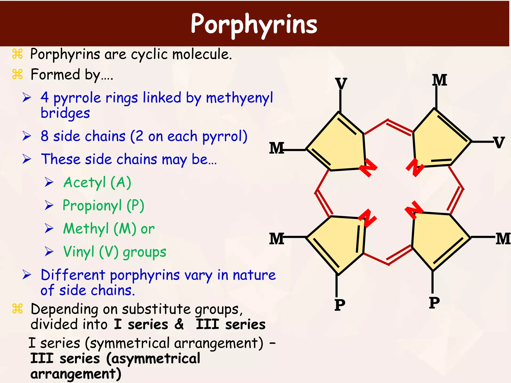
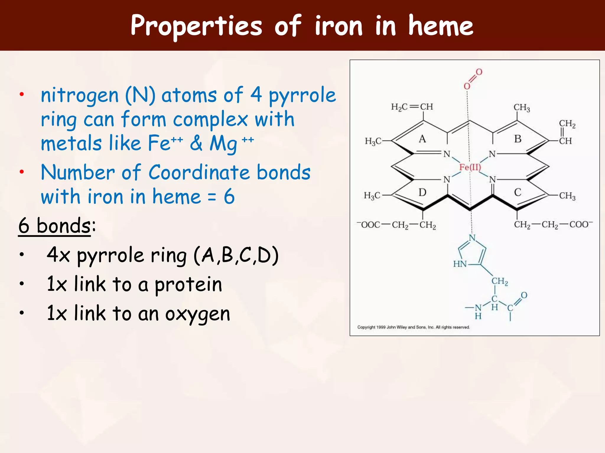
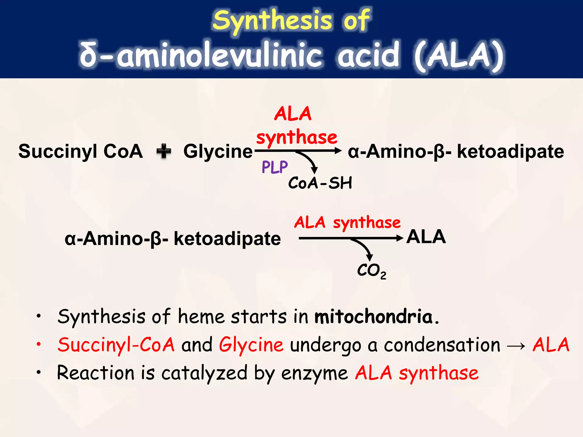
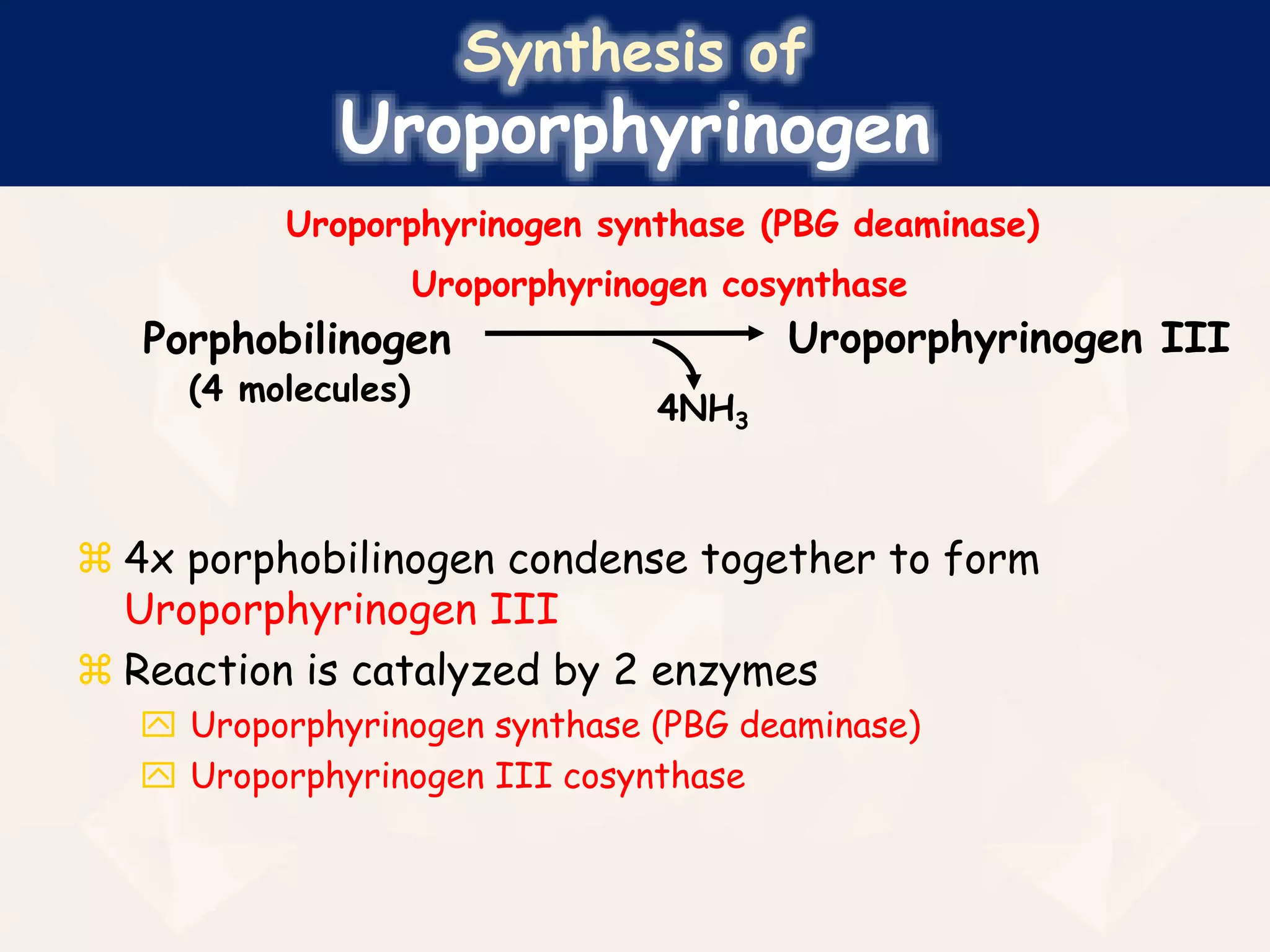
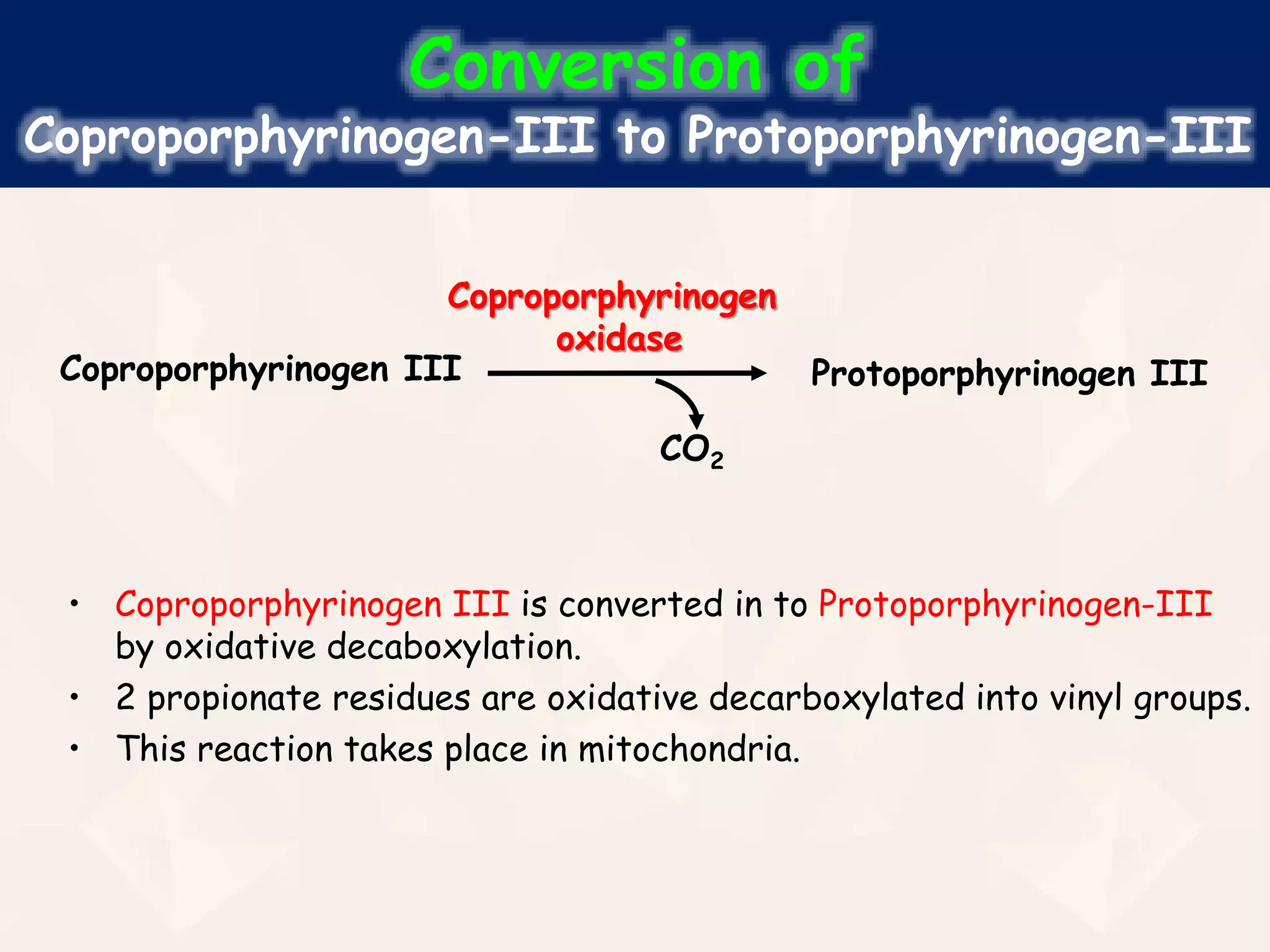
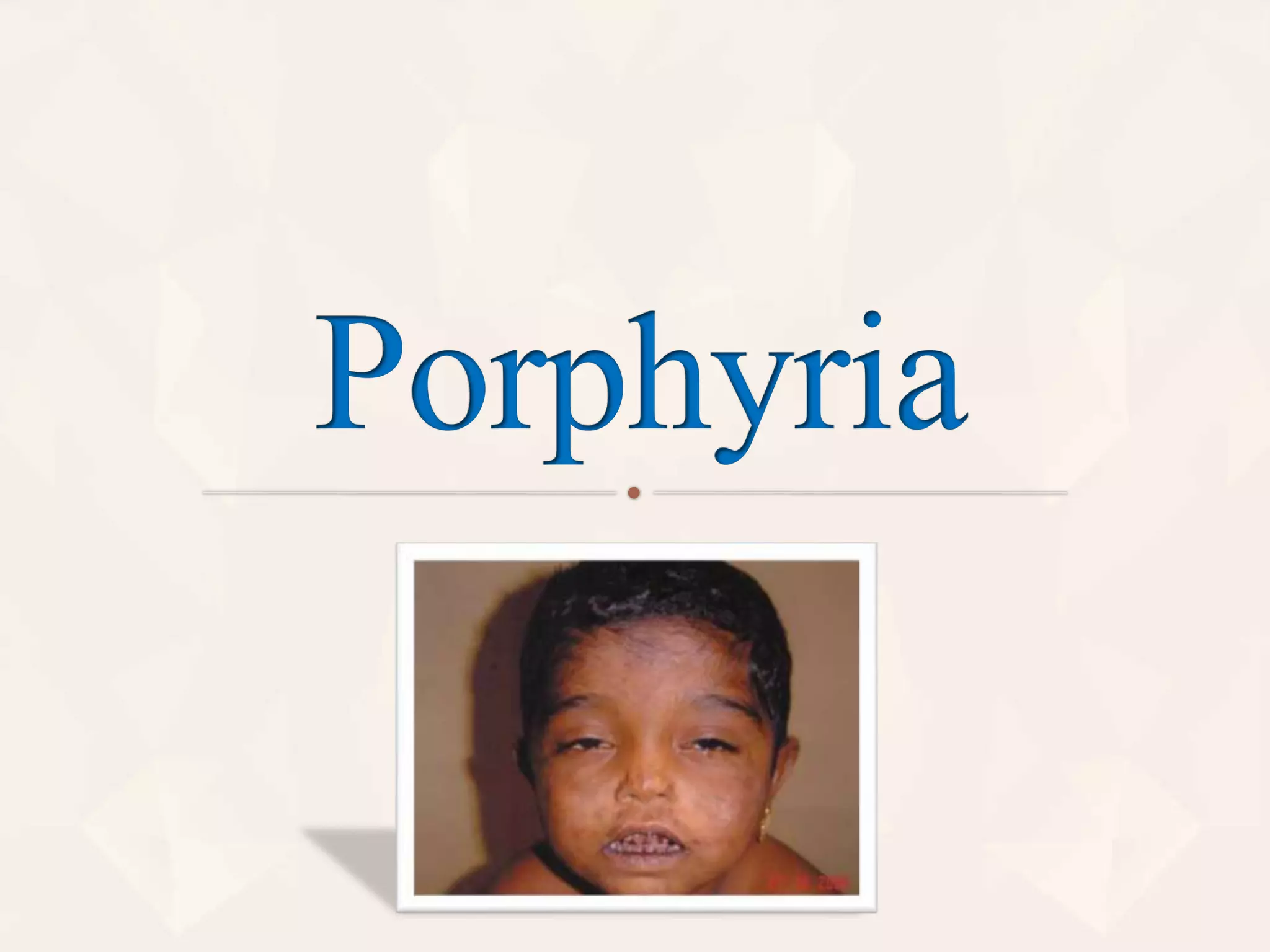
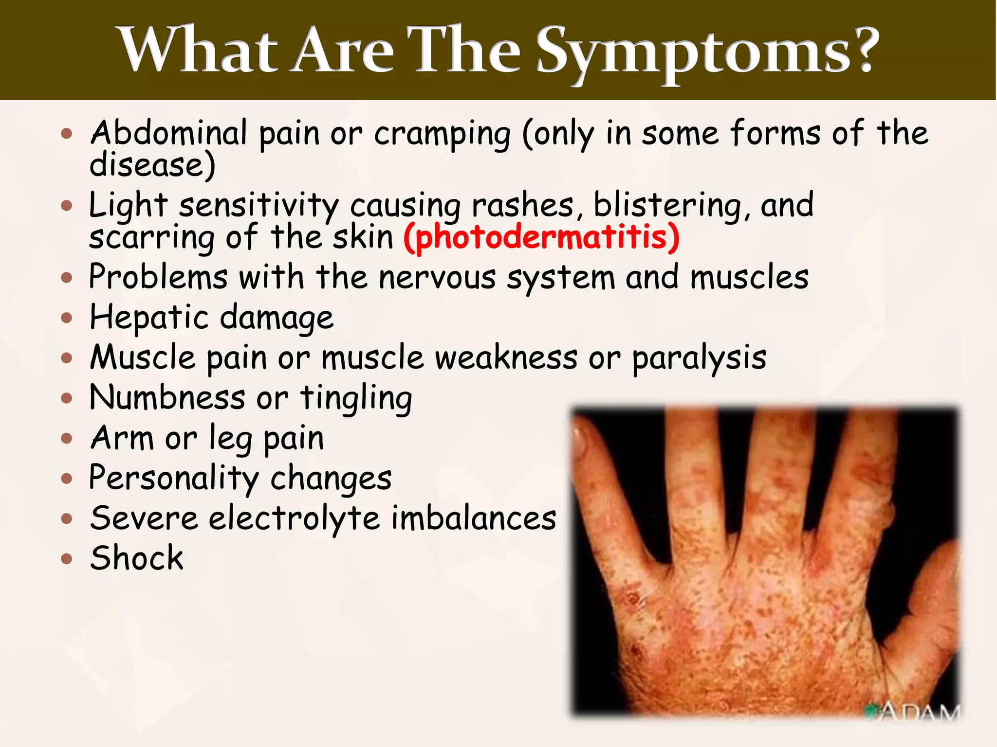
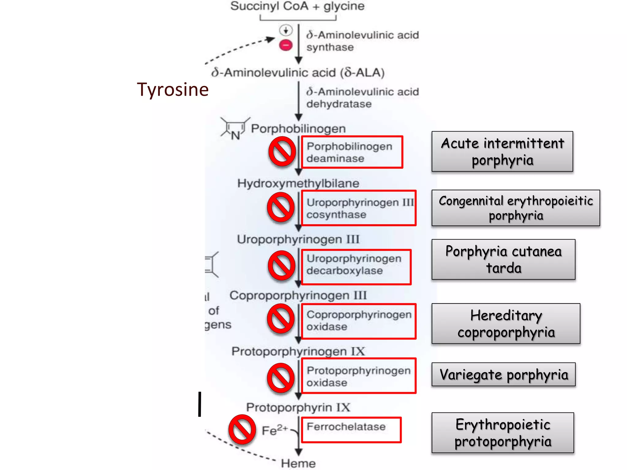
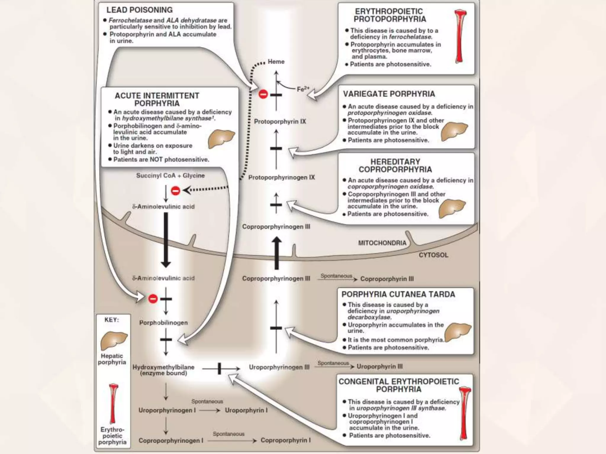
1 of 58
Hemoglobin (Hb)
Is a hemoprotein (Conjugated protein)
Only found in the cytoplasm of erythrocytes (RBC)
Transports O2 & CO2 between lungs and various
tissues
Normal concentration of Hb in the blood:
Adult males 13.5 – 16.5 gm/dl
Adult females 12.0 – 14.8 gm/dl
Structure of hemoglobin
 Hb is a spherical molecule consisting of 4 peptide subunits
(globins) = quartenary structure
 Hb of adults (Hb A) is a tetramer consisting of 2 α & 2 β-
globins → each globin contains 1 heme group with a
central Fe2+ ion (ferrous ion)
Heme structure
(cyclic tetrapyrrole)
Heme is a
metaloporphyrine
It contains:
 Porphyrin
 1 iron cation (Fe2+)
V
M
M M
V
M
PP
Fe++
Porphyrins
 Porphyrins are cyclic molecule.
 Formed by….
 4 pyrrole rings linked by methyenyl
bridges
 8 side chains (2 on each pyrrol)
 These side chains may be…
 Acetyl (A)
 Propionyl (P)
 Methyl (M) or
 Vinyl (V) groups
 Different porphyrins vary in nature
of side chains.
 Depending on substitute groups,
divided into I series & III series
I series (symmetrical arrangement) –
III series (asymmetrical
arrangement)
V
M
M M
V
M
PP
Properties of iron in heme
• nitrogen (N) atoms of 4 pyrrole
ring can form complex with
metals like Fe++ & Mg ++
• Number of Coordinate bonds
with iron in heme = 6
6 bonds:
• 4x pyrrole ring (A,B,C,D)
• 1x link to a protein
• 1x link to an oxygen
In which compounds can we find a heme group?
Hemoproteins
• Hemoglobin (Hb)
• Myoglobin (Mb)
• Cytochromes
• Catalases
(decomposition of 2 H2O2
to 2 H2O and O2)
• Peroxidases
• Tryptophan pyrrolase.
V
M
M M
V
M
PP
Fe++
Heme biosynthesis
 In bone marrow (85% of Hb) & liver (cytochromes)
 Cell location: mitochondria/cytoplasm
 Substrates: succinyl-CoA + glycine
Important intermediates:
-aminolevulinic acid (5-aminolevulinic acid, ALA)
Porphobilinogen (PBG) (Pyrrole derivate)
Uroporphyrinogen III (porphyrinogen – heme
precursor)
protoporphyrin IX (= direct heme precursor)
key regulatory enzyme: ALA synthase
Synthesis of
δ-aminolevulinic acid (ALA)
• Synthesis of heme starts in mitochondria.
• Succinyl-CoA and Glycine undergo a condensation → ALA
• Reaction is catalyzed by enzyme ALA synthase
Succinyl CoA Glycine α-Amino-β- ketoadipate
ALAα-Amino-β- ketoadipate
PLP
ALA
synthase
ALA synthase
CoA-SH
CO2
Synthesis of
Porphobilinogen (PBG)
 ALA leaves the mitochondria → cytoplasm
 2x ALA condense together to form
porphobilinogen
 Reaction is catalyzed by porphobilinogen
synthase (ALA dehydratase)
δ-aminolevulinate Porphobilinogen
(2 molecules)
2H2O
ALA dehydratase
Synthesis of
Uroporphyrinogen
 4x porphobilinogen condense together to form
Uroporphyrinogen III
 Reaction is catalyzed by 2 enzymes
 Uroporphyrinogen synthase (PBG deaminase)
 Uroporphyrinogen III cosynthase
Uroporphyrinogen III
(4 molecules)
4NH3
Uroporphyrinogen synthase (PBG deaminase)
Porphobilinogen
Uroporphyrinogen cosynthase
• Uroporphyrinogen III is converted in to
Coproporphyrinogen III by decarboxylation.
• 4 acetate residues are decarboxylated into methyl groups.
• This reaction takes place in cytosol.
• Coproporphyrinogen III returns to the mitochondria again
Conversion of
Uroporphyrinogen to Coproporphyrinogen III
Coproporphyrinogen III
CO2
Uroporphyrinogen
decarboxylase
Uroporphyrinogen III
• Coproporphyrinogen III is converted in to Protoporphyrinogen-III
by oxidative decaboxylation.
• 2 propionate residues are oxidative decarboxylated into vinyl groups.
• This reaction takes place in mitochondria.
Conversion of
Coproporphyrinogen-III to Protoporphyrinogen-III
Protoporphyrinogen III
CO2
Coproporphyrinogen
oxidase
Coproporphyrinogen III
• Oxidation of protoporphyrinogen-III produces protoporphyrin III
• Enzyme protoporphyrinogen oxidase catalyses this reaction.
Conversion of
Protoporphyrinogen III to Protoporphyrin III
Protoporphyrin-III
Protoporphyrinogen
oxidase
Protoporphyrinogen III
Fe2+ is incorporated into protoporphyrin-III
Reaction is catalyzed by enzyme ferrochelatase/heme
synthase
F o r m a t i o n o f
Heme
Heme
Ferrochelatase
Protoporphyrin-III
Fe++
Pathway
Glycine
4 NH3
Succinyl-CoA
+ PLP
ALA
synthase
δ ALA
δ-Aminolevulinic
acid (ALA)
Porphobilinogen (PBG)
Hydroxymethylbilane
(linear tetrapyrrol)
Uroporphyrinogen-III
Uroporphyrinogen-I
Coproporphyrinogen-III
Protoporphyrinogen-III
Protoporphyrin-III
Heme
H2O
2x
4x
ALA dehydratase
Uroporphyrinogen-I
synthase
Uroporphyrinogen-III
cosynthase Spontaneous
Uroporphyrinogen
decarboxylaseCO2
Coproporphyrinogen
oxidase
Protoporphyrinogen
oxidase
Ferrochelatase
(heme synthase)
ALA synthase is a key regulatory enzyme
it is an allosteric enzyme that is inhibited by an end product - heme
(feedback inhibition)
Regulation occurs at the level of enzyme synthesis.
Increased levels of heme represses the synthesis of enzyme ALA
synthase.
The iron atom itself may regulate the heme synthesis.
Several substances induce the synthesis of hepatic
ALA synthase. e.g.. steroid hormones, ethanol, certain
drugs like barbiturates, etc.
Most of the drugs are metabolized in the liver by
cytochrome P450. so, maximum amount of heme is
utilized for the formation of cyto. P450, which in turn
diminishes the intracellular heme conc.
Regulation of Heme biosynthesis
Regulation of
Heme
biosynthesis
Group of rare genetic disorders
Resulting from deficiency of enzymes in biosynthesis
of heme.
This results in accumulation & increased excretion of
porphyrins or
porphyrin precursors (ALA & PBG).
Porphyria directly affects
Nervous system and
The skin,
but is possible to only affect one of the two.
This disease is often inherited from a parent, but
environmental factors may initiate the disease.
Autosomal dominant trait (with exception, i.e,
congenital erythropoitic)
 Arises from disruption of the production of heme.
 Autosomal dominant pattern inherited form parent
 These gene defects cause one or more of the
enzymes involved in the process of converting
porphyrins to heme to be abnormal.
 In specific environments, your brain triggers for
your body to make more heme, overwhelming the
enzyme and triggering the disease.
 Triggers include: Alcohol, smoking, drugs, and
excessive fasting
 Abdominal pain or cramping (only in some forms of the
disease)
 Light sensitivity causing rashes, blistering, and
scarring of the skin (photodermatitis)
 Problems with the nervous system and muscles
 Hepatic damage
 Muscle pain or muscle weakness or paralysis
 Numbness or tingling
 Arm or leg pain
 Personality changes
 Severe electrolyte imbalances
 Shock
Acute intermittent
porphyria
Congennital erythropoieitic
porphyria
Porphyria cutanea
tarda
Hereditary
coproporphyria
Variegate porphyria
Erythropoietic
protoporphyria
Tyrosine
….

Heme Biosynthesis and Its disorders (Porphyria)

  • 1.
  • 2.
    Hemoglobin (Hb) Is ahemoprotein (Conjugated protein) Only found in the cytoplasm of erythrocytes (RBC) Transports O2 & CO2 between lungs and various tissues Normal concentration of Hb in the blood: Adult males 13.5 – 16.5 gm/dl Adult females 12.0 – 14.8 gm/dl
  • 3.
    Structure of hemoglobin Hb is a spherical molecule consisting of 4 peptide subunits (globins) = quartenary structure  Hb of adults (Hb A) is a tetramer consisting of 2 α & 2 β- globins → each globin contains 1 heme group with a central Fe2+ ion (ferrous ion)
  • 4.
    Heme structure (cyclic tetrapyrrole) Hemeis a metaloporphyrine It contains:  Porphyrin  1 iron cation (Fe2+) V M M M V M PP Fe++
  • 5.
    Porphyrins  Porphyrins arecyclic molecule.  Formed by….  4 pyrrole rings linked by methyenyl bridges  8 side chains (2 on each pyrrol)  These side chains may be…  Acetyl (A)  Propionyl (P)  Methyl (M) or  Vinyl (V) groups  Different porphyrins vary in nature of side chains.  Depending on substitute groups, divided into I series & III series I series (symmetrical arrangement) – III series (asymmetrical arrangement) V M M M V M PP
  • 6.
    Properties of ironin heme • nitrogen (N) atoms of 4 pyrrole ring can form complex with metals like Fe++ & Mg ++ • Number of Coordinate bonds with iron in heme = 6 6 bonds: • 4x pyrrole ring (A,B,C,D) • 1x link to a protein • 1x link to an oxygen
  • 7.
    In which compoundscan we find a heme group? Hemoproteins • Hemoglobin (Hb) • Myoglobin (Mb) • Cytochromes • Catalases (decomposition of 2 H2O2 to 2 H2O and O2) • Peroxidases • Tryptophan pyrrolase. V M M M V M PP Fe++
  • 8.
    Heme biosynthesis  Inbone marrow (85% of Hb) & liver (cytochromes)  Cell location: mitochondria/cytoplasm  Substrates: succinyl-CoA + glycine Important intermediates: -aminolevulinic acid (5-aminolevulinic acid, ALA) Porphobilinogen (PBG) (Pyrrole derivate) Uroporphyrinogen III (porphyrinogen – heme precursor) protoporphyrin IX (= direct heme precursor) key regulatory enzyme: ALA synthase
  • 9.
    Synthesis of δ-aminolevulinic acid(ALA) • Synthesis of heme starts in mitochondria. • Succinyl-CoA and Glycine undergo a condensation → ALA • Reaction is catalyzed by enzyme ALA synthase Succinyl CoA Glycine α-Amino-β- ketoadipate ALAα-Amino-β- ketoadipate PLP ALA synthase ALA synthase CoA-SH CO2
  • 10.
    Synthesis of Porphobilinogen (PBG) ALA leaves the mitochondria → cytoplasm  2x ALA condense together to form porphobilinogen  Reaction is catalyzed by porphobilinogen synthase (ALA dehydratase) δ-aminolevulinate Porphobilinogen (2 molecules) 2H2O ALA dehydratase
  • 11.
    Synthesis of Uroporphyrinogen  4xporphobilinogen condense together to form Uroporphyrinogen III  Reaction is catalyzed by 2 enzymes  Uroporphyrinogen synthase (PBG deaminase)  Uroporphyrinogen III cosynthase Uroporphyrinogen III (4 molecules) 4NH3 Uroporphyrinogen synthase (PBG deaminase) Porphobilinogen Uroporphyrinogen cosynthase
  • 12.
    • Uroporphyrinogen IIIis converted in to Coproporphyrinogen III by decarboxylation. • 4 acetate residues are decarboxylated into methyl groups. • This reaction takes place in cytosol. • Coproporphyrinogen III returns to the mitochondria again Conversion of Uroporphyrinogen to Coproporphyrinogen III Coproporphyrinogen III CO2 Uroporphyrinogen decarboxylase Uroporphyrinogen III
  • 13.
    • Coproporphyrinogen IIIis converted in to Protoporphyrinogen-III by oxidative decaboxylation. • 2 propionate residues are oxidative decarboxylated into vinyl groups. • This reaction takes place in mitochondria. Conversion of Coproporphyrinogen-III to Protoporphyrinogen-III Protoporphyrinogen III CO2 Coproporphyrinogen oxidase Coproporphyrinogen III
  • 14.
    • Oxidation ofprotoporphyrinogen-III produces protoporphyrin III • Enzyme protoporphyrinogen oxidase catalyses this reaction. Conversion of Protoporphyrinogen III to Protoporphyrin III Protoporphyrin-III Protoporphyrinogen oxidase Protoporphyrinogen III
  • 15.
    Fe2+ is incorporatedinto protoporphyrin-III Reaction is catalyzed by enzyme ferrochelatase/heme synthase F o r m a t i o n o f Heme Heme Ferrochelatase Protoporphyrin-III Fe++
  • 16.
    Pathway Glycine 4 NH3 Succinyl-CoA + PLP ALA synthase δALA δ-Aminolevulinic acid (ALA) Porphobilinogen (PBG) Hydroxymethylbilane (linear tetrapyrrol) Uroporphyrinogen-III Uroporphyrinogen-I Coproporphyrinogen-III Protoporphyrinogen-III Protoporphyrin-III Heme H2O 2x 4x ALA dehydratase Uroporphyrinogen-I synthase Uroporphyrinogen-III cosynthase Spontaneous Uroporphyrinogen decarboxylaseCO2 Coproporphyrinogen oxidase Protoporphyrinogen oxidase Ferrochelatase (heme synthase)
  • 17.
    ALA synthase isa key regulatory enzyme it is an allosteric enzyme that is inhibited by an end product - heme (feedback inhibition) Regulation occurs at the level of enzyme synthesis. Increased levels of heme represses the synthesis of enzyme ALA synthase. The iron atom itself may regulate the heme synthesis. Several substances induce the synthesis of hepatic ALA synthase. e.g.. steroid hormones, ethanol, certain drugs like barbiturates, etc. Most of the drugs are metabolized in the liver by cytochrome P450. so, maximum amount of heme is utilized for the formation of cyto. P450, which in turn diminishes the intracellular heme conc. Regulation of Heme biosynthesis
  • 18.
  • 21.
    Group of raregenetic disorders Resulting from deficiency of enzymes in biosynthesis of heme. This results in accumulation & increased excretion of porphyrins or porphyrin precursors (ALA & PBG). Porphyria directly affects Nervous system and The skin, but is possible to only affect one of the two. This disease is often inherited from a parent, but environmental factors may initiate the disease. Autosomal dominant trait (with exception, i.e, congenital erythropoitic)
  • 22.
     Arises fromdisruption of the production of heme.  Autosomal dominant pattern inherited form parent  These gene defects cause one or more of the enzymes involved in the process of converting porphyrins to heme to be abnormal.  In specific environments, your brain triggers for your body to make more heme, overwhelming the enzyme and triggering the disease.  Triggers include: Alcohol, smoking, drugs, and excessive fasting
  • 23.
     Abdominal painor cramping (only in some forms of the disease)  Light sensitivity causing rashes, blistering, and scarring of the skin (photodermatitis)  Problems with the nervous system and muscles  Hepatic damage  Muscle pain or muscle weakness or paralysis  Numbness or tingling  Arm or leg pain  Personality changes  Severe electrolyte imbalances  Shock
  • 24.
    Acute intermittent porphyria Congennital erythropoieitic porphyria Porphyriacutanea tarda Hereditary coproporphyria Variegate porphyria Erythropoietic protoporphyria Tyrosine
  • 26.