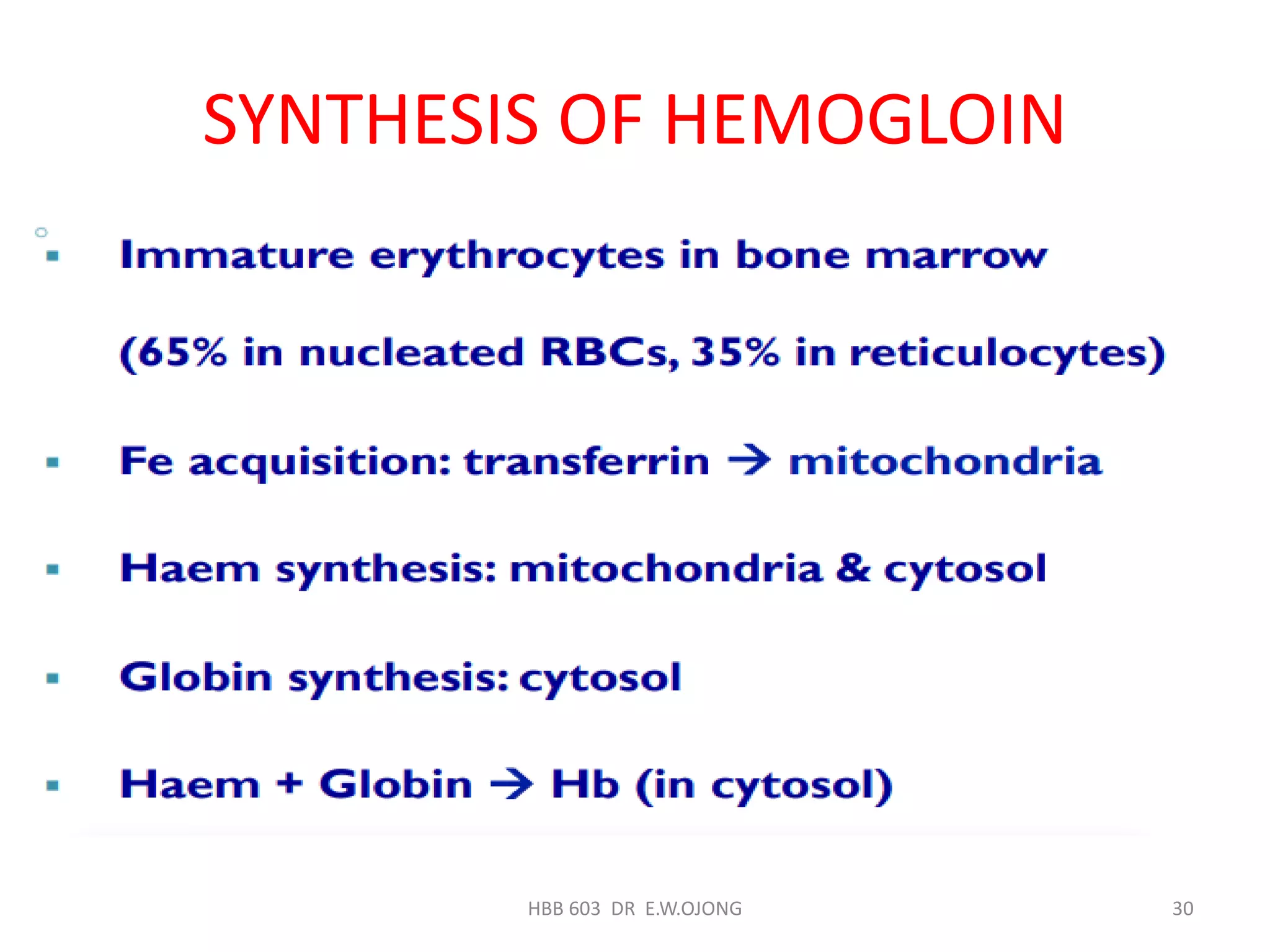
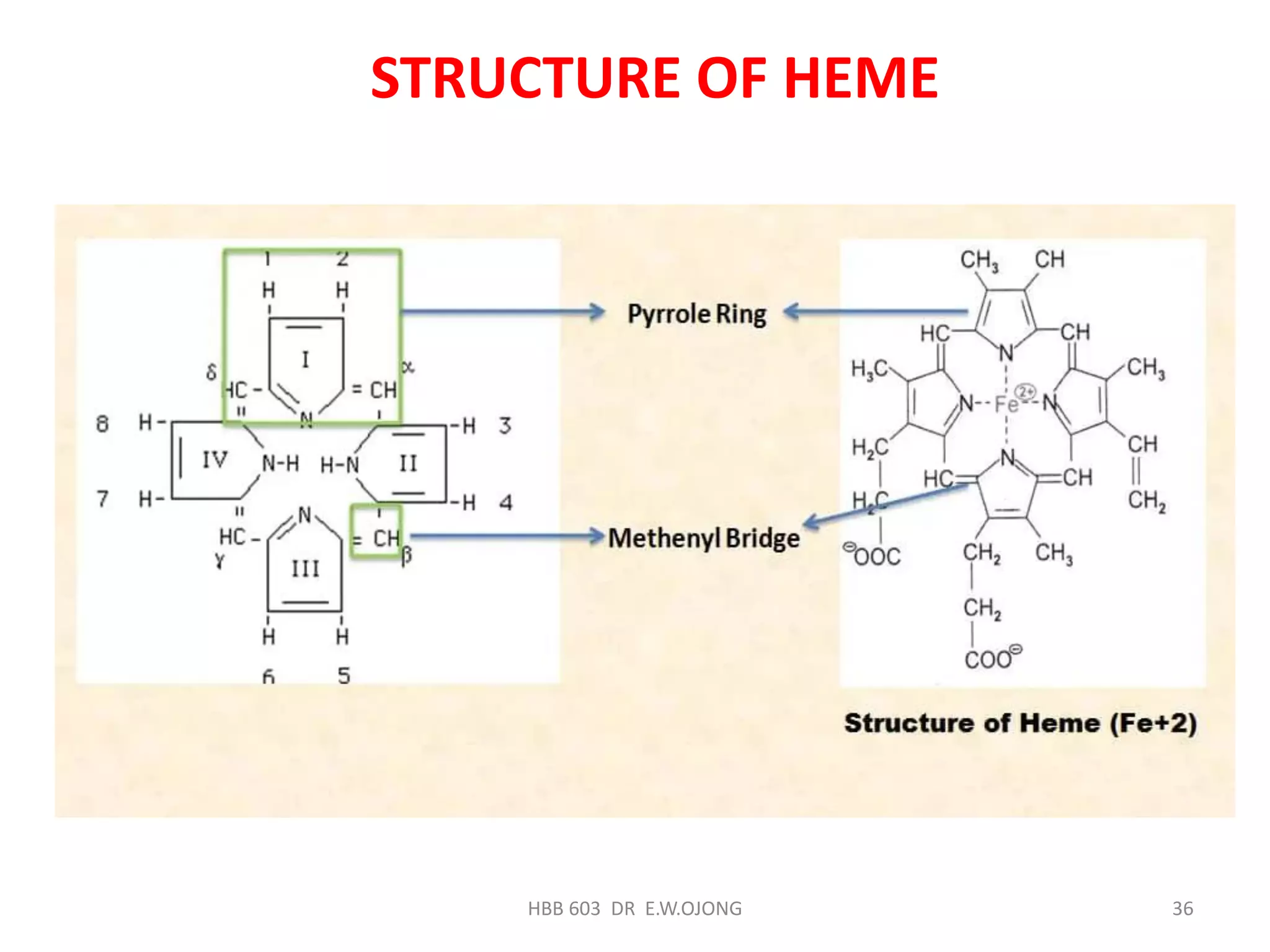
The document discusses hemoglobin synthesis, which consists of two parts: globin synthesis and heme synthesis. Globin is synthesized through transcription and translation of genes on chromosomes 16 and 11. Heme is synthesized through the Shemin cycle, where glycine and succinyl-CoA are condensed to form delta-aminolevulinic acid, which undergoes further modifications to ultimately form protoporphyrin IX. Protoporphyrin then binds iron to form heme. Heme synthesis is regulated by feedback inhibition of delta-aminolevulinic acid synthase, and both heme and globin are required for the formation of functional hemoglobin.
This document provides an overview of red blood cell structure and physiology. It discusses that blood volume is calculated based on weight, and lists the functions of blood. It describes hematopoiesis, the process of blood cell formation, and focuses on erythropoiesis, the development of red blood cells. Key aspects of red blood cell structure are summarized, including the lipid bilayer membrane, cytoskeleton, integral and peripheral proteins. Hemoglobin structure and function in oxygen transport is also covered. The roles of erythropoietin and iron metabolism in red blood cell production are highlighted.
Haemoglobin is a protein in red blood cells that transports oxygen from the lungs to tissues and carbon dioxide from tissues back to the lungs. It is composed of haem and globin subunits. Globin is produced in polyribosomes while haem is produced in mitochondria and the two subunits combine to form functional haemoglobin. Abnormalities in haemoglobin structure or levels can impair oxygen delivery and cause anemia.
GANDHAM RAJEEV-BIOCHEMISTRY IMPORTANT QUESTIONS-RGUHS-2017YESANNA
The document outlines the syllabus for biochemistry exams for MBBS/Phase 1 students at Rajiv Gandhi University of Health Sciences in Bangalore, India. It includes the topics that will be covered in Paper 1 and Paper 2, along with sample long essays, short essays, and short answer questions for each topic. For Paper 1, the topics include cell structures, carbohydrate metabolism, lipid metabolism, amino acid and protein chemistry, and more. Paper 2 topics include nucleic acid chemistry, vitamins, minerals, molecular genetics, and other subjects. The document provides a detailed outline of the material and questions that will be covered on the exams.
This document discusses plasma proteins, including their functions, measurement, classification, and major types. It notes that plasma contains thousands of proteins like albumin, globulins, antibodies, enzymes, and transport proteins. Total plasma protein concentration is normally 7-7.5 g/dL. Major classes of globulins include alpha, beta, and gamma globulins. Key plasma proteins discussed in more detail include albumin, immunoglobulins, haptoglobin, transferrin, ceruloplasmin, alpha-1 antitrypsin, and alpha-2 macroglobulin.
Hemoglobin is the protein in red blood cells that carries oxygen. It is made up of heme and globin subunits produced in different parts of the cell. Hemoglobin binds to oxygen molecules in the lungs and transports oxygen to tissues throughout the body, where it releases the oxygen to tissues and picks up carbon dioxide to carry back to the lungs. Each red blood cell contains over 640 million hemoglobin molecules to facilitate this gas exchange process.
Hemoglobin is an oxygen-binding protein in red blood cells. It is composed of four polypeptide subunits - two alpha chains and two beta chains - as well as a heme group containing iron. The heme group gives hemoglobin its red color and allows it to carry oxygen from the lungs to tissues and carbon dioxide from tissues back to the lungs. Mutations in hemoglobin genes can lead to hemoglobinopathies like thalassemias, sickle cell anemia, and hemoglobin M disease. These disorders disrupt hemoglobin's ability to carry oxygen and can cause anemia.
The document discusses the functions of blood and its components. It states that blood has three main cell types - red blood cells, white blood cells, and platelets. Red blood cells contain hemoglobin and transport oxygen, white blood cells aid in immunity, and platelets help with clotting. The document also explains that plasma transports nutrients, waste, hormones, and dissolved proteins throughout the body. It provides details on how red blood cells pick up oxygen in the lungs and release it to tissues, as well as transporting carbon dioxide to the lungs to be released.
The document discusses the history and structure of hemoglobin. It describes key discoveries such as the identification of red blood cells in 1665, the isolation of hemoglobin in 1862, and the determination of hemoglobin's role in oxygen transport in 1904. The document then provides details on the structure of hemoglobin, including that it is composed of heme and globin. Hemoglobin contains four heme groups, each containing an iron ion, and it exists as an alpha-2 beta-2 tetramer in its main form HbA. The document also reviews factors that affect hemoglobin's oxygen binding such as pH, temperature, and 2,3-BPG levels.
1. The document discusses hematopoiesis, the formation and maturation of white blood cells (WBCs) including granulocytes, monocytes, and lymphocytes from stem cells in the bone marrow.
2. It describes the stages of development for granulocytes and monocytes, from stem cell to mature cell, including the changes in physical characteristics at each stage.
3. Lymphocytes develop from stem cells in the bone marrow and various lymph tissues, with T cells maturing in the thymus and B cells maturing in lymph nodes and spleen. Mature lymphocytes circulate in the blood and lymphatic systems.
Hemoglobin (Hb) is an iron-containing protein in red blood cells that carries oxygen and carbon dioxide throughout the body. Hb is composed of globin proteins attached to heme groups. It transports oxygen from the lungs to tissues, and carbon dioxide from tissues back to the lungs. The average Hb content in the blood is 14-16 g/dL, though this can vary by age, sex, and other factors. Hb also acts as a buffer to help maintain acid-base balance in the blood and tissues.
This document discusses erythropoiesis, the process of red blood cell formation. It covers the sites of hematopoiesis, blood cell precursors, the stages of erythropoiesis from pronormoblast to reticulocyte, and the factors that regulate and are necessary for erythropoiesis. Key factors discussed include erythropoietin, which stimulates red blood cell production; vitamin B12 and folic acid, which are required for DNA synthesis and cell maturation; and intrinsic factor, which is needed for vitamin B12 absorption.
1. Red blood cells (erythrocytes) constitute 99% of blood cells and function to transport oxygen to tissues and carbon dioxide from tissues to the lungs.
2. Erythrocytes contain hemoglobin which gives blood its red color and allows for oxygen transport. They are biconcave discs that are able to deform to pass through small blood vessels.
3. The erythrocyte membrane is selectively permeable and regulates ion content. It contains proteins, lipids, and carbohydrates that maintain the biconcave shape and cellular integrity of erythrocytes.
This document summarizes plasma proteins and their functions. It discusses the three major types of plasma proteins - albumin, globulins, and fibrinogen. Albumin provides colloidal osmotic pressure, globulins are responsible for immunity, and fibrinogen forms blood clots. The liver produces most plasma proteins at a high rate of up to 30g per day to replace losses. Conditions like burns or kidney disease that cause major protein losses can be compensated by the liver. Plasma proteins also act as carriers for hormones and provide a source of amino acids for tissues.
The red blood cell membrane consists of 50% protein, 20% phospholipid, 20% cholesterol, and 10% carbohydrate. It has three basic components: a lipid bilayer, integral membrane proteins, and a membrane cytoskeleton. The cytoskeleton is formed by structural proteins including spectrin, actin, ankyrin, and proteins 4.1 and 4.2. It interacts with the lipid bilayer and maintains the biconcave shape of red blood cells. Defects in membrane proteins can cause hereditary disorders like hereditary spherocytosis or elliptocytosis, which are inherited hemolytic anemias.
This document provides information about plasma proteins including their composition, classification, properties, functions, and changes in health and disease. It discusses that plasma is composed primarily of water but also contains proteins, organic/inorganic molecules, and gases. The major plasma proteins are albumin, globulins, and fibrinogen. Various methods can separate and analyze the different plasma protein fractions, including precipitation, electrophoresis, immunoelectrophoresis, and ultracentrifugation. The proteins have important functions such as maintaining osmotic pressure and blood viscosity, nutrient transport, blood clotting, and immune defense. Abnormalities in plasma protein levels can occur in various diseases.
Hemoglobin is synthesized in proerythroblasts and reticulocytes through a process involving succinyl-CoA, glycine, protoporphyrin IX, iron, and globin chains. Each hemoglobin molecule binds four oxygen molecules and transports them throughout the body. Hemoglobin is then broken down, with iron recycled and bilirubin produced and excreted. Abnormal hemoglobins can cause conditions like sickle cell anemia. Jaundice results from high bilirubin levels causing a yellowish skin color.
Normal & abnormal hemoglobin derivativesrohini sane
Comprehensive presentation on Normal & abnormal hemoglobin derivatives for medical ,dental ,biotechnology & pharmacology students Comparison of molecular aspects & absorption spectra of normal & Meth-Hb are illustrated. Congenital & acquired Meth hemoglobinemia is described. briefly.Treatment of Meth-hemoglobinemia is presented along with its biochemical basis.Formation & clinical manifestations of Carboxy-hemoglobinemia is illustrated.Identification of Carboxy-hemoglobin in a diagnostic laboratory has been described for perusal of technologists.Google images are used to convey the aspect in a lucid way.
Hemoglobin is a protein in red blood cells that transports oxygen throughout the body. It is composed of four polypeptide subunits, each containing an iron-containing heme group that reversibly binds oxygen. The subunits are made of two alpha and two beta chains. Hemoglobin transports oxygen from the lungs to tissues and carbon dioxide from tissues back to the lungs for exhalation. Erythropoietin is a hormone that regulates red blood cell production and hemoglobin synthesis, and its release is stimulated by low oxygen levels in order to increase oxygen delivery capacity to tissues.
Haemoglobin transports oxygen from the lungs to tissues and carbon dioxide from tissues back to the lungs. It exists as either oxygenated oxyhaemoglobin or deoxygenated haemoglobin. There are four main types of haemoglobin - embryonic, fetal, and two adult forms. Variants like haemoglobin S cause sickle cell disease. Laboratory tests like complete blood counts and blood smears evaluate red blood cells to diagnose haemoglobinopathies. Genetic testing identifies mutations in globin genes.
Erythropoiesis is the process where red blood cells are produced. It occurs in multiple stages starting with stem cells in the bone marrow that differentiate into erythroid progenitor cells and progress through normoblast stages as the cells take up hemoglobin and lose their nuclei to become reticulocytes and then mature red blood cells. Key regulators of erythropoiesis include erythropoietin, iron, vitamin B12 and folic acid which promote red blood cell production and maturation. Hypoxia is the main stimulant for increased erythropoietin secretion from the kidneys which then acts to accelerate the production of red blood cells from progenitor cells in the bone marrow.
Hemoglobin is the iron-containing protein in red blood cells that carries oxygen from the lungs to tissues and carbon dioxide from tissues back to the lungs. It consists of four polypeptide chains and a heme group, which gives blood its red color. Sickle cell anemia is a genetic blood disorder caused by a mutation in the hemoglobin gene, where glutamic acid is replaced by valine in the beta chain. This causes hemoglobin S to polymerize under low oxygen conditions, distorting red blood cells into a sickle shape and blocking blood vessels.
1. Bile pigments like bilirubin are formed through the breakdown of hemoglobin from senescent red blood cells.
2. Bilirubin is transported through the blood bound to albumin and taken up by the liver, where it is conjugated with glucuronic acid.
3. The conjugated bilirubin is excreted into the intestine through bile, where bacteria convert some of it into urobilinogen, which undergoes further changes and is excreted in urine and feces, completing the enterohepatic circulation.
Plasma proteins have important functions including transport, osmotic regulation, catalytic functions, and protective functions. The major plasma proteins are albumin, globulins, and fibrinogen. Albumin is the most abundant plasma protein and serves important roles such as transporting metabolites, maintaining colloid osmotic pressure, and buffering. Electrophoresis is used to separate plasma proteins into fractions including albumin, alpha-1-globulin, alpha-2-globulin, beta-globulin, and gamma-globulin. Abnormal protein levels can provide clues for various clinical diseases.
Hemoglubin is are carrier protein for oxygen and CO2. it a pigmented and globular protein present within the red blood cell, its structure, synthesis, and how it function in the transportation of oxygen and CO2 are given in this presentation
Heme synthesis is the biochemical pathway that produces heme, an iron-containing molecule that is an essential part of hemoglobin. The pathway has many steps that occur in both the cytosol and mitochondria of cells. A deficiency in any of the enzymes or substrates involved can cause a condition called porphyria. The first reaction is the rate-limiting step of condensing glycine and succinyl-CoA to form delta-aminolevulinic acid (ALA). Subsequent steps modify ALA and its derivatives to ultimately form protoporphyrin IX. The last step is the insertion of an iron ion into protoporphyrin IX by the enzyme ferrochelatase to complete heme synthesis.
The document discusses heme metabolism and catabolism. It describes how heme is synthesized through a series of steps starting with glycine and succinyl CoA and ending with the addition of iron to protoporphyrin. Heme contains an iron atom bound to a protoporphyrin ring and functions as a prosthetic group in hemoglobin, myoglobin, cytochromes and other proteins. The catabolism of heme yields bilirubin which is conjugated and excreted in bile. Disorders can occur if there are defects in heme synthesis or catabolism.
Hemoglobin is the iron-containing protein in red blood cells that transports oxygen and carbon dioxide throughout the body. It consists of a protein component called globin and an iron-containing heme group. Hemoglobin exists in two forms - adult hemoglobin which contains two alpha and two beta chains, and fetal hemoglobin which contains two alpha and two gamma chains. Fetal hemoglobin has a higher affinity for oxygen to meet the needs of the developing fetus. Hemoglobin is synthesized through a series of steps involving the mitochondria and cytoplasm, where heme is produced from succinyl-CoA and glycine and globin chains are produced from the combination of alpha, beta, gamma and delta polypeptide chains in the ribosomes
This document discusses hemoglobin metabolism and heme synthesis. It notes that hemoglobin is made up of heme and globin, with heme containing an iron atom bound to a porphyrin ring. Heme synthesis occurs in multiple steps, starting with the formation of alpha-aminolevulinate from succinyl-CoA and glycine in the mitochondria. Several other reactions then occur to ultimately form protoporphyrin, into which iron is inserted by ferrochelatase to form heme. Heme is broken down to bilirubin, which is excreted in bile. Key enzymes involved in heme synthesis and the regulation of the process are also outlined.
Heme Structure. synthesis and porphyrias Ravi Kiran
This document discusses the structure of hemoglobin. It begins by stating that hemoglobin is a globular protein in red blood cells with a normal level of 14-16 g/dL in males and 13-15 g/dL in females. Hemoglobin is made up of four subunits, with each subunit containing one heme group and one globin polypeptide chain. The four subunits can either be two alpha and two beta chains (HbA), two alpha and two gamma chains (HbF), or two alpha and two delta chains (HbA2). The document then describes the structure of heme and how it attaches to the globin chain in each subunit to form hemoglobin.
Porphyrins serve as prosthetic groups for proteins involved in oxygen transport and other functions. Hemoglobin contains heme, an iron-containing porphyrin, at the center of each subunit. Heme synthesis involves several steps beginning with the condensation of glycine and succinyl-CoA to form 5-aminolevulinate. Regulation occurs through feedback inhibition by heme. Hemoglobin transports oxygen through a sigmoidal binding curve conferred by cooperativity between subunits. Pathologies can result from oxidation, genetic mutations like sickle cell anemia, or defects in subunit synthesis as seen in thalassemias.
Hemoglobin is an iron-containing protein in red blood cells that carries oxygen and carbon dioxide throughout the body. It makes up about 30-34% of the wet weight of red blood cells. Hemoglobin is composed of protein globin and heme, which contains iron. In adults, hemoglobin levels are typically 14-16 g/dL. Hemoglobin transports oxygen as oxyhemoglobin and carbon dioxide as carbhemoglobin. It also acts as a buffer in the blood. There are two main types of hemoglobin - adult hemoglobin and fetal hemoglobin, which differs in structure and oxygen affinity. Abnormal hemoglobin can also occur due to genetic disorders or environmental factors. Iron is also important for hemoglobin synthesis and is
Hemoglobin is composed of four globin molecules and four heme groups that reversibly bind oxygen. Each heme group contains an iron atom held at the center of a porphyrin ring. Hemoglobin transports oxygen from the lungs to tissues via oxygen binding to the heme groups. Insulin is a hormone produced by the pancreas that regulates blood glucose levels. It is composed of two polypeptide chains that form a hexamer structure. Insulin causes cells to uptake glucose from the blood for energy storage.
Hemoglobin is a protein in red blood cells that carries oxygen throughout the body. It is composed of heme and globin. There are over 500 hemoglobin variants but all have the same basic structure of four polypeptide chains, each with a heme group. Hemoglobin transports oxygen from the lungs to tissues and carbon dioxide from tissues back to the lungs. The oxygen affinity of hemoglobin is affected by factors like pH, temperature, 2,3-DPG levels, and hemoglobin variants. Hemoglobin is broken down at the end of the red blood cell lifespan, with iron and amino acids being recycled and heme being broken down to bilirubin and excreted.
complete information related to hemoglobin , its structure, functions etc.
oxygen - hemoglobin dissociation curve, items essential for the synthesis of hemoglobin , destruction of hemoglobin into heme & globin portion, abormal derivatives of hemoglobin .
iron its metabolism,absorption,storage etc is also given
applied physiology.
Porphyrins are organic compounds containing four pyrrole rings linked together with methenyl bridges. Biologically important porphyrins are usually conjugated with proteins. Important porphyrin-containing compounds include hemoglobin, myoglobin, cytochromes, catalase, and peroxidase. Hemoglobin is a conjugated protein made of four subunits, each containing a heme group with an iron ion. Hemoglobin can reversibly bind oxygen and transport it throughout the body. Porphyrin synthesis begins with the condensation of succinyl-CoA and glycine, and ultimately results in the formation of heme, which is critical for the function of various heme proteins.
Hemoglobin is red color blood pigment, present in red blood cells (erythrocytes).
It is a chromoprotein, containing heme as the prosthetic group & globin as the protein part-apoprotein.
It is a tetrameric protein & molecular weight about 67,000 dalton.
Each gram of Hb contains 3.4 mg of iron.
The document discusses the molecular basis of hemoglobin and hemoglobinopathies. It describes the structures and functions of myoglobin and hemoglobin, comparing their oxygen binding properties. Hemoglobin has a sigmoidal oxygen binding curve due to its allosteric properties arising from quaternary structure. Effectors like CO2, H+, chloride ions, and 2,3-BPG regulate hemoglobin's affinity for oxygen. The molecular basis of sickle cell anemia is explained, with the point mutation causing hemoglobin tetramers to aggregate and deform red blood cells, clogging capillaries. ELISA principles and importance for serodiagnosis of infectious diseases is briefly outlined.
Hemoglobin is the metalloprotein in red blood cells that carries oxygen throughout the body. It is a tetramer composed of two alpha and two beta or gamma globin chains. Different hemoglobins are produced during development and adulthood. Hemoglobin transports oxygen via a heme group that binds oxygen molecules. Erythropoiesis is regulated by erythropoietin which stimulates red blood cell production in response to tissue oxygen needs. Hemoglobin releases oxygen in tissues via cooperativity and factors like pH and 2,3-BPG that influence its oxygen binding affinity.
1. Hemoglobin is an oxygen-carrying protein found in red blood cells. It is a tetramer composed of two alpha and two beta subunits that allows for cooperative oxygen binding.
2. Variants of hemoglobin can result in hemoglobinopathies like sickle cell anemia and thalassemia. Sickle cell anemia is caused by a single amino acid substitution that causes hemoglobin to polymerize under low oxygen conditions.
3. Thalassemia results from reduced or absent globin chain production leading to imbalanced globin synthesis and anemia. Haptoglobin is a protein that binds free hemoglobin in the bloodstream. It has alpha chain variants in humans that are the
The document summarizes the structure and function of hemoglobin. Hemoglobin is a protein in red blood cells that carries oxygen from the lungs to tissues and carbon dioxide from tissues back to the lungs. It is composed of heme and globin proteins. Heme contains iron and is produced in mitochondria, while globin chains are produced by ribosomes and combine with heme to form hemoglobin. The main types of hemoglobin in humans are fetal hemoglobin during development and adult hemoglobin after birth. Hemoglobin transports oxygen via an oxygen-binding reaction that allows it to efficiently deliver oxygen to tissues and receive carbon dioxide.
Erythropoiesis is the process of red blood cell production in the bone marrow. Mature red blood cells are derived from committed progenitor cells through mitotic division and maturation phases stimulated by erythropoietin. Key events in erythropoiesis include proliferation and differentiation of red blood cell precursors in the bone marrow, entry of mature red blood cells into circulation, and removal of aged red blood cells by the spleen. Proper red blood cell structure and function relies on normal hemoglobin synthesis, membrane integrity, and cellular energetics.
Hemoglobin is a metalloprotein in red blood cells that transports oxygen from the lungs to tissues and carbon dioxide from tissues back to the lungs. It is composed of four polypeptide chains and a heme group containing iron. Hemoglobin is synthesized in maturing red blood cells in the bone marrow. It facilitates oxygen delivery, removes waste carbon dioxide, and helps maintain blood pH. Variations in hemoglobin types occur during development and in some disease states.
1. Haem is synthesized through 8 enzymatic steps involving 8 molecules of succinyl CoA and glycine.
2. The pathway involves both cytosolic and mitochondrial enzymes with the rate-limiting step being ALA synthase which is inhibited by haem and glucose.
3. Deficiencies in the enzymes can lead to various porphyrias like acute intermittent porphyria or erythropoietic protoporphyria.
This document discusses the mechanism and regulation of hydrochloric acid secretion in the stomach. It contains the following key points:
1) Hydrochloric acid is secreted by parietal cells in the stomach through an energy-requiring process that transports protons against a concentration gradient. This involves the K+-ATPase pump and generation of hydrogen ions from carbonic acid.
2) Gastrin and histamine are the main stimulators of acid secretion. Gastrin secretion is inhibited by acidic pH in a feedback loop.
3) Tests of gastric function include analysis of resting gastric contents, pentagastrin testing to measure maximal acid output, and insulin-induced hypoglycemia testing.
The document discusses peptic ulcer disease, specifically focusing on the role of Helicobacter pylori infection as the main cause of gastric and duodenal ulcers. It details how H. pylori infection increases the risk of gastric cancer and peptic ulcers. The document then discusses various diagnostic tests for H. pylori infection and peptic ulcer disease, including biopsy, urea breath tests, and stool antigen tests. It also discusses the recommended treatment of peptic ulcer disease when patients are infected with H. pylori.
This document discusses peptic ulcer disease and its main cause, Helicobacter pylori bacteria. It notes that H. pylori infection is present in about half of the world's population and increases the risk of gastric cancer and peptic ulcers. At least 95% of duodenal ulcer patients are infected with H. pylori. The document discusses various tests used to diagnose H. pylori infection, including urea breath tests and biopsy tests. Eradication of H. pylori through treatment is recommended for patients with peptic ulcers who test positive for the infection.
Spectrophotometry and colorimetry are analytical techniques that use light to determine properties of substances. Spectrophotometry measures how much light is absorbed by a solution and can be used to determine concentration. It works by passing light through a sample and measuring the absorption at specific wavelengths. Many factors can affect the measurements, including concentration, path length, and calibration. Spectrophotometry has wide applications in fields like chemistry, medicine, food science and more.
This document discusses the principles and applications of nephelometry and turbidimetry. Both methods measure the scattering of light by particles in solution, but differ in how the scattered light is measured. Nephelometry measures scattered light at an angle, usually 90 degrees, to the incident light beam. Turbidimetry measures light transmitted through the solution in the direction of the incident beam. Factors that affect scattering include particle concentration, size, shape, wavelength of light, and refractive indices of particles and solvent. Applications include determining concentrations of substances like proteins, sulfate, and ammonia in biochemical and environmental analysis.
This document discusses the structure and function of haemoglobin and the transport of gases in the blood. It provides a history of discoveries about haemoglobin dating back to the 17th century. Key points covered include the tetrameric structure of haemoglobin, with each subunit binding one heme group and iron atom. Haemoglobin is able to efficiently transport oxygen and carbon dioxide via changes in its quaternary structure and binding of effectors like hydrogen ions, carbon dioxide and 2,3-BPG. The sigmoidal oxygen dissociation curve illustrates haemoglobin's ability to load and unload oxygen in the lungs and tissues respectively. Factors like pH, temperature and organic phosphates influence the curve.
This document discusses pilot studies and pretesting of surveys. It defines a pilot study as a small-scale trial of the research process to assess feasibility, while pretesting specifically refers to testing survey questions to ensure they are clear and understood by respondents. The key objectives of pilot studies and pretesting are to validate research tools and methods, identify any issues, and improve the quality and rigor of the full research project. Sample sizes of 10-30% of the planned study are typically used for pilot testing.
This document provides an overview of proteins and amino acids. It discusses the 20 standard amino acids that make up proteins, how they are joined by peptide bonds, and how amino acids are classified. It also outlines several important roles of proteins in biological processes, including enzymatic catalysis, transport, mechanical support, and growth regulation.
This document discusses carbohydrates, including their classification, roles, and functions. Carbohydrates can be classified as monosaccharides, disaccharides, oligosaccharides, or polysaccharides based on the number of sugar units present. Examples of monosaccharides are glucose and galactose. Important polysaccharides include glycogen, starch, and cellulose. Carbohydrates serve important structural and metabolic roles, such as providing energy, storing energy as glycogen, detoxifying the body, and being components of genetic material and cellular structures.
A workshop hosted by the South African Journal of Science aimed at postgraduate students and early career researchers with little or no experience in writing and publishing journal articles.
This slide is special for master students (MIBS & MIFB) in UUM. Also useful for readers who are interested in the topic of contemporary Islamic banking.
How to Setup Warehouse & Location in Odoo 17 InventoryCeline George
In this slide, we'll explore how to set up warehouses and locations in Odoo 17 Inventory. This will help us manage our stock effectively, track inventory levels, and streamline warehouse operations.
ISO/IEC 27001, ISO/IEC 42001, and GDPR: Best Practices for Implementation and...PECB
Denis is a dynamic and results-driven Chief Information Officer (CIO) with a distinguished career spanning information systems analysis and technical project management. With a proven track record of spearheading the design and delivery of cutting-edge Information Management solutions, he has consistently elevated business operations, streamlined reporting functions, and maximized process efficiency.
Certified as an ISO/IEC 27001: Information Security Management Systems (ISMS) Lead Implementer, Data Protection Officer, and Cyber Risks Analyst, Denis brings a heightened focus on data security, privacy, and cyber resilience to every endeavor.
His expertise extends across a diverse spectrum of reporting, database, and web development applications, underpinned by an exceptional grasp of data storage and virtualization technologies. His proficiency in application testing, database administration, and data cleansing ensures seamless execution of complex projects.
What sets Denis apart is his comprehensive understanding of Business and Systems Analysis technologies, honed through involvement in all phases of the Software Development Lifecycle (SDLC). From meticulous requirements gathering to precise analysis, innovative design, rigorous development, thorough testing, and successful implementation, he has consistently delivered exceptional results.
Throughout his career, he has taken on multifaceted roles, from leading technical project management teams to owning solutions that drive operational excellence. His conscientious and proactive approach is unwavering, whether he is working independently or collaboratively within a team. His ability to connect with colleagues on a personal level underscores his commitment to fostering a harmonious and productive workplace environment.
Date: May 29, 2024
Tags: Information Security, ISO/IEC 27001, ISO/IEC 42001, Artificial Intelligence, GDPR
-------------------------------------------------------------------------------
Find out more about ISO training and certification services
Training: ISO/IEC 27001 Information Security Management System - EN | PECB
ISO/IEC 42001 Artificial Intelligence Management System - EN | PECB
General Data Protection Regulation (GDPR) - Training Courses - EN | PECB
Webinars: https://pecb.com/webinars
Article: https://pecb.com/article
-------------------------------------------------------------------------------
For more information about PECB:
Website: https://pecb.com/
LinkedIn: https://www.linkedin.com/company/pecb/
Facebook: https://www.facebook.com/PECBInternational/
Slideshare: http://www.slideshare.net/PECBCERTIFICATION
How to Add Chatter in the odoo 17 ERP ModuleCeline George
In Odoo, the chatter is like a chat tool that helps you work together on records. You can leave notes and track things, making it easier to talk with your team and partners. Inside chatter, all communication history, activity, and changes will be displayed.
LAND USE LAND COVER AND NDVI OF MIRZAPUR DISTRICT, UPRAHUL
This Dissertation explores the particular circumstances of Mirzapur, a region located in the
core of India. Mirzapur, with its varied terrains and abundant biodiversity, offers an optimal
environment for investigating the changes in vegetation cover dynamics. Our study utilizes
advanced technologies such as GIS (Geographic Information Systems) and Remote sensing to
analyze the transformations that have taken place over the course of a decade.
The complex relationship between human activities and the environment has been the focus
of extensive research and worry. As the global community grapples with swift urbanization,
population expansion, and economic progress, the effects on natural ecosystems are becoming
more evident. A crucial element of this impact is the alteration of vegetation cover, which plays a
significant role in maintaining the ecological equilibrium of our planet.Land serves as the foundation for all human activities and provides the necessary materials for
these activities. As the most crucial natural resource, its utilization by humans results in different
'Land uses,' which are determined by both human activities and the physical characteristics of the
land.
The utilization of land is impacted by human needs and environmental factors. In countries
like India, rapid population growth and the emphasis on extensive resource exploitation can lead
to significant land degradation, adversely affecting the region's land cover.
Therefore, human intervention has significantly influenced land use patterns over many
centuries, evolving its structure over time and space. In the present era, these changes have
accelerated due to factors such as agriculture and urbanization. Information regarding land use and
cover is essential for various planning and management tasks related to the Earth's surface,
providing crucial environmental data for scientific, resource management, policy purposes, and
diverse human activities.
Accurate understanding of land use and cover is imperative for the development planning
of any area. Consequently, a wide range of professionals, including earth system scientists, land
and water managers, and urban planners, are interested in obtaining data on land use and cover
changes, conversion trends, and other related patterns. The spatial dimensions of land use and
cover support policymakers and scientists in making well-informed decisions, as alterations in
these patterns indicate shifts in economic and social conditions. Monitoring such changes with the
help of Advanced technologies like Remote Sensing and Geographic Information Systems is
crucial for coordinated efforts across different administrative levels. Advanced technologies like
Remote Sensing and Geographic Information Systems
9
Changes in vegetation cover refer to variations in the distribution, composition, and overall
structure of plant communities across different temporal and spatial scales. These changes can
occur natural.
How to Manage Your Lost Opportunities in Odoo 17 CRMCeline George
Odoo 17 CRM allows us to track why we lose sales opportunities with "Lost Reasons." This helps analyze our sales process and identify areas for improvement. Here's how to configure lost reasons in Odoo 17 CRM
Walmart Business+ and Spark Good for Nonprofits.pdfTechSoup
"Learn about all the ways Walmart supports nonprofit organizations.
You will hear from Liz Willett, the Head of Nonprofits, and hear about what Walmart is doing to help nonprofits, including Walmart Business and Spark Good. Walmart Business+ is a new offer for nonprofits that offers discounts and also streamlines nonprofits order and expense tracking, saving time and money.
The webinar may also give some examples on how nonprofits can best leverage Walmart Business+.
The event will cover the following::
Walmart Business + (https://business.walmart.com/plus) is a new shopping experience for nonprofits, schools, and local business customers that connects an exclusive online shopping experience to stores. Benefits include free delivery and shipping, a 'Spend Analytics” feature, special discounts, deals and tax-exempt shopping.
Special TechSoup offer for a free 180 days membership, and up to $150 in discounts on eligible orders.
Spark Good (walmart.com/sparkgood) is a charitable platform that enables nonprofits to receive donations directly from customers and associates.
Answers about how you can do more with Walmart!"
Strategies for Effective Upskilling is a presentation by Chinwendu Peace in a Your Skill Boost Masterclass organisation by the Excellence Foundation for South Sudan on 08th and 09th June 2024 from 1 PM to 3 PM on each day.
3. INTRODUCTION
Hemoglobin is an oxygen-binding protein found in
erythrocytes that transports oxygen from the lungs to tissues.
Each hemoglobin molecule is a tetramer made of four
polypeptide globin chains.
Hemoglobin (average molecular weight 64,000) consists of
heme (iron and protoporphyrin) and globin (two polypeptide
chains).
Haem & globin produced at two different sites in the cells.
HBB 603 DR E.W.OJONG 3
4. INTRODUCTION
The iron molecule in each heme moiety can bind
and unbind oxygen, allowing for oxygen transport
in the body.
The most common type of hemoglobin in the adult
is HbA, which comprises two alpha-globin and two
beta-globin subunits.
Different globin genes encode each type of globin
subunit.
HBB 603 DR E.W.OJONG 4
14. MECHANISM OF ACTION OF HAEMOGLOBIN BUFFER SYSTEM
(KHb/H Hb and KHbO2/HHbO2)
HBB 603 DR E.W.OJONG 14
15. INTRODUCTION
Porphyrins are a class of water-soluble,
nitrogenous biological pigments (biochromes),
derivatives of which include the hemoproteins.
A class of pigments (including haem and
chlorophyll) whose molecule contain a flat ring of
four linked heterocyclic groups, with a central
metal atom.
HBB 603 DR E.W.OJONG 15
16. STRUCTURE OF PORPHYRINS
Porphyrins are cyclic compounds (complex cyclic
compounds).
Each porphyrin molecule is made up of four
pyrrole rings.
Each pyrrole ring is a five membered close
structure containing four carbon atoms and one
nitrogen.
The rings are linked by methylene (=HC-) bridge.
Porphyrins are found in heme and chlorophyll.
HBB 603 DR E.W.OJONG 16
21. SYNTHESIS OF PORPHYRINS (SHEMIN CYCLE)
Shemin cycle (also called glycine-succinate cycle).
A series of metabolic steps in which glycine is
condensed with succinyl-CoA and is then oxidised
to CO2 and H2O with regeneration of the succinyl–
coA; important in the synthesis of d-
aminolevulinic acid and in the metabolism of red
blood cells.
HBB 603 DR E.W.OJONG 21
22. SYNTHESIS OF PORPHYRINS (SHEMIN CYCLE)
The committed step for porphyrin biosynthesis is the
formation of δ-aminolevulinic acid (δ-ALA, 5-ALA or
dALA) by the reaction of the amino acid glycine with
succinyl-CoA from the citric acid cycle.
Two molecules of dALA are then combined by
porphobilinogen synthase to give porphobilinogen
(PBG), which contains a pyrrole ring.
Four PBGs are then combined through deamination
into hydroxymethyl bilane (HMB), which is hydrolysed
to form the circular tetrapyrrole uroporphyrinogen III.
HBB 603 DR E.W.OJONG 22
23. SYNTHESIS OF PORPHYRINS (SHEMIN CYCLE)
This molecule undergoes a number of further
modifications.
Intermediates are used in different species to
form particular substances, but, in humans, the
main end-product protoporphyrin IX is combined
with iron to form heme.
Porphyrias arise as a result of deficiency of
enzymes in the biosynthetic pathway
HBB 603 DR E.W.OJONG 23
27. SYNTHESIS OF HEMOGLOIN
• Synthesis of hemoglobin actually starts in the
proerythroblastic stage.
• However, hemoglobin appears in the intermediate
noroblastic stage.
• Production of hemoglobin continues until the
stage of reticulocyte (65% at erythroblast stage
and 35% at reticulocyte stage).
• The heme portion of the hemoglobin is synthesized
in the mitochondria and the globin part is
synthesized in the ribosomes (cytosol).
HBB 603 DR E.W.OJONG 27
28. SYNTHESIS OF HEMOGLOIN
The two main components of hemoglobin
synthesis are globin production and heme
synthesis.
Globin chain production occurs in the cytosol
(polyribosomes) of erythrocytes and occurs by
genetic transcription and translation.
Many studies have shown that the presence of
heme induces globin gene transcription.
HBB 603 DR E.W.OJONG 28
29. SYNTHESIS OF HEMOGLOIN
Genes for the alpha chain are on chromosome 16,
and genes for the beta chain are on chromosome
11.
Heme synthesis occurs in both the cytosol and the
mitochondria of erythrocytes.
It begins with glycine and succinyl coenzyme A and
ends with the production of a protoporphyrin IX
ring.
The binding of the protoporphyrin to a Fe2+ ion
forms the final heme molecule.
HBB 603 DR E.W.OJONG 29
34. INTRODUCTION
Heme is an iron containing porphyrin
Heme is the prosthetic group of several proteins and enzymes like:
I. Hemoglobin: Important for oxygen transport
II. Myoglobin: Acts as a storage of oxygen in the tissues
III. Cytochromes: Plays important role in the electron transport chain
IV. Cytochrome P450: Important for detoxification of drugs in the liver
V. Catalase: Antioxidant enzyme
VI. Peroxidase: Antioxidant enzyme
HBB 603 DR E.W.OJONG 34
35. STRUCTURE OF HEME
Heme is formed from four pyrole rings joined
together with the help of methenyl bridges
The pyrole rings can be numbered in roman
numbers I, ii, iii, iv
The methenyl bridges can be numbered as alpha,
beta, gamma and delta
Type III porphyrin rings are commonly seen in
humnas
HBB 603 DR E.W.OJONG 35
38. SITE OF HEME SYNTHESIS
It takes place in all cells (except
mature RBCs).
To a major extent heme synthesis
takes place in the liver and bone
marrow.
HBB 603 DR E.W.OJONG 38
39. MAIN PATHWAY OF HEME SYNTHESIS
The first step in the synthesis of
heme occurs in the mitochodria and
it is the rate limiting step.
There is condensation of glycine with
succinyl-CoA (from the TCA cycle)
catalysed by ALA synthase (rate
limiting enzyme).
Deficiency of PLP leads to anaemia.
HBB 603 DR E.W.OJONG 39
40. MAIN PATHWAY OF HEME SYNTHESIS
The next step occurs in the cytosol.
ALA dehydratase and ferrochelatase are lead
sensitive enzymes(inhibited by lead).
Delta – ALA is detected in the urine of patients
suspected of having lead poisoning.
Coproporphyronogen-III enters the mitochondrion
where it is oxidatively decarboxylated to
protoporphyrinogen-IX.
Heme inhibits the first step of this pathway by
feedback inhibition.
HBB 603 DR E.W.OJONG 40
42. REGULATION OF HEME SYNTHESIS
Heme synthesis is mainly regulated by the rate limiting step
enzyme, ALA synthase.
This enzyme is allosterically regulated with the help of heme
Heme inhibits the transport of ALA synthase from the cytoplasm
into the mitochondrial matrix
Heme decreases the translation of ALA Synthase
It represses ALA synthase gene
ALA synthase I enzyme in the hepatocytes is induced by drugs like
barbiturates, phenytoin etc.
Availability of PLP is important for heme synthesis as deficiency will
lead to microcytic hypochromic anaemia
ALA synthase I activity is increased during fasting and starvation
ALA synthase II is regulated entirely by the availability of iron
HBB 603 DR E.W.OJONG 42
46. REGULATION OF HEME SYNTHESIS
Carbohydrate diet and infusion of glucose reduces ALA synthase
activity because it works by PPAR alpha.
ALA synthase exists in two forms, type I and type II. Type I ALA
synthase is present in all cells of the body but type II is present in
erythroid precursor cells. The gene for ALA synthase II is on X
chromosome
Erythroid cells contribute 85% of heme synthesis
The other 15% heme synthesis takes place in other tissues but
chiefly in the liver.
Steroids and barbiturates will lead to the stimulation of
cytochrome P450. This will lead to stimulation of heme synthesis.
The compartmentalisation of enzymes also plays an important role
in the regulation of heme biosynthesis.
HBB 603 DR E.W.OJONG 46
49. SYNTHESIS OF GLOBIN
HBB 603 DR E.W.OJONG 49
The alpha and beta globin chains are made via the process of
protein synthesis. The information about these polypeptide
chains is present in the DNA of the red blood cells.
The synthesis of the protein part of hemoglobin involves two
steps;
Copying of the information present in the gene in the form of
messenger RNA. The mRNA copies of the gene are made by
RNA polymerase 2 in a process called transcription. Once the
mRNA molecule has undergone post-transcriptional changes,
it moves out of the nucleus into the cytoplasm of the cell.
Here, it is acted upon by the protein manufacturing
machinery of the cell.
50. SYNTHESIS OF GLOBIN
HBB 603 DR E.W.OJONG 50
The second step involves translating the information
present in the mRNA to the sequence of amino acids in
the polypeptide chains. The codons on the mRNA are
read by the tRNA on the surface of the ribosomes and
complementary amino acids are added making a
polypeptide chain. Once a termination codon is reached,
the polypeptide chain is released from the translation
complex.
The genes for alpha-globin chains are present on
chromosome 16 while those for beta-globin chains are
present on chromosome 11. There are two alpha genes
on chromosome 16 while only one beta gene is present
on chromosome 11.
53. SYNTHESIS OF GLOBIN
HBB 603 DR E.W.OJONG 53
• The alpha and beta chains formed by the above
process undergo further structural modifications.
• Normally, different types of hemoglobins are
present during embryonic life, fetal life, infancy,
and adulthood.
• Globin synthesis, starts at 3rd week of gestation
56. FACTORS REQUIRED FOR THE SYNTHESIS OF
HEMOGLOBIN
First class proteins i.e. proteins of high biological
value are necessary for Hb synthesis. e.g. Histidine,
Phynyl alanine, Leucine etc.
Vitamins : Vit B12, Vit C are helpful in synthesis.
Folic acid, riboflavin, nicotinic acid, pantothenic
acid play important role. Vit B12 and Folic acid are
called maturation factors and are required for
multiplication and maturation of RBC.
HBB 603 DR E.W.OJONG 56
57. FACTORS REQUIRED FOR THE SYNTHESIS OF
HEMOGLOBIN
Endocrine Secretions: Thyroid hormones are
required for synthesis.
Thyroxine is important.
Iron is required in ferrous state for synthesis of the
haem of Hb and deficiency of Fe causes
microcytic and hypocromic anemia.
Copper is required for biosynthesis of Hb.
Cobalt and Manganese is required in very small
quantities.
HBB 603 DR E.W.OJONG 57
58. FACTORS WHICH AFFECT HEMOGLOBIN LEVELS
Age
Sex
Diurnal variation
Altitude
Exercise
Excitement
Adrenaline
HBB 603 DR E.W.OJONG 58