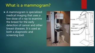
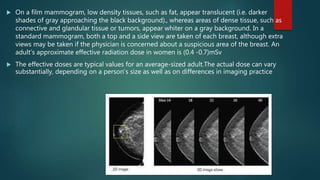
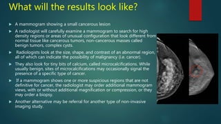
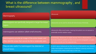
Mammography uses low-dose x-rays to examine the breast for early cancer detection. It has advanced from film to digital mammography and tomosynthesis, which creates 3D breast images. Computer-aided detection highlights abnormal areas. Screening mammograms aim to detect cancer in asymptomatic women, while diagnostic mammograms investigate symptoms. Benefits include early detection, but limitations include false positives and negatives due to breast density. Yearly mammograms after age 40 are recommended for breast cancer screening. Ultrasound provides localized breast images without radiation but cannot screen whole breasts. MRI is superior for dense breasts but has no radiation risk.
I have include all the contain about mammography like introduction,principle,anatomy,general views ,mammography physics (x-ray tube, housing,filter ,collimator and generator) and different advance technology about mammography.
Hope it will help your queries.
Thank you....!!
Definition of Mammography
Types of Mammography
Indications of Mammography
Contraindications of Mammography
Mammography Views
Mammogram
Mammography Unit
Additional Views of Mammography
Presentation contains info on Radiation Protection in Pregnancy - radiodiagnosis, MRI, nuclear medicine and radiotherapy. Including notes on foetal dose, shielding techniques in radiotherapy and precaution for pregnant radiographers/medical physicists.
Learn Barium Meal & Follow Through for the beginners from a Radiology Resident.For some image description please go through the text book "David Sutton" because i have described these image during my presentation Verbally..There are many animations used inside this presentation so to see all the pictures which are placed layer by layer with the help of animations you simple need to download this presentation first.... Thanx.
1. PRESENTING BY: SUMREEN KOSAR, ASSISTANT PROFESSOR DESH BHAGAT UNIVERSITY PUNJAB
2. over view: Introduction to mammography. Anatomy. Breast cancer. Imaging modalities. Role of a mammographic technologist. Mammographic views. Reporting system. Additional information for a mammography technologist. Artifacts. Male mammography . Conclusion.
For more information, visit https://www.timberlandmedical.com
Timberland Medical Centre is a private hospital that has been in operation since 1994. We are strategically located at the 3rd Mile roundabout on Jalan Rock, Kuching, Sarawak, East Malaysia. Our hospital is 10 minutes from the Kuching International Airport and 15 minutes from the Central Bus Terminal. We continually seek to improve and upgrade our services and facilities, as we strive to provide the best medical care for our patients and customers.
Here in these slides we have discuss about the Mammogram Screening with the help of which one can get the X-ray image of the breast to detect the breast cancer and to check it without having symptoms.
These simplified slides by Dr. Sidra Arshad present an overview of the non-respiratory functions of the respiratory tract.
Learning objectives:
1. Enlist the non-respiratory functions of the respiratory tract
2. Briefly explain how these functions are carried out
3. Discuss the significance of dead space
4. Differentiate between minute ventilation and alveolar ventilation
5. Describe the cough and sneeze reflexes
Study Resources:
1. Chapter 39, Guyton and Hall Textbook of Medical Physiology, 14th edition
2. Chapter 34, Ganong’s Review of Medical Physiology, 26th edition
3. Chapter 17, Human Physiology by Lauralee Sherwood, 9th edition
4. Non-respiratory functions of the lungs https://academic.oup.com/bjaed/article/13/3/98/278874
MANAGEMENT OF ATRIOVENTRICULAR CONDUCTION BLOCK.pdfJim Jacob Roy
Cardiac conduction defects can occur due to various causes.
Atrioventricular conduction blocks ( AV blocks ) are classified into 3 types.
This document describes the acute management of AV block.
Title: Sense of Taste
Presenter: Dr. Faiza, Assistant Professor of Physiology
Qualifications:
MBBS (Best Graduate, AIMC Lahore)
FCPS Physiology
ICMT, CHPE, DHPE (STMU)
MPH (GC University, Faisalabad)
MBA (Virtual University of Pakistan)
Learning Objectives:
Describe the structure and function of taste buds.
Describe the relationship between the taste threshold and taste index of common substances.
Explain the chemical basis and signal transduction of taste perception for each type of primary taste sensation.
Recognize different abnormalities of taste perception and their causes.
Key Topics:
Significance of Taste Sensation:
Differentiation between pleasant and harmful food
Influence on behavior
Selection of food based on metabolic needs
Receptors of Taste:
Taste buds on the tongue
Influence of sense of smell, texture of food, and pain stimulation (e.g., by pepper)
Primary and Secondary Taste Sensations:
Primary taste sensations: Sweet, Sour, Salty, Bitter, Umami
Chemical basis and signal transduction mechanisms for each taste
Taste Threshold and Index:
Taste threshold values for Sweet (sucrose), Salty (NaCl), Sour (HCl), and Bitter (Quinine)
Taste index relationship: Inversely proportional to taste threshold
Taste Blindness:
Inability to taste certain substances, particularly thiourea compounds
Example: Phenylthiocarbamide
Structure and Function of Taste Buds:
Composition: Epithelial cells, Sustentacular/Supporting cells, Taste cells, Basal cells
Features: Taste pores, Taste hairs/microvilli, and Taste nerve fibers
Location of Taste Buds:
Found in papillae of the tongue (Fungiform, Circumvallate, Foliate)
Also present on the palate, tonsillar pillars, epiglottis, and proximal esophagus
Mechanism of Taste Stimulation:
Interaction of taste substances with receptors on microvilli
Signal transduction pathways for Umami, Sweet, Bitter, Sour, and Salty tastes
Taste Sensitivity and Adaptation:
Decrease in sensitivity with age
Rapid adaptation of taste sensation
Role of Saliva in Taste:
Dissolution of tastants to reach receptors
Washing away the stimulus
Taste Preferences and Aversions:
Mechanisms behind taste preference and aversion
Influence of receptors and neural pathways
Impact of Sensory Nerve Damage:
Degeneration of taste buds if the sensory nerve fiber is cut
Abnormalities of Taste Detection:
Conditions: Ageusia, Hypogeusia, Dysgeusia (parageusia)
Causes: Nerve damage, neurological disorders, infections, poor oral hygiene, adverse drug effects, deficiencies, aging, tobacco use, altered neurotransmitter levels
Neurotransmitters and Taste Threshold:
Effects of serotonin (5-HT) and norepinephrine (NE) on taste sensitivity
Supertasters:
25% of the population with heightened sensitivity to taste, especially bitterness
Increased number of fungiform papillae
ARTIFICIAL INTELLIGENCE IN HEALTHCARE.pdfAnujkumaranit
Artificial intelligence (AI) refers to the simulation of human intelligence processes by machines, especially computer systems. It encompasses tasks such as learning, reasoning, problem-solving, perception, and language understanding. AI technologies are revolutionizing various fields, from healthcare to finance, by enabling machines to perform tasks that typically require human intelligence.
TEST BANK for Operations Management, 14th Edition by William J. Stevenson, Ve...kevinkariuki227
TEST BANK for Operations Management, 14th Edition by William J. Stevenson, Verified Chapters 1 - 19, Complete Newest Version.pdf
Ethanol (CH3CH2OH), or beverage alcohol, is a two-carbon alcohol
that is rapidly distributed in the body and brain. Ethanol alters many
neurochemical systems and has rewarding and addictive properties. It
is the oldest recreational drug and likely contributes to more morbidity,
mortality, and public health costs than all illicit drugs combined. The
5th edition of the Diagnostic and Statistical Manual of Mental Disorders
(DSM-5) integrates alcohol abuse and alcohol dependence into a single
disorder called alcohol use disorder (AUD), with mild, moderate,
and severe subclassifications (American Psychiatric Association, 2013).
In the DSM-5, all types of substance abuse and dependence have been
combined into a single substance use disorder (SUD) on a continuum
from mild to severe. A diagnosis of AUD requires that at least two of
the 11 DSM-5 behaviors be present within a 12-month period (mild
AUD: 2–3 criteria; moderate AUD: 4–5 criteria; severe AUD: 6–11 criteria).
The four main behavioral effects of AUD are impaired control over
drinking, negative social consequences, risky use, and altered physiological
effects (tolerance, withdrawal). This chapter presents an overview
of the prevalence and harmful consequences of AUD in the U.S.,
the systemic nature of the disease, neurocircuitry and stages of AUD,
comorbidities, fetal alcohol spectrum disorders, genetic risk factors, and
pharmacotherapies for AUD.
Tom Selleck Health: A Comprehensive Look at the Iconic Actor’s Wellness Journeygreendigital
Tom Selleck, an enduring figure in Hollywood. has captivated audiences for decades with his rugged charm, iconic moustache. and memorable roles in television and film. From his breakout role as Thomas Magnum in Magnum P.I. to his current portrayal of Frank Reagan in Blue Bloods. Selleck's career has spanned over 50 years. But beyond his professional achievements. fans have often been curious about Tom Selleck Health. especially as he has aged in the public eye.
Follow us on: Pinterest
Introduction
Many have been interested in Tom Selleck health. not only because of his enduring presence on screen but also because of the challenges. and lifestyle choices he has faced and made over the years. This article delves into the various aspects of Tom Selleck health. exploring his fitness regimen, diet, mental health. and the challenges he has encountered as he ages. We'll look at how he maintains his well-being. the health issues he has faced, and his approach to ageing .
Early Life and Career
Childhood and Athletic Beginnings
Tom Selleck was born on January 29, 1945, in Detroit, Michigan, and grew up in Sherman Oaks, California. From an early age, he was involved in sports, particularly basketball. which played a significant role in his physical development. His athletic pursuits continued into college. where he attended the University of Southern California (USC) on a basketball scholarship. This early involvement in sports laid a strong foundation for his physical health and disciplined lifestyle.
Transition to Acting
Selleck's transition from an athlete to an actor came with its physical demands. His first significant role in "Magnum P.I." required him to perform various stunts and maintain a fit appearance. This role, which he played from 1980 to 1988. necessitated a rigorous fitness routine to meet the show's demands. setting the stage for his long-term commitment to health and wellness.
Fitness Regimen
Workout Routine
Tom Selleck health and fitness regimen has evolved. adapting to his changing roles and age. During his "Magnum, P.I." days. Selleck's workouts were intense and focused on building and maintaining muscle mass. His routine included weightlifting, cardiovascular exercises. and specific training for the stunts he performed on the show.
Selleck adjusted his fitness routine as he aged to suit his body's needs. Today, his workouts focus on maintaining flexibility, strength, and cardiovascular health. He incorporates low-impact exercises such as swimming, walking, and light weightlifting. This balanced approach helps him stay fit without putting undue strain on his joints and muscles.
Importance of Flexibility and Mobility
In recent years, Selleck has emphasized the importance of flexibility and mobility in his fitness regimen. Understanding the natural decline in muscle mass and joint flexibility with age. he includes stretching and yoga in his routine. These practices help prevent injuries, improve posture, and maintain mobilit
Lung Cancer: Artificial Intelligence, Synergetics, Complex System Analysis, S...Oleg Kshivets
RESULTS: Overall life span (LS) was 2252.1±1742.5 days and cumulative 5-year survival (5YS) reached 73.2%, 10 years – 64.8%, 20 years – 42.5%. 513 LCP lived more than 5 years (LS=3124.6±1525.6 days), 148 LCP – more than 10 years (LS=5054.4±1504.1 days).199 LCP died because of LC (LS=562.7±374.5 days). 5YS of LCP after bi/lobectomies was significantly superior in comparison with LCP after pneumonectomies (78.1% vs.63.7%, P=0.00001 by log-rank test). AT significantly improved 5YS (66.3% vs. 34.8%) (P=0.00000 by log-rank test) only for LCP with N1-2. Cox modeling displayed that 5YS of LCP significantly depended on: phase transition (PT) early-invasive LC in terms of synergetics, PT N0—N12, cell ratio factors (ratio between cancer cells- CC and blood cells subpopulations), G1-3, histology, glucose, AT, blood cell circuit, prothrombin index, heparin tolerance, recalcification time (P=0.000-0.038). Neural networks, genetic algorithm selection and bootstrap simulation revealed relationships between 5YS and PT early-invasive LC (rank=1), PT N0—N12 (rank=2), thrombocytes/CC (3), erythrocytes/CC (4), eosinophils/CC (5), healthy cells/CC (6), lymphocytes/CC (7), segmented neutrophils/CC (8), stick neutrophils/CC (9), monocytes/CC (10); leucocytes/CC (11). Correct prediction of 5YS was 100% by neural networks computing (area under ROC curve=1.0; error=0.0).
CONCLUSIONS: 5YS of LCP after radical procedures significantly depended on: 1) PT early-invasive cancer; 2) PT N0--N12; 3) cell ratio factors; 4) blood cell circuit; 5) biochemical factors; 6) hemostasis system; 7) AT; 8) LC characteristics; 9) LC cell dynamics; 10) surgery type: lobectomy/pneumonectomy; 11) anthropometric data. Optimal diagnosis and treatment strategies for LC are: 1) screening and early detection of LC; 2) availability of experienced thoracic surgeons because of complexity of radical procedures; 3) aggressive en block surgery and adequate lymph node dissection for completeness; 4) precise prediction; 5) adjuvant chemoimmunoradiotherapy for LCP with unfavorable prognosis.
Pulmonary Thromboembolism - etilogy, types, medical- Surgical and nursing man...VarunMahajani
Disruption of blood supply to lung alveoli due to blockage of one or more pulmonary blood vessels is called as Pulmonary thromboembolism. In this presentation we will discuss its causes, types and its management in depth.
New Directions in Targeted Therapeutic Approaches for Older Adults With Mantl...i3 Health
i3 Health is pleased to make the speaker slides from this activity available for use as a non-accredited self-study or teaching resource.
This slide deck presented by Dr. Kami Maddocks, Professor-Clinical in the Division of Hematology and
Associate Division Director for Ambulatory Operations
The Ohio State University Comprehensive Cancer Center, will provide insight into new directions in targeted therapeutic approaches for older adults with mantle cell lymphoma.
STATEMENT OF NEED
Mantle cell lymphoma (MCL) is a rare, aggressive B-cell non-Hodgkin lymphoma (NHL) accounting for 5% to 7% of all lymphomas. Its prognosis ranges from indolent disease that does not require treatment for years to very aggressive disease, which is associated with poor survival (Silkenstedt et al, 2021). Typically, MCL is diagnosed at advanced stage and in older patients who cannot tolerate intensive therapy (NCCN, 2022). Although recent advances have slightly increased remission rates, recurrence and relapse remain very common, leading to a median overall survival between 3 and 6 years (LLS, 2021). Though there are several effective options, progress is still needed towards establishing an accepted frontline approach for MCL (Castellino et al, 2022). Treatment selection and management of MCL are complicated by the heterogeneity of prognosis, advanced age and comorbidities of patients, and lack of an established standard approach for treatment, making it vital that clinicians be familiar with the latest research and advances in this area. In this activity chaired by Michael Wang, MD, Professor in the Department of Lymphoma & Myeloma at MD Anderson Cancer Center, expert faculty will discuss prognostic factors informing treatment, the promising results of recent trials in new therapeutic approaches, and the implications of treatment resistance in therapeutic selection for MCL.
Target Audience
Hematology/oncology fellows, attending faculty, and other health care professionals involved in the treatment of patients with mantle cell lymphoma (MCL).
Learning Objectives
1.) Identify clinical and biological prognostic factors that can guide treatment decision making for older adults with MCL
2.) Evaluate emerging data on targeted therapeutic approaches for treatment-naive and relapsed/refractory MCL and their applicability to older adults
3.) Assess mechanisms of resistance to targeted therapies for MCL and their implications for treatment selection
Factory Supply Best Quality Pmk Oil CAS 28578–16–7 PMK Powder in Stockrebeccabio
Factory Supply Best Quality Pmk Oil CAS 28578–16–7 PMK Powder in Stock
Telegram: bmksupplier
signal: +85264872720
threema: TUD4A6YC
You can contact me on Telegram or Threema
Communicate promptly and reply
Free of customs clearance, Double Clearance 100% pass delivery to USA, Canada, Spain, Germany, Netherland, Poland, Italy, Sweden, UK, Czech Republic, Australia, Mexico, Russia, Ukraine, Kazakhstan.Door to door service
Hot Selling Organic intermediates
2. What is a mammogram?
A mammogram is specialized
medical imaging that uses a
low-dose of x-ray to examine
the breast for the early
detection of cancer and other
breast diseases. It is used as
both a diagnostic and
screening tool.
3. History and advances
Mid 1950s – Jacob Gershon Cohen uses
mammography to screen healthy women for
breast cancer. Late 1950s – Robert Egan
developed a new method of screening
mammography. He published his results in a
paper in 1959 and in a book in 1964. 1960s –
Mammography became a widely used diagnostic
tool.
Three recent advances in mammography include
digital mammography, computer-aided detection
and breast tomosynthesis
4. Digital mammography
also called full-field digital mammography (FFDM),
in which the x-ray film is replaced by electronics that convert x-
rays into mammographic pictures of the breast .
These detectors convert the x-rays that pass through them into
electronic signals that are sent to a computer.
The computer then converts these electronic signals into images
that can be displayed on a monitor and also stored for later use.
5.
6. Breast tomosynthesis
in which x-rays of the breast are taken at different angles to generate thin cross-sections
while In 2D mammograms, take images only from the front and side, this may create images
with overlapping breast tissue . The 3D representation of the breast is similar to the 3d
images created by standard CT technology.
Tomosynthesis differs from CT technology in that significantly fewer x-ray beams are
projected through the breast
Breast tomosynthesis may also result in:
* earlier detection of small breast cancers that may be hidden on a conventional
mammogram
* greater accuracy in pinpointing the size, shape and location of breast abnormalities
* fewer unnecessary biopsies or additional tests
* detecting multiple breast tumors
clearer images of abnormalities
7. Computer-aided detection (CAD)
CAD techniques developed recently for breast cancer
The im3D CAD digital tomosythesis system allows
detection of both masses and microcalcification
clusters at DBT examination(three-dimensional digital
breast tomosynthesis) .
including detection of abnormal areas of density,
mass, or calcification that may indicate the presence
of cancer.
The CAD system highlights these areas on the images
.Therefore, (CAD) system is still in developing to help
reduce reading time and prevent errors.
8. What are the types of mammograms?
Mammograms are used as :
1-as a Screening Mammography to detect early
breast cancer in women experiencing no symptoms.
2- as Diagnostic Mammography breast disease in
women experiencing symptoms such as a lump, pain,
skin dimpling or nipple discharge
9. How are screening and diagnostic
mammograms different?
The same machines are used for both types of mammograms.
However, diagnostic mammography takes longer to perform
than screening mammography and the total dose of radiation
is higher because more x-ray images are needed to obtain
views of the breast from several angles.
The technologist may magnify a suspicious area to produce a
detailed picture that can help the doctor make an accurate
diagnosis.
10. How should patients prepare for
mammography?
* Schedule your mammogram when your breasts are not tender or swollen to help reduce
discomfort and get good pictures.
* Do not wear deodorant, talcum powder or lotion under your arms or on your breasts on the
day of the exam. These can appear on the mammogram as calcium spots.
* Describe any breast symptoms or problems to the technologist performing the exam.
* Obtain your prior mammograms and make them available to the radiologist if they were
done at a different location. This is needed for comparison with your current exam.
* Ask when your results will be available; do not assume the results
11. How does the procedure work?
During a mammogram, a patient’s breast is placed on a flat support plate and compressed with a
parallel plate called a paddle.
An x-ray machine produces a small burst of x-rays that pass through the breast to a detector
located on the opposite side.
The detector can be either a photographic film plate, which captures the x-ray image on film, or a
solid-state detector, which transmits electronic signals to a computer to form a digital image.
The images produced are called mammograms.
12.
13. On a film mammogram, low density tissues, such as fat, appear translucent (i.e. darker
shades of gray approaching the black background)., whereas areas of dense tissue, such as
connective and glandular tissue or tumors, appear whiter on a gray background. In a
standard mammogram, both a top and a side view are taken of each breast, although extra
views may be taken if the physician is concerned about a suspicious area of the breast. An
adult’s approximate effective radiation dose in women is (0.4 -0.7)mSv
The effective doses are typical values for an average-sized adult.The actual dose can vary
substantially, depending on a person's size as well as on differences in imaging practice
14. Why does the breast need to be
compressed?
* Compression holds the breast in place to
minimize blurring of the x-ray image that can be
caused by patient motion.
Also, compression evens out the shape of the
breast so that the x-rays can travel through a
shorter path to reach the detector. This reduces the
radiation dose and improves the quality of the x-
ray image.
Finally, compression allows all the tissues to be
visualized in a single plane so that small
abnormalities are less likely to be obscured by
overlying breast tissue.
15. What will the results look like?
A mammogram showing a small cancerous lesion
A radiologist will carefully examine a mammogram to search for high
density regions or areas of unusual configuration that look different from
normal tissue like cancerous tumors, non-cancerous masses called
benign tumors, complex cysts.
Radiologists look at the size, shape, and contrast of an abnormal region,
all of which can indicate the possibility of malignancy (i.e. cancer).
They also look for tiny bits of calcium, called microcalcifications. While
usually benign, sites of microcalcifications may occasionally signal the
presence of a specific type of cancer.
If a mammogram shows one or more suspicious regions that are not
definitive for cancer, the radiologist may order additional mammogram
views, with or without additional magnification or compression, or they
may order a biopsy.
Another alternative may be referral for another type of non-invasive
imaging study.
16.
17.
18.
19. What are the benefits vs. risks?
Benefits
* detect small tumors When cancers are small,
the woman has more treatment options.
* The use of screening mammography increases the detection of small abnormal tissue growths confined to the milk ducts in the breast . It is also useful for detecting all types of breast cancer,
including invasive ductal and invasive lobular cancer.
* No radiation remains in a patient's body after
an x-ray examination.
* X-rays usually have no side effects in the
typical diagnostic range for this exam.
Risks
* There is always a slight chance of cancer from excessive exposure to radiation.
* If you are pregnant or suspect that you may be pregnant, you should notify your health care provider. Radiation exposure during pregnancy may lead to birth defects.
* Mammograms may be more difficult to interpret in women younger than 30 years of age, due to the increased density of their breast tissue.
* Mammography cannot prove that an abnormal area is cancer, but if it raises a significant suspicion of cancer, tissue will be removed for a biopsy
20. Limitations of Mammograms
Mammograms are the best breast cancer screening tests we have at this time. But
mammograms have their limits. For example, they aren’t 100% accurate in showing if a
woman has breast cancer:
* A false-negative mammogram looks normal even though breast cancer is present.
* A false-positive mammogram looks abnormal even though there’s no cancer in the
breast.
False-negative results
A false-negative mammogram looks normal even though breast cancer is present.
Overall, screening mammograms do not find about 1 in 5 breast cancers.
* Women with dense breasts have more false-negative Limitations of Mammograms
21. False-positive results
A false-positive mammogram looks abnormal even though no cancer is actually present.
Abnormal mammograms require extra testing (diagnostic mammograms, ultrasound, and
sometimes MRI or even a breast biopsy) to find out if the change is cancer.
* False-positive results are more common in women who are younger, have dense breasts,
have had breast biopsies, have breast cancer in the family, or are taking estrogen.
* About half of the women getting annual mammograms over a 10-year period will have a
false-positive finding.
* The odds of a false-positive finding are highest for the first mammogram. Women who
have past mammograms available for comparison reduce their odds of a false-positive
finding by about 50%.
* False-positive mammograms can cause anxiety. They can also lead to extra tests to be sure
cancer isn’t there, which cost time and money and maybe even physical discomfort.
22. How often should I get mammography?
You should do a breast self exam (BSE) every month if you
are over the age of 20 and it's a good idea to have a
complete breast exam every 3 years as well. If you are
over 40 years old then you should get a mammogram
every year.
23. What is the difference between mammography , and
breast ultrasound?
Mammography
Mammograms are specifically designed to target the breast
region,
mammograms use radiation (albeit small amounts),
mammograms provide an image of the entire breast, and
often identify lumps that cannot be felt or externally seen.
They are also useful if a mammogram has detected an
unusual lesion,
Ultrasounds
ultrasounds can be used for almost all internal areas of the body.
ultrasounds utilise sound waves, meaning that patients are not exposed to
potentially harmful radiation waves.
Contrastingly, ultrasounds are highly directed. That is, ultrasounds are
extremely useful if a patient can feel a lump and the sonographer can place
the camera directly over the suspected area.
in which case an ultrasound can then hone in on that specific area. However,
unlike mammograms, ultrasounds are not effective screening mechanisms,
and rarely do they detect small lumps on their own
24. What is the difference between
mammography and MRI?
Mammography
mammograms use radiation (albeit small amounts),
mammograms provide an image of the entire breast, and
often identify lumps that cannot be felt or externally seen.
They are also useful if a mammogram has detected an
unusual lesion,
Breast MRI
There is no risk of radiation exposure because MRIs use
magnetic fields to create images
MRIs are more effective in detecting breast cancer in patients
with dense breasts and patients with breast implants
Its ability is better to detect small breast lesions that are
sometimes missed on a mammography machine