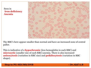
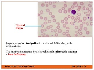
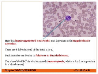
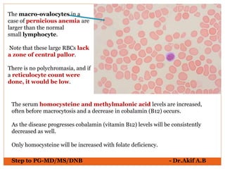
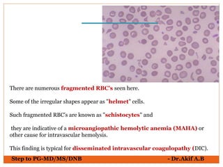
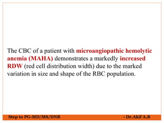
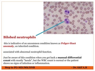
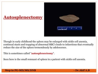
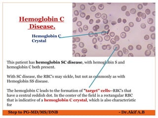
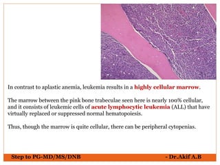
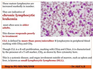
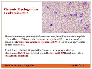
حمزة الحرايزة
جامعة العلوم والتكنولوجيا الاردنية - اربد
كلية التمريض..قسم تمريض صحة الاطفال
مناقشة مرض الثلاسيميا والانيميا
اعداد حمزة الحرايزة
الدكتورة هدى غرايبة
about the red blood cell.helpful for slide presentation.thank you all.
Ayub Ali
B,pharm (Hons.)
State University Of Bangladesh
ayubthecriminalgenious@gmail.com
Sickle cell Anemia: A worldwide popular blood disorder, basically a inheritable disease. This document provides you with basic introduction to blood, Anemia its general considerations, signs and symptoms and lastly about Sickle cell Anemia in detail.
Exercise Testing in Cardiology : Dr. Akif Baigakifab93
The testing modality and protocol should be selected in accordance with the patient’s estimated functional capacity based on age, estimated physical fitness from the patient’s history, and underlying disease
Several exercise test protocols are available for both treadmill and stationary cycle ergometers
Patients who have low estimated fitness levels or are deemed to be at higher risk because of underlying disease (e.g., recent MI, heart failure) should be tested with a less aggressive exercise protocol
Treadmill and cycle ergometers may use stepped or continuous ramp protocols
Work rate increments (stages) during stepped protocols can vary from 1 to 2.5 METs
Ramp protocols are designed with stages that are no longer than 1 minute and for the patient to attain peak effort within 8 to 12 minutes
The natriuretic peptide system works antagonistically to the RAAS and has favorable effects on the pathogenesis of heart failure
Natriuretic peptides are broken down by an enzyme called neprilysin
Neprilysin is also responsible for the breakdown of other substances, including bradykinin and angiotensin II
Sacubitril/valsartan is a combination product
Sacubitril is a pro-drug that, upon activation, acts as a neprilysin inhibitor
It works by blocking the action of neprilysin, thus preventing the breakdown of natriuretic peptides
This leads to a prolonged duration of the favorable effects of these peptides
Coronary heart disease (CHD) remains a leading cause of death worldwide, accounting for 16% of total deaths globally .
Atherosclerosis plays a central role, with early fatty streaks progressing to late complex atheromas
Vascular calcification, the pathogenic and process of ectopic bone production, specifically was shown to strongly correlate with degree of atherosclerosis (both calcified and noncalcified)
Vascular calcification was shown independently to predict cardiovascular morbidity and mortality
These associations, combined with the radio-opaque appearance of calcium hydroxyappatite on CT images, have led to extensive investigation of the quantification, or scoring, of coronary artery calcium (CAC).
CAC scoring has emerged as a widely available and powerful tool for stratifying cardiovascular risk, predicting patient outcomes, and guiding preventive therapy
A coronary bifurcation consists of a flow divider (carina) and three vessel segments:
The proximal main vessel (PMV)
The distal main vessel (DMV) and
The side branch (SB).
A bifurcation lesion is a major epicardial coronary artery stenosis next to and/or including the ostium of a significant side branch
A significant SB is a branch whose severe narrowing or acute occlusion before or during intervention can cause considerable ischemia or a new infarction area that will worsen the clinical course of a particular patient.
Other important elements to consider that are not inherent in the bifurcation classifications include:
Extent of disease on the SB (limited to the ostium or involving the vessel beyond the ostium)
Its size (over 2.5mm in reference diameter)
Bifurcation angle, and
Disease distribution
Left ventricular (LV) dysfunction remains one of the
best prognostic determinants of survival in patients
with coronary artery disease (CAD)
⚫ It was originally thought that dysfunctional
myocardium after an infarction was irreversibly
damaged
⚫ However, it was later recognized that some of the
involved tissue remained viable and contractility may
be restored with revascularization
HCM is a common genetic heart disease reported in populations globally
Inherited in an autosomal dominant pattern
The distribution of HCM is equal by sex, although women are diagnosed less commonly than men
The prevalence of unexplained asymptomatic hypertrophy in young adults has been reported to range from 1:200 to 1:500
Tetralogy of Fallot (TOF) is a congenital heart defect, which has four anatomical components:
Anterior malalignment ventricular septal defect (VSD)
Aortic override over the muscular septum
Variable degrees of subvalvar, valvar, and supravalvar pulmonary stenosis
Right ventricular (RV) infundibular narrowing and RV hypertrophy
Ventricular septal defects occur either as an isolated defect or as a component of a more complex lesion
It occurs in 50 percent of all children with CHD and in 20 to 30 percent as an isolated lesion
Most common congenital cardiac anomaly in children
Second most common congenital abnormality in adults, second only to bicuspid aortic valves
They are more common in premature infants and those born with low weight
VSDs are slightly more common in females (56%)
Patients with peripheral artery disease who have undergone lower-extremity revascularization are at high risk for major adverse limb and cardiovascular events
The efficacy and safety of rivaroxaban in this context are uncertain
Most common cyanotic heart defect seen in children beyond infancy, accounting for a third of all congenital heart disease (CHD) in this age group
Tetralogy of Fallot (TOF) is a congenital heart defect, which has four anatomical components:
Anterior malalignment ventricular septal defect (VSD)
Aortic override over the muscular septum
Variable degrees of subvalvar, valvar, and supravalvar pulmonary stenosis
Right ventricular (RV) infundibular narrowing and RV hypertrophy
Bentracimab (also known as PB2452) is a neutralizing recombinant human immunoglobulin G1 monoclonal antibody antigen-binding fragment that binds ticagrelor and its major active circulating metabolite with high affinity and specificity
Chlorthalidone for hypertension in advanced ckdakifab93
Chlorthalidone, a thiazide-like diuretic, reduces cardiovascular morbidity, such as the incidence of stroke and heart failure, and cardiovascular mortality
However, its efficacy and safety among patients with advanced chronic kidney disease remain poorly understood
An acute illness caused by an autoimmune response to infection with group A Streptococcus, leading to a range of possible symptoms and signs affecting any or all of heart, joints, brain, skin and subcutaneous tissues
Amyloidosis is a group of protein-folding disorders in which >1 organ is infiltrated by proteinaceous deposits known as amyloid. Amyloid involvement of the heart (cardiac amyloidosis) carries the worst prognosis of any involved organ, and light-chain (AL) amyloidosis is the most serious form of the disease
263778731218 Abortion Clinic /Pills In Harare ,sisternakatoto
263778731218 Abortion Clinic /Pills In Harare ,ABORTION WOMEN’S CLINIC +27730423979 IN women clinic we believe that every woman should be able to make choices in her pregnancy. Our job is to provide compassionate care, safety,affordable and confidential services. That’s why we have won the trust from all generations of women all over the world. we use non surgical method(Abortion pills) to terminate…Dr.LISA +27730423979women Clinic is committed to providing the highest quality of obstetrical and gynecological care to women of all ages. Our dedicated staff aim to treat each patient and her health concerns with compassion and respect.Our dedicated group ABORTION WOMEN’S CLINIC +27730423979 IN women clinic we believe that every woman should be able to make choices in her pregnancy. Our job is to provide compassionate care, safety,affordable and confidential services. That’s why we have won the trust from all generations of women all over the world. we use non surgical method(Abortion pills) to terminate…Dr.LISA +27730423979women Clinic is committed to providing the highest quality of obstetrical and gynecological care to women of all ages. Our dedicated staff aim to treat each patient and her health concerns with compassion and respect.Our dedicated group of receptionists, nurses, and physicians have worked together as a teamof receptionists, nurses, and physicians have worked together as a team wwww.lisywomensclinic.co.za/
The Gram stain is a fundamental technique in microbiology used to classify bacteria based on their cell wall structure. It provides a quick and simple method to distinguish between Gram-positive and Gram-negative bacteria, which have different susceptibilities to antibiotics
Muktapishti is a traditional Ayurvedic preparation made from Shoditha Mukta (Purified Pearl), is believed to help regulate thyroid function and reduce symptoms of hyperthyroidism due to its cooling and balancing properties. Clinical evidence on its efficacy remains limited, necessitating further research to validate its therapeutic benefits.
- Video recording of this lecture in English language: https://youtu.be/lK81BzxMqdo
- Video recording of this lecture in Arabic language: https://youtu.be/Ve4P0COk9OI
- Link to download the book free: https://nephrotube.blogspot.com/p/nephrotube-nephrology-books.html
- Link to NephroTube website: www.NephroTube.com
- Link to NephroTube social media accounts: https://nephrotube.blogspot.com/p/join-nephrotube-on-social-media.html
Best Ayurvedic medicine for Gas and IndigestionSwastikAyurveda
Here is the updated list of Top Best Ayurvedic medicine for Gas and Indigestion and those are Gas-O-Go Syp for Dyspepsia | Lavizyme Syrup for Acidity | Yumzyme Hepatoprotective Capsules etc
Basavarajeeyam is a Sreshta Sangraha grantha (Compiled book ), written by Neelkanta kotturu Basavaraja Virachita. It contains 25 Prakaranas, First 24 Chapters related to Rogas& 25th to Rasadravyas.
Ozempic: Preoperative Management of Patients on GLP-1 Receptor Agonists Saeid Safari
Preoperative Management of Patients on GLP-1 Receptor Agonists like Ozempic and Semiglutide
ASA GUIDELINE
NYSORA Guideline
2 Case Reports of Gastric Ultrasound
CDSCO and Phamacovigilance {Regulatory body in India}NEHA GUPTA
The Central Drugs Standard Control Organization (CDSCO) is India's national regulatory body for pharmaceuticals and medical devices. Operating under the Directorate General of Health Services, Ministry of Health & Family Welfare, Government of India, the CDSCO is responsible for approving new drugs, conducting clinical trials, setting standards for drugs, controlling the quality of imported drugs, and coordinating the activities of State Drug Control Organizations by providing expert advice.
Pharmacovigilance, on the other hand, is the science and activities related to the detection, assessment, understanding, and prevention of adverse effects or any other drug-related problems. The primary aim of pharmacovigilance is to ensure the safety and efficacy of medicines, thereby protecting public health.
In India, pharmacovigilance activities are monitored by the Pharmacovigilance Programme of India (PvPI), which works closely with CDSCO to collect, analyze, and act upon data regarding adverse drug reactions (ADRs). Together, they play a critical role in ensuring that the benefits of drugs outweigh their risks, maintaining high standards of patient safety, and promoting the rational use of medicines.
Basavarajeeyam is an important text for ayurvedic physician belonging to andhra pradehs. It is a popular compendium in various parts of our country as well as in andhra pradesh. The content of the text was presented in sanskrit and telugu language (Bilingual). One of the most famous book in ayurvedic pharmaceutics and therapeutics. This book contains 25 chapters called as prakaranas. Many rasaoushadis were explained, pioneer of dhatu druti, nadi pareeksha, mutra pareeksha etc. Belongs to the period of 15-16 century. New diseases like upadamsha, phiranga rogas are explained.
2. Explaining full hematology in pdf is not possible
So I will just try to help u with the peripheral smear pictures of Various conditions
Rest u can follow in whichever book u r reading………….
3. Step to PG-MD/MS/DNB - Dr.Akif A.B
The red blood cells here are
normal RBC's.
They have a zone of central
pallor about 1/3 the size of the
RBC.
The RBC's demonstrate minimal
variation in size (anisocytosis) and
shape (poikilocytosis).
A few small fuzzy
blue platelets are seen.
Segmented
neutrophil
Band
neutrophil
4. Step to PG-MD/MS/DNB - Dr.Akif A.B
A normal mature
lymphocyte with a
single large nucleus
Segmented
neutrophil
5. Step to PG-MD/MS/DNB - Dr.Akif A.B
Here is a monocyte.
It is slightly larger than a lymphocyte and
has a folded nucleus.
6. Step to PG-MD/MS/DNB - Dr.Akif A.B
In the center of the field is an eosinophil with a bilobed nucleus and
numerous bright red cytoplasmic granules.
Just underneath it is a small lymphocyte.
Eosinophil
small
lymphocyte
7. Step to PG-MD/MS/DNB - Dr.Akif A.B
There is a basophil in the center of the field that has a multi-lobed nucleus (like
PMN's) and numerous coarse, dark blue granules in the cytoplasm, just like tissue
mast cells.
A band neutrophil is seen on the left, and a large, activated lymphocyte on the
right.
basophil
band
neutrophil
8. Step to PG-MD/MS/DNB - Dr.Akif A.B
The RBC's here have stacked together in long chains.
This is known as "rouleaux formation" and it happens with increased
serum proteins, particularly fibrinogen and globulins.
Such long chains of RBC's sediment more readily.
This is the mechanism for the sedimentation rate, which increases non-
specifically with inflammation and increased "acute phase" serum proteins.
Seen in Multiple Myeloma
9. Step to PG-MD/MS/DNB - Dr.Akif A.B
The RBC's here appear smaller than normal and have an increased zone of central
pallor.
This is indicative of a hypochromic (less hemoglobin in each RBC) and
microcytic (smaller size of each RBC) anemia. There is also increased
anisocytosis (variation in RBC size) and poikilocytosis (variation in RBC
shape).
Seen in
Iron deficiency
Anemia
10. Step to PG-MD/MS/DNB - Dr.Akif A.B
larger zones of central pallor in these small RBCs, along with
poikilocytosis.
The most common cause for a hypochromic microcytic anemia
is iron deficiency.
Central
Pallor
11. Step to PG-MD/MS/DNB - Dr.Akif A.B
Microcytosis is indicated by the low MCV
(mean corpuscular volume).
Hypochromia correlates here with the low
MCH (mean corpuscular hemoglobin).
12. Step to PG-MD/MS/DNB - Dr.Akif A.B
The peripheral blood smear shown here appears normal, because it is.
A normal neutrophil and lymphocyte are present. Only a CBC will
demonstrate anemia.
This is a normochromic, normocytic anemia.
13. Step to PG-MD/MS/DNB - Dr.Akif A.B
There is impaired utilization of iron, without either an absolute deficiency or
an excess of iron.
There is cytokine-mediated blockage in transfer of iron from the storage pool
to the erythroid precursors in the bone marrow.
The defect is either inability to free the iron from macrophages or to load it onto
transferrin.
Inflammatory cytokines also depress erythropoiesis
Inflammatory conditions release cytokines such as interleukins 1 and 6 (IL1,
IL6) that stimulate hepatic production of hepcidin, which reduces iron
absorption and decreases release of iron from stores in macrophages.
Anemia of chronic disease is addressed by treating the underlying
condition.
15. Step to PG-MD/MS/DNB - Dr.Akif A.B
Here is a hypersegmented neutrophil that is present with megaloblastic
anemias.
There are 8 lobes instead of the usual 3 or 4.
Such anemias can be due to folate or to B12 deficiency.
The size of the RBC's is also increased (macrocytosis, which is hard to appreciate
in a blood smear)
16. Step to PG-MD/MS/DNB - Dr.Akif A.B
The macro-ovalocytes in a
case of pernicious anemia are
larger than the normal
small lymphocyte.
Note that these large RBCs lack
a zone of central pallor.
There is no polychromasia, and if
a reticulocyte count were
done, it would be low.
The serum homocysteine and methylmalonic acid levels are increased,
often before macrocytosis and a decrease in cobalamin (B12) occurs.
As the disease progresses cobalamin (vitamin B12) levels will be consistently
decreased as well.
Only homocysteine will be increased with folate deficiency.
17. Step to PG-MD/MS/DNB - Dr.Akif A.B
There are numerous fragmented RBC's seen here.
Some of the irregular shapes appear as "helmet" cells.
Such fragmented RBC's are known as "schistocytes" and
they are indicative of a microangiopathic hemolytic anemia (MAHA) or
other cause for intravascular hemolysis.
This finding is typical for disseminated intravascular coagulopathy (DIC).
18. Step to PG-MD/MS/DNB - Dr.Akif A.B
The CBC of a patient with microangiopathic hemolytic
anemia (MAHA) demonstrates a markedly increased
RDW (red cell distribution width) due to the marked
variation in size and shape of the RBC population.
19. Step to PG-MD/MS/DNB - Dr.Akif A.B
The RBC in the center of the field contains several Howell-Jolly bodies, or
inclusions of nuclear chromatin remnants.
There is also a nucleated RBC just beneath this RBC. Abnormal and aged RBC's
are typically removed by the spleen.
The appearance of increased poikilocytosis, anisocytosis, and RBC inclusions
suggests that a spleen is not present.(Asplenia)
Nucleated
RBC
Howell-Jolly
bodies
20. Step to PG-MD/MS/DNB - Dr.Akif A.B
The size of many of these RBC's is
quite small, with lack of the central
zone of pallor.
These RBC's are spherocytes.
In hereditary spherocytosis,
there is a lack of spectrin, a key
RBC cytoskeletal membrane
protein.
This produces membrane
instability that forces the cell to the
smallest volume--a sphere.
In the laboratory, this is shown by
increased osmotic fragility.
The spherocytes do not survive
as long as normal RBC's.
21. Step to PG-MD/MS/DNB - Dr.Akif A.B
The sinusoids are packed with
RBC's in this case of hereditary
spherocytosis.
The osmotic fragility of spherocytes
is increased, because the RBC's
have decreased surface area per
unit volume.
The major clinical features are
anemia, splenomegaly, and
jaundice.
An aplastic crisis may occur with
parvovirus infection.
22. Step to PG-MD/MS/DNB - Dr.Akif A.B
increased central pallor,
consistent with hypochromic
microcytic anemia. The most
common cause is iron deficiency.
RBC's are large and lack central pallor,
consistent with macro-ovalocytes.
The most common cause is
megaloblastic anemia, from vitamin
B12 or folate deficiency.
RBC's are small and lack central pallor,
consistent with spherocytes lacking
central pallor. Hereditary
spherocytosis.
23. Step to PG-MD/MS/DNB - Dr.Akif A.B
The nucleated RBCs contain basophilic stippling of the cytoplasm.
This suggests a toxic injury to the bone marrow, such as lead poisoning.
Such stippling may also appear with severe anemia, such as a megaloblastic
anemia.
24. Step to PG-MD/MS/DNB - Dr.Akif A.B
Atypical lymphocytes are shown here.
These WBC's are "atypical" because they are larger (more cytoplasm) and have
nucleoli in their nuclei.
Such atypical lymphocytes are often associated with infectious mononucleosis
from Epstein-Barr virus (EBV) infection.
25. Step to PG-MD/MS/DNB - Dr.Akif A.B
Bilobed neutrophils
this is indicative of an uncommon condition known as Pelger-Huet
anomaly, an inherited condition.
associated with abnormal neutrophil function.
Just be aware of this condition when you get back a manual differential
count with mostly "bands", but the WBC count is normal or the patient
shows no signs of infection or inflammation.
26. Step to PG-MD/MS/DNB - Dr.Akif A.B
This is sickle cell anemia
- Autosomal Recessive Disease
27. Step to PG-MD/MS/DNB - Dr.Akif A.B
Though in early childhood the spleen may be enlarged with sickle cell anemia,
continual stasis and trapping of abnormal RBC's leads to infarctions that eventually
reduce the size of the spleen tremendously by adolescence.
This is sometimes called "autosplenectomy".
Seen here is the small remnant of spleen in a patient with sickle cell anemia.
Autosplenectomy
28. Step to PG-MD/MS/DNB - Dr.Akif A.B
Severe, chronic anemias (such as thalassemias and sickle cell
anemia) can increase the bone marrow response to form RBC's.
This drive for erythropoiesis may increase the mass of marrow and
lead to increase in marrow in places, such as the skull seen here, that is
not normally found.
Such an increase in marrow in skull may lead to "frontal bossing" or
forehead prominence because of the skull shape change.
29. Step to PG-MD/MS/DNB - Dr.Akif A.B
This patient has hemoglobin SC disease, with hemoglobin S and
hemoglobin C both present.
With SC disease, the RBC's may sickle, but not as commonly as with
Hemoglobin SS disease.
The hemoglobin C leads to the formation of "target" cells--RBC's that
have a central reddish dot. In the center of the field is a rectangular RBC
that is indicative of a hemoglobin C crystal, which is also characteristic
for
Hemoglobin C
Disease.
Hemoglobin C
Crystal
30. Step to PG-MD/MS/DNB - Dr.Akif A.B
Hematopoietic elements in this bone marrow biopsy are markedly
reduced.
This is a case of aplastic anemia.
Aplastic Anemia
31. Step to PG-MD/MS/DNB - Dr.Akif A.B
In contrast to aplastic anemia, leukemia results in a highly cellular marrow.
The marrow between the pink bone trabeculae seen here is nearly 100% cellular,
and it consists of leukemic cells of acute lymphocytic leukemia (ALL) that have
virtually replaced or suppressed normal hematopoiesis.
Thus, though the marrow is quite cellular, there can be peripheral cytopenias.
32. The WBC's seen here are lymphocytes, but they are blasts--very immature cells
with scant cytoplasm and large nuclei that contain nucleoli.
Such abnormal lymphocytes are indicative of acute lymphoblastic
leukemia (ALL).
ALL is more common in children than adults.
Blast Cells
Step to PG-MD/MS/DNB - Dr.Akif A.B
33. These mature lymphocytes are
increased markedly in number.
They are indicative of
chronic lymphocytic
leukemia
most often seen in older
adults.
This disease responds poorly
to treatment
CLL is defined by more than 5000/microliter B lymphocytes in peripheral blood
marking with CD23 and CD5.
Though CLL is a B cell proliferation, marking with CD19 and CD20, it is characterized
by the presence of a T cell marker, CD5, as shown by flow cytometry here.
This is a systemic disease, and organ involement outside of marrow, such as spleen and
liver, is known as small lymphocytic lymphoma (SLL).
Step to PG-MD/MS/DNB - Dr.Akif A.B
34. -very large, immature myeloblasts with many nucleoli.
- "Auer rod" composed of crystallized granules.
-These findings are typical for acute myelogenous leukemia (AML)
- most prevalent in young adults.
Acute Myelogenous
Leukemia (AML)
Auer rod
Step to PG-MD/MS/DNB - Dr.Akif A.B
35. There are numerous granulocytic forms seen here, including immature myeloid
cells and bands. This condition is one of the myeloproliferative states and is
known as chronic myelogenous leukemia (CML) that is most prevalent in
middle-aged adults.
A useful test to help distinguish this disease is the leukocyte alkaline
phosphatase (LAP) score, which should be low with CML and high with a
leukemoid reaction.
Chronic Myelogenous
Leukemia (CML)
Step to PG-MD/MS/DNB - Dr.Akif A.B
36. Myeloid cells of CML are also characterized by the Philadelphia
chromosome (Ph1) on karyotyping.
This is a translocation of a portion of the q arm of chromosome 22 to
the q arm of chromosome 9, designated t(9:22).
Step to PG-MD/MS/DNB - Dr.Akif A.B
37. Here is another view of a peripheral blood smear in a patient with CML.
Often, the numbers of basophils and eosinophils, as well as bands and more
immature myeloid cells (metamyelocytes and myelocytes) are increased.
Unlike AML, there are not many blasts with CML.
Step to PG-MD/MS/DNB - Dr.Akif A.B
38. The structure of a lymph node is
diagrammed above:
A - Afferent lymphatic channels
B - Subcapsular sinus
C - Follicle
D - Sinuses
E - Paracortical region
F - Medullary cords
G - Efferent lymphatic channel
Step to PG-MD/MS/DNB - Dr.Akif A.B
39. Here is the normal appearance of a benign reactive lymph node.
Lymphatics that drain tissues peripheral to the node enter the subcapsular sinus
and lymph percolates around and into lymphoid follicles of variable size and
having a surrounding mantle zone that is surrounding a pale germinal
center in which the immune responses are often generated with a predominance
of B cells..
Mantle Zone
Germinal Center
Step to PG-MD/MS/DNB - Dr.Akif A.B