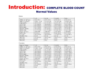
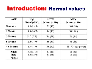
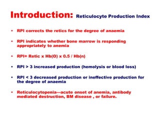
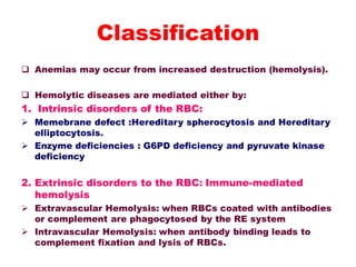
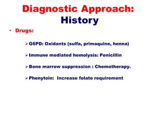
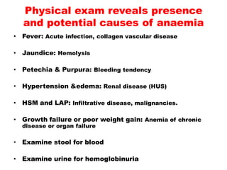
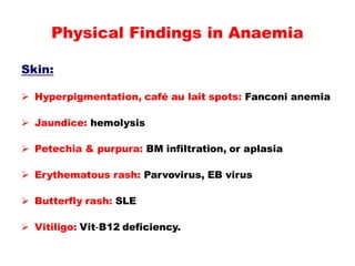
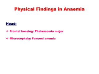
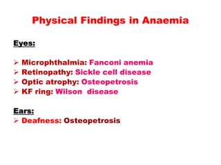
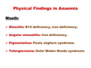
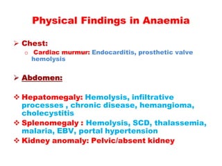
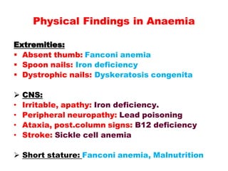
The Anemias
Professor Dr. Salah Al-Zuhairy, MD.
Introduction
 The study of the blood has a long history.
 Humankind probably always has been
interested in the blood because it is likely
that even primitive peoples realized that loss
of blood, if sufficiently great, was associated
with death.
 In biblical references, to shed blood meant to
kill.
Introduction
Anemia :
 It is a reduction of the RBC volume or hemoglobin
concentration below the range of values occurring in
healthy persons.
 It is a reduction of the RBC volume or hemoglobin
concentration below -2SD for age, race and sex.
 In certain pathologic states, anemia may be present
when the hemoglobin level is within the normal range:
 cyanotic cardiac diseases
 pulmonary disease
 hemoglobin with an abnormally high affinity for oxygen
is present.
 A reduction in the amount of circulating
hemoglobin decreases the oxygen-carrying
capacity of the blood
 But few clinical disturbances occur until the
hemoglobin level < 7–8 g/dL.
 Anemia is not a specific entity
 It is the result of many underlying pathologic
processes.
 Anemia is a common complication of many
disorders and may accentuate organ
dysfunction.
Introduction
Introduction
It is important for pediatricians to recognize
that:
 RBC size changes with age
 MCV variations in childhood
 It is essential to review the appearance of
RBCs on a peripheral blood smear
 The presence of polychromatophilia, which
usually correlates with the degree of
reticulocytosis, indicates that the marrow is
able to respond to RBC loss or destruction.
Introduction:COMPLETE BLOOD COUNT
Hb Concentration (g/dl)
Hematocrit (PCV) (%)
MCV (fl)
MCH (pg)
MCHC (g/dl)
RBC Count (…x106/ mm3 )
RDW (%)
WBC Count (…x103/ mm3 )
Neutrophil ( % )
lymphocyte ( % )
Monocyte ( % )
Basophil ( % )
Eosinophil ( % )
Platelet Count (…x103/ mm3 )
Reticulocyte Count ( % )
Introduction: COMPLETE BLOOD COUNT
Normal Values
Introduction: Normal values
Introduction: Reticulocyte Production Index
• RPI corrects the retics for the degree of anaemia
• RPI indicates whether bone marrow is responding
appropriately to anemia
• RPI= Retic x Hb(O) x 0.5 / Hb(n)
• RPI > 3 increased production (hemolysis or blood loss)
• RPI < 3 decreased production or ineffective production for
the degree of anaemia
• Reticulocytopenia—acute onset of anemia, antibody
mediated destruction, BM disease , or failure.
Anemias are classified based on the size and hemoglobin
content of the cells
1. Hypochromic, microcytic anemia: is caused by an
inadequate production of hemoglobin.
 The most common causes of this type of anemia are
iron deficiency and thalassemia.
2. Normocytic anemia: are associated with a systemic
illness that impairs adequate marrow synthesis of
RBCs.
3. Macrocytic anemia: are associated with vitamin B12
and folic acid deficiencies
Classification
 Anemias may occur from increased destruction (hemolysis).
 Hemolytic diseases are mediated either by:
1. Intrinsic disorders of the RBC:
 Memebrane defect :Hereditary spherocytosis and Hereditary
elliptocytosis.
 Enzyme deficiencies : G6PD deficiency and pyruvate kinase
deficiency
2. Extrinsic disorders to the RBC: Immune-mediated
hemolysis
 Extravascular Hemolysis: when RBCs coated with antibodies
or complement are phagocytosed by the RE system
 Intravascular Hemolysis: when antibody binding leads to
complement fixation and lysis of RBCs.
Classification
• Age : Iron deficiency is rare without
blood loss before 6 mo in term infants.
• Family History & Genetics:
1. X-linked: G6PD deficiency.
2. Autosomal dominant: Spherocytosis
3. Autosomal recessive : Sickle cell diseases ,
Fanconi anemia
4. Family member with early age of
cholecystectomy or splenectomy
5. Ethnicity: Thalassemia; G6PD deficiency.
Diagnostic Approach:
History
• Diarrhoea:
Malabsorption of Vitamin B12 / E /Fe.
Inflammatory bowel diseases, and
anemia of chronic disease with or
without blood loss.
Milk protein intolerance induced blood
loss.
Intestinal resection: Vitamin B12
deficiency.
Diagnostic Approach:
History
• Infections:
Giardia: iron malabsorption (IDA)
Intestinal bacterial overgrowth: Vitamin B12
deficiency.
EBV, CMV, Parvovirus: Bone marrow suppression.
Mycoplasma, Malaria: Hemolysis.
Hepatitis: Aplastic anemia.
Endocarditis, HIV: Anemia of chronic disease, or
hemolysis.
Diagnostic Approach:
History
• Nutrition:
Cows milk diet: Iron deficiency
Strict vegetarian: B12 deficiency.
Goats milk: Folate deficiency.
Pica: Plumbism, Iron deficiency.
Cholestasis, malabsorption: Vitamin E
deficiency.
Diagnostic Approach:
History
• Drugs:
G6PD: Oxidants (sulfa, primaquine, henna)
Immune mediated hemolysis: Penicillin
Bone marrow suppression : Chemotherapy.
Phenytoin: Increase folate requirement
Diagnostic Approach:
History
• Fever: Acute infection, collagen vascular disease
• Jaundice: Hemolysis
• Petechia & Purpura: Bleeding tendency
• Hypertension &edema: Renal disease (HUS)
• HSM and LAP: Infiltrative disease, malignancies.
• Growth failure or poor weight gain: Anemia of chronic
disease or organ failure
• Examine stool for blood
• Examine urine for hemoglobinuria
Physical exam reveals presence
and potential causes of anaemia
Skin:
 Hyperpigmentation, café au lait spots: Fanconi anemia
 Jaundice: hemolysis
 Petechia & purpura: BM infiltration, or aplasia
 Erythematous rash: Parvovirus, EB virus
 Butterfly rash: SLE
 Vitiligo: Vit-B12 deficiency.
Physical Findings in Anaemia
Head:
 Frontal bossing: Thalassemia major
 Microcephaly: Fanconi anemia
Physical Findings in Anaemia
Eyes:
 Microphthalmia: Fanconi anemia
 Retinopathy: Sickle cell disease
 Optic atrophy: Osteopetrosis
 KF ring: Wilson disease
Ears:
 Deafness: Osteopetrosis
Physical Findings in Anaemia
Mouth:
 Glossitis: B12 deficiency, iron deficiency.
 Angular stomatitis: Iron deficiency.
 Pigmentation: Peutz Jeghers syndrome.
 Telangiectasia: Osler Weber Rendu syndrome
Physical Findings in Anaemia
 Chest:
o Cardiac murmur: Endocarditis, prosthetic valve
hemolysis
 Abdomen:
 Hepatomegaly: Hemolysis, infiltrative
processes , chronic disease, hemangioma,
cholecystitis
 Splenomegaly : Hemolysis, SCD, thalassemia,
malaria, EBV, portal hypertension
 Kidney anomaly: Pelvic/absent kidney
Physical Findings in Anaemia
Extremities:
 Absent thumb: Fanconi anemia
 Spoon nails: Iron deficiency
 Dystrophic nails: Dyskeratosis congenita
 CNS:
• Irritable, apathy: Iron deficiency.
• Peripheral neuropathy: Lead poisoning
• Ataxia, post.column signs: B12 deficiency
• Stroke: Sickle cell anemia
 Short stature: Fanconi anemia, Malnutrition
Physical Findings in Anaemia
Thank You

Anemia Ped 5th yr1 (1).pdf

  • 1.
    The Anemias Professor Dr.Salah Al-Zuhairy, MD.
  • 2.
    Introduction  The studyof the blood has a long history.  Humankind probably always has been interested in the blood because it is likely that even primitive peoples realized that loss of blood, if sufficiently great, was associated with death.  In biblical references, to shed blood meant to kill.
  • 3.
    Introduction Anemia :  Itis a reduction of the RBC volume or hemoglobin concentration below the range of values occurring in healthy persons.  It is a reduction of the RBC volume or hemoglobin concentration below -2SD for age, race and sex.  In certain pathologic states, anemia may be present when the hemoglobin level is within the normal range:  cyanotic cardiac diseases  pulmonary disease  hemoglobin with an abnormally high affinity for oxygen is present.
  • 4.
     A reductionin the amount of circulating hemoglobin decreases the oxygen-carrying capacity of the blood  But few clinical disturbances occur until the hemoglobin level < 7–8 g/dL.  Anemia is not a specific entity  It is the result of many underlying pathologic processes.  Anemia is a common complication of many disorders and may accentuate organ dysfunction. Introduction
  • 5.
    Introduction It is importantfor pediatricians to recognize that:  RBC size changes with age  MCV variations in childhood  It is essential to review the appearance of RBCs on a peripheral blood smear  The presence of polychromatophilia, which usually correlates with the degree of reticulocytosis, indicates that the marrow is able to respond to RBC loss or destruction.
  • 6.
    Introduction:COMPLETE BLOOD COUNT HbConcentration (g/dl) Hematocrit (PCV) (%) MCV (fl) MCH (pg) MCHC (g/dl) RBC Count (…x106/ mm3 ) RDW (%) WBC Count (…x103/ mm3 ) Neutrophil ( % ) lymphocyte ( % ) Monocyte ( % ) Basophil ( % ) Eosinophil ( % ) Platelet Count (…x103/ mm3 ) Reticulocyte Count ( % )
  • 7.
    Introduction: COMPLETE BLOODCOUNT Normal Values
  • 8.
  • 9.
    Introduction: Reticulocyte ProductionIndex • RPI corrects the retics for the degree of anaemia • RPI indicates whether bone marrow is responding appropriately to anemia • RPI= Retic x Hb(O) x 0.5 / Hb(n) • RPI > 3 increased production (hemolysis or blood loss) • RPI < 3 decreased production or ineffective production for the degree of anaemia • Reticulocytopenia—acute onset of anemia, antibody mediated destruction, BM disease , or failure.
  • 10.
    Anemias are classifiedbased on the size and hemoglobin content of the cells 1. Hypochromic, microcytic anemia: is caused by an inadequate production of hemoglobin.  The most common causes of this type of anemia are iron deficiency and thalassemia. 2. Normocytic anemia: are associated with a systemic illness that impairs adequate marrow synthesis of RBCs. 3. Macrocytic anemia: are associated with vitamin B12 and folic acid deficiencies Classification
  • 11.
     Anemias mayoccur from increased destruction (hemolysis).  Hemolytic diseases are mediated either by: 1. Intrinsic disorders of the RBC:  Memebrane defect :Hereditary spherocytosis and Hereditary elliptocytosis.  Enzyme deficiencies : G6PD deficiency and pyruvate kinase deficiency 2. Extrinsic disorders to the RBC: Immune-mediated hemolysis  Extravascular Hemolysis: when RBCs coated with antibodies or complement are phagocytosed by the RE system  Intravascular Hemolysis: when antibody binding leads to complement fixation and lysis of RBCs. Classification
  • 12.
    • Age :Iron deficiency is rare without blood loss before 6 mo in term infants. • Family History & Genetics: 1. X-linked: G6PD deficiency. 2. Autosomal dominant: Spherocytosis 3. Autosomal recessive : Sickle cell diseases , Fanconi anemia 4. Family member with early age of cholecystectomy or splenectomy 5. Ethnicity: Thalassemia; G6PD deficiency. Diagnostic Approach: History
  • 13.
    • Diarrhoea: Malabsorption ofVitamin B12 / E /Fe. Inflammatory bowel diseases, and anemia of chronic disease with or without blood loss. Milk protein intolerance induced blood loss. Intestinal resection: Vitamin B12 deficiency. Diagnostic Approach: History
  • 14.
    • Infections: Giardia: ironmalabsorption (IDA) Intestinal bacterial overgrowth: Vitamin B12 deficiency. EBV, CMV, Parvovirus: Bone marrow suppression. Mycoplasma, Malaria: Hemolysis. Hepatitis: Aplastic anemia. Endocarditis, HIV: Anemia of chronic disease, or hemolysis. Diagnostic Approach: History
  • 15.
    • Nutrition: Cows milkdiet: Iron deficiency Strict vegetarian: B12 deficiency. Goats milk: Folate deficiency. Pica: Plumbism, Iron deficiency. Cholestasis, malabsorption: Vitamin E deficiency. Diagnostic Approach: History
  • 16.
    • Drugs: G6PD: Oxidants(sulfa, primaquine, henna) Immune mediated hemolysis: Penicillin Bone marrow suppression : Chemotherapy. Phenytoin: Increase folate requirement Diagnostic Approach: History
  • 17.
    • Fever: Acuteinfection, collagen vascular disease • Jaundice: Hemolysis • Petechia & Purpura: Bleeding tendency • Hypertension &edema: Renal disease (HUS) • HSM and LAP: Infiltrative disease, malignancies. • Growth failure or poor weight gain: Anemia of chronic disease or organ failure • Examine stool for blood • Examine urine for hemoglobinuria Physical exam reveals presence and potential causes of anaemia
  • 18.
    Skin:  Hyperpigmentation, caféau lait spots: Fanconi anemia  Jaundice: hemolysis  Petechia & purpura: BM infiltration, or aplasia  Erythematous rash: Parvovirus, EB virus  Butterfly rash: SLE  Vitiligo: Vit-B12 deficiency. Physical Findings in Anaemia
  • 19.
    Head:  Frontal bossing:Thalassemia major  Microcephaly: Fanconi anemia Physical Findings in Anaemia
  • 20.
    Eyes:  Microphthalmia: Fanconianemia  Retinopathy: Sickle cell disease  Optic atrophy: Osteopetrosis  KF ring: Wilson disease Ears:  Deafness: Osteopetrosis Physical Findings in Anaemia
  • 21.
    Mouth:  Glossitis: B12deficiency, iron deficiency.  Angular stomatitis: Iron deficiency.  Pigmentation: Peutz Jeghers syndrome.  Telangiectasia: Osler Weber Rendu syndrome Physical Findings in Anaemia
  • 22.
     Chest: o Cardiacmurmur: Endocarditis, prosthetic valve hemolysis  Abdomen:  Hepatomegaly: Hemolysis, infiltrative processes , chronic disease, hemangioma, cholecystitis  Splenomegaly : Hemolysis, SCD, thalassemia, malaria, EBV, portal hypertension  Kidney anomaly: Pelvic/absent kidney Physical Findings in Anaemia
  • 23.
    Extremities:  Absent thumb:Fanconi anemia  Spoon nails: Iron deficiency  Dystrophic nails: Dyskeratosis congenita  CNS: • Irritable, apathy: Iron deficiency. • Peripheral neuropathy: Lead poisoning • Ataxia, post.column signs: B12 deficiency • Stroke: Sickle cell anemia  Short stature: Fanconi anemia, Malnutrition Physical Findings in Anaemia
  • 24.