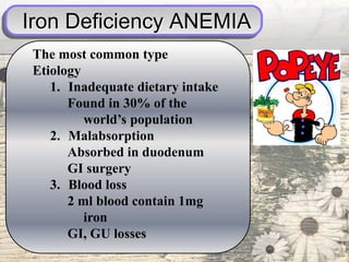
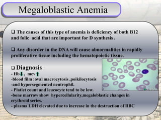
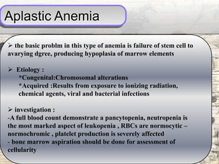
Done by :
Dr. Nawras
PALLOR
 pallor is a pale color of the skin and mucous membrane
due to reduce in amount of oxyhemoglobin
 there are many causes of pallor :
- Anemia : which is the most common cause
-Leukemia
-Heart disease
-Sleep deprivation
- Shock : whether it is septic , Anaphylactic, Cardiogenic,
Neurogenic and hypovolemic shock
- syncope =decrease in the blood supply to the
brain=hypotension leading to pallor
.
PALLOR
-Also in case of endocrine defect including:
1. long steady diabetic patient lead to keratin
deposition in the skin
2. Hypothyroidism with or without anemia
3. hypopituitarism leads to decrease in the
melanin stimulatory hormone
 Pallor is seen in :
* Palm creases
* Conjunctiva.
* And mucous membranes
ANEMIA
is a reduction in the number of RBCs, the quantity of
hemoglobin, or the volume of RBCs
Hb level below than 10 g/dl is considered as a significant level
to assess anemia
 Because the main function of RBCs is oxygenation, anemia
results in varying degrees of hypoxia depending on the degree of
anemia and the rapidity of development but its independent on
the cause.
 symptom of anemia : like fatigue ,headache ,dizziness
Palpitation , dimness of vision and others
 signs : the most important is pallor and others like :
Tachycardia, cardiac dilation, edema and murmurs
physical finding of ANEMIA
Severe Pallor
Koilonychia
Smooth shiny tongue
Types of ANEMIA
Anemia
Iron def.
anemia
Megaloplasti
c anemia
Hemolyti
c anemia
aplastic
anemia
Anemia of
chronic dis.
Iron Deficiency ANEMIA
The most common type
Etiology
1. Inadequate dietary intake
Found in 30% of the
world’s population
2. Malabsorption
Absorbed in duodenum
GI surgery
3. Blood loss
2 ml blood contain 1mg
iron
GI, GU losses
Investigation
Blood film : Hemoglobin level reduced, mean
corpuscular volume reduced ,blood film:
hypochromic microcytic,poikilocytosis
bone marrow aspiration : iron stores is empty
plasma iron
 serum ferritin reduced and the total binding
capacity increased.
gastrointestinal tract endoscopy.
patients with intravascular red cell destruction
should have urine test for hemosiderin
Lack of iron in
bone marrow aspirate
normal
Megaloblastic Anemia
 The causes of this type of anemia is deficiency of both B12
and folic acid that are important for D synthesis .
 Any disorder in the DNA will cause abnormalities in rapidly
proliferative tissue including the hematopoietic tissue.
 Diagnosis :
- Hb , mcv
-blood film :oval macrocytosis ,poikilocytosis
-and hypersegmented neutrophil.
- Platlet count and leucocyte tend to be low.
-bone marrow show hypercellularity,megaloblastic changes in
erythroid series.
- plasma LDH elevated due to increase in the destruction of RBC
Megaloblastic Anemia
 Common forms of megaloblastic anemia is :
1. Cobalamin deficiency ( vitamin B12) known as
pernicious anemia
-Inadequate diet ,intrinsic factor deficiency (congenital or
due to gastoctomy
-disease of the terminal ileum(e.g. Crohn’s disease),
- may be removed from gut by bacterial proliferation and
arasite
2. folate deficiency
- Poor intake
- malabsorption(Coeliac disease)
- increase demand(pregnancy)or drugs.
Hemolytic Anemia
o Various abnormalities lead to decrease in the life span of
the RBC & development of anemia when the marrow output
no longer compensates
o increasing in BM actiivity will be reflected as increasing in
reticulocye count in periphral blood
othe catabolic pathways for heamoglobin degredation are
overloaded and a modest increase in unconjucated
bilirubine in the blood and increase absorption urobilinogen
from the gut that excreted in urine
othere is no colour change in urine sine it is color less also
jaundice may developed .
Hemolytic Anemia
CAUSES OF HEMOLYTIC ANEMIA
* CONGENITAL
* AQUIRED
-membrane abnormalities : hereditory spherocytosis Or
elliptocytosis
-hemoglobinopathies: in lack of Hb chain synthesis
(thalassemia) or in amino acid substitution or alternation of
Hb chain like in sickle cell anemia
- membrane defect : G6PD def.
- IMMUNE : Autoimmune disease like SLA
- NON IMMUNE : like : burn , artificial cardiac valve , infections ,
malaria , drugs , dyserthropoietic like in paroxysmal nocturnal
hemoglobinuria
Aplastic Anemia
 the basic problm in this type of anemia is failure of stem cell to
avarying dgree, producing hypoplasia of marrow elements
 Etiology :
*Congenital:Chromosomal alterations
*Acquired :Results from exposure to ionizing radiation,
chemical agents, viral and bacterial infections
 investigation :
-A full blood count demonstrate a pancytopenia, neutropenia is
the most marked aspect of leukopenia , RBCs are normocytic –
normochromic , platelet production is severely affected
- bone marrow aspiration should be done for assessment of
cellularity
Anemia of chronic disease
 This is a commen type of anemia ,especially in hospital
papulation characterized by :
- Anemia occurs in he setting of chronic infection ,
inflammation , or neoplasia
- Anemia is not related o bleeding , hemolysis or marrow
infiltration
- Anemia is mild , with normal MCV and normocytic –
noromchromic RBC
- the serum iron is low but iron stors is normal or increased
Anemia of chronic disease
* Correlates with non specific symptoms of CRF
*several mechanism are implicated including:
- Relative deficiency of erythropoietin
- diminished erythropoiesis due to toxic effect of uremia on marrow
precursor cell.
- reduce red cell survival.
- increased blood loss due to capillary fragility and poor platelet
formation.
- reduced dietary intake and absorption of iron.
Example is anemia due to chronic renal failure :
Pallor

Pallor

  • 1.
  • 2.
    PALLOR  pallor isa pale color of the skin and mucous membrane due to reduce in amount of oxyhemoglobin  there are many causes of pallor : - Anemia : which is the most common cause -Leukemia -Heart disease -Sleep deprivation - Shock : whether it is septic , Anaphylactic, Cardiogenic, Neurogenic and hypovolemic shock - syncope =decrease in the blood supply to the brain=hypotension leading to pallor .
  • 3.
    PALLOR -Also in caseof endocrine defect including: 1. long steady diabetic patient lead to keratin deposition in the skin 2. Hypothyroidism with or without anemia 3. hypopituitarism leads to decrease in the melanin stimulatory hormone  Pallor is seen in : * Palm creases * Conjunctiva. * And mucous membranes
  • 4.
    ANEMIA is a reductionin the number of RBCs, the quantity of hemoglobin, or the volume of RBCs Hb level below than 10 g/dl is considered as a significant level to assess anemia  Because the main function of RBCs is oxygenation, anemia results in varying degrees of hypoxia depending on the degree of anemia and the rapidity of development but its independent on the cause.  symptom of anemia : like fatigue ,headache ,dizziness Palpitation , dimness of vision and others  signs : the most important is pallor and others like : Tachycardia, cardiac dilation, edema and murmurs
  • 5.
    physical finding ofANEMIA Severe Pallor
  • 6.
  • 7.
  • 8.
    Types of ANEMIA Anemia Irondef. anemia Megaloplasti c anemia Hemolyti c anemia aplastic anemia Anemia of chronic dis.
  • 9.
    Iron Deficiency ANEMIA Themost common type Etiology 1. Inadequate dietary intake Found in 30% of the world’s population 2. Malabsorption Absorbed in duodenum GI surgery 3. Blood loss 2 ml blood contain 1mg iron GI, GU losses
  • 10.
    Investigation Blood film :Hemoglobin level reduced, mean corpuscular volume reduced ,blood film: hypochromic microcytic,poikilocytosis bone marrow aspiration : iron stores is empty plasma iron  serum ferritin reduced and the total binding capacity increased. gastrointestinal tract endoscopy. patients with intravascular red cell destruction should have urine test for hemosiderin Lack of iron in bone marrow aspirate normal
  • 11.
    Megaloblastic Anemia  Thecauses of this type of anemia is deficiency of both B12 and folic acid that are important for D synthesis .  Any disorder in the DNA will cause abnormalities in rapidly proliferative tissue including the hematopoietic tissue.  Diagnosis : - Hb , mcv -blood film :oval macrocytosis ,poikilocytosis -and hypersegmented neutrophil. - Platlet count and leucocyte tend to be low. -bone marrow show hypercellularity,megaloblastic changes in erythroid series. - plasma LDH elevated due to increase in the destruction of RBC
  • 12.
    Megaloblastic Anemia  Commonforms of megaloblastic anemia is : 1. Cobalamin deficiency ( vitamin B12) known as pernicious anemia -Inadequate diet ,intrinsic factor deficiency (congenital or due to gastoctomy -disease of the terminal ileum(e.g. Crohn’s disease), - may be removed from gut by bacterial proliferation and arasite 2. folate deficiency - Poor intake - malabsorption(Coeliac disease) - increase demand(pregnancy)or drugs.
  • 13.
    Hemolytic Anemia o Variousabnormalities lead to decrease in the life span of the RBC & development of anemia when the marrow output no longer compensates o increasing in BM actiivity will be reflected as increasing in reticulocye count in periphral blood othe catabolic pathways for heamoglobin degredation are overloaded and a modest increase in unconjucated bilirubine in the blood and increase absorption urobilinogen from the gut that excreted in urine othere is no colour change in urine sine it is color less also jaundice may developed .
  • 14.
    Hemolytic Anemia CAUSES OFHEMOLYTIC ANEMIA * CONGENITAL * AQUIRED -membrane abnormalities : hereditory spherocytosis Or elliptocytosis -hemoglobinopathies: in lack of Hb chain synthesis (thalassemia) or in amino acid substitution or alternation of Hb chain like in sickle cell anemia - membrane defect : G6PD def. - IMMUNE : Autoimmune disease like SLA - NON IMMUNE : like : burn , artificial cardiac valve , infections , malaria , drugs , dyserthropoietic like in paroxysmal nocturnal hemoglobinuria
  • 15.
    Aplastic Anemia  thebasic problm in this type of anemia is failure of stem cell to avarying dgree, producing hypoplasia of marrow elements  Etiology : *Congenital:Chromosomal alterations *Acquired :Results from exposure to ionizing radiation, chemical agents, viral and bacterial infections  investigation : -A full blood count demonstrate a pancytopenia, neutropenia is the most marked aspect of leukopenia , RBCs are normocytic – normochromic , platelet production is severely affected - bone marrow aspiration should be done for assessment of cellularity
  • 16.
    Anemia of chronicdisease  This is a commen type of anemia ,especially in hospital papulation characterized by : - Anemia occurs in he setting of chronic infection , inflammation , or neoplasia - Anemia is not related o bleeding , hemolysis or marrow infiltration - Anemia is mild , with normal MCV and normocytic – noromchromic RBC - the serum iron is low but iron stors is normal or increased
  • 17.
    Anemia of chronicdisease * Correlates with non specific symptoms of CRF *several mechanism are implicated including: - Relative deficiency of erythropoietin - diminished erythropoiesis due to toxic effect of uremia on marrow precursor cell. - reduce red cell survival. - increased blood loss due to capillary fragility and poor platelet formation. - reduced dietary intake and absorption of iron. Example is anemia due to chronic renal failure :