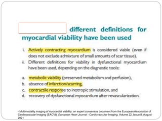
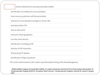
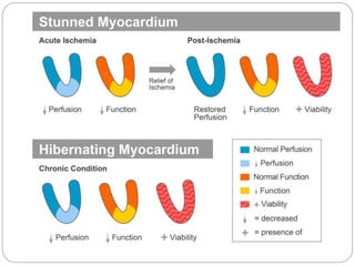
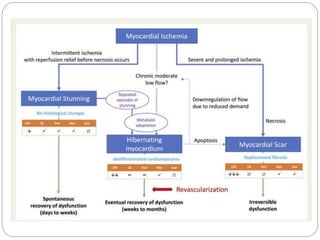
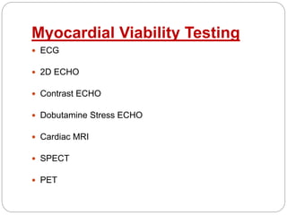
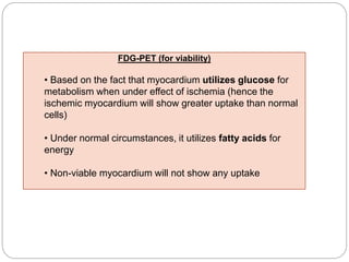
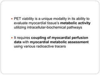
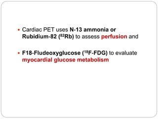
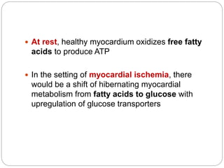
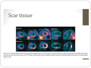
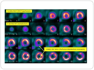
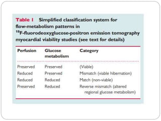
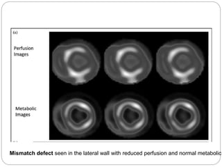
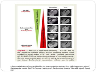
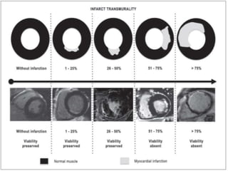
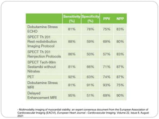
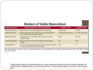
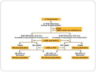
Left ventricular (LV) dysfunction remains one of the
best prognostic determinants of survival in patients
with coronary artery disease (CAD)
⚫ It was originally thought that dysfunctional
myocardium after an infarction was irreversibly
damaged
⚫ However, it was later recognized that some of the
involved tissue remained viable and contractility may
be restored with revascularization
preop TEE assessment of atrial septal defect is very important for making decision for device closure, properly assessed adequate rims of ASD will reduce risk of device embolization to almost nil.
A lecture on the echocardiographic evaluation of hypertrophic cardiomyopathy. Starts with an overview of the topic then a systematic approach to diagnosis and then a differential diagnosis followed by take-home messages and conclusion.
IVUS may not be clinically warranted in all interventions, and should be seen as an adjunct to angiography. IVUS provides information about vessel morphology, plaque topography, and therapeutic outcomes that is often either equivocal or unavailable in angiographic images.
There are 3 situations in which IVUS has the most clinical utility:
Small vessel stenting: Studies have shown that post-stent restenosis rates are higher in small vessels. This is particularly true for vessels with diameters of 3.0mm or less, wherein small increases in stent diameter have been shown to significantly decrease the rate of restenosis. A study by Moussa et al showed that, as measured by IVUS, the incidence of restenosis has an inverse relationship to the post-procedure in-stent lumen CSA1.
In-Stent restenosis: In these cases, IVUS helps to determine whether the restenosis is due to inadequate stent deployment (underexpansion or incomplete apposition) due to intimal hyperplasia. IVUS will also help you select the proper device size for treatment of the stented area.
Difficult to assess lesions: At times, images of a lesion and the adjacent reference segment are often hazy. IVUS should be used to identify whether the angiographic appearance is due to dissection, thrombus, residual plaque, or is benign.
Reverse Takotsubo Cardiomyopathy Following General AnaesthesiaPremier Publishers
Reverse takotsubo cardiomyopathy(r-TTC) is a rare condition in which regional wall motion abnormalities affect the basal segments of left ventricle in absence of significant coronary artery disease. The Diagnosis is established by characteristic echocardiographic findings, clinical manifestations, and laboratory features. In this report we demonstrate a case of general anaesthesia induced cardiomyopathy in 21 years old female.
Refers to cardiac muscle that is alive, not dead presence of cellular, metabolic, and microscopic contractile function Clinically LV systolic dysfunction in ischemic heart disease does not always represent irreversible damage and dysfunctional but viable myocardium has the potential to improve its systolic function after revascularization Two basic mechanisms of reversible ischemic dysfunction myocardial stunning myocardial hibernation
Abstract | In clinical guidelines, drugs for symptomatic angina are classified as being first choice
(β‑blockers, calcium-channel blockers, short-acting nitrates) or second choice (ivabradine,
nicorandil, ranolazine, trimetazidine), with the recommendation to reserve second-choice
medications for patients who have contraindications to first-choice agents, do not tolerate them,
or remain symptomatic. No direct comparisons between first-choice and second-choice
treatments have demonstrated the superiority of one group of drugs over the other.
Meta-analyses show that all antianginal drugs have similar efficacy in reducing symptoms,
but provide no evidence for improvement in survival. The newer, second-choice drugs have more
evidence-based clinical data that are more contemporary than is available for traditional
first-choice drugs. Considering some drugs, but not others, to be first choice is, therefore,
difficult. Moreover, double or triple therapy is often needed to control angina. Patients with
angina can have several comorbidities, and symptoms can result from various underlying
pathophysiologies. Some agents, in addition to having antianginal effects, have properties that
could be useful depending on the comorbidities present and the mechanisms of angina, but the
guidelines do not provide recommendations on the optimal combinations of drugs. In this
Consensus Statement, we propose an individualized approach to angina treatment, which takes
into consideration the patient, their comorbidities, and the underlying mechanism of disease
Exercise Testing in Cardiology : Dr. Akif Baigakifab93
The testing modality and protocol should be selected in accordance with the patient’s estimated functional capacity based on age, estimated physical fitness from the patient’s history, and underlying disease
Several exercise test protocols are available for both treadmill and stationary cycle ergometers
Patients who have low estimated fitness levels or are deemed to be at higher risk because of underlying disease (e.g., recent MI, heart failure) should be tested with a less aggressive exercise protocol
Treadmill and cycle ergometers may use stepped or continuous ramp protocols
Work rate increments (stages) during stepped protocols can vary from 1 to 2.5 METs
Ramp protocols are designed with stages that are no longer than 1 minute and for the patient to attain peak effort within 8 to 12 minutes
The natriuretic peptide system works antagonistically to the RAAS and has favorable effects on the pathogenesis of heart failure
Natriuretic peptides are broken down by an enzyme called neprilysin
Neprilysin is also responsible for the breakdown of other substances, including bradykinin and angiotensin II
Sacubitril/valsartan is a combination product
Sacubitril is a pro-drug that, upon activation, acts as a neprilysin inhibitor
It works by blocking the action of neprilysin, thus preventing the breakdown of natriuretic peptides
This leads to a prolonged duration of the favorable effects of these peptides
Coronary heart disease (CHD) remains a leading cause of death worldwide, accounting for 16% of total deaths globally .
Atherosclerosis plays a central role, with early fatty streaks progressing to late complex atheromas
Vascular calcification, the pathogenic and process of ectopic bone production, specifically was shown to strongly correlate with degree of atherosclerosis (both calcified and noncalcified)
Vascular calcification was shown independently to predict cardiovascular morbidity and mortality
These associations, combined with the radio-opaque appearance of calcium hydroxyappatite on CT images, have led to extensive investigation of the quantification, or scoring, of coronary artery calcium (CAC).
CAC scoring has emerged as a widely available and powerful tool for stratifying cardiovascular risk, predicting patient outcomes, and guiding preventive therapy
A coronary bifurcation consists of a flow divider (carina) and three vessel segments:
The proximal main vessel (PMV)
The distal main vessel (DMV) and
The side branch (SB).
A bifurcation lesion is a major epicardial coronary artery stenosis next to and/or including the ostium of a significant side branch
A significant SB is a branch whose severe narrowing or acute occlusion before or during intervention can cause considerable ischemia or a new infarction area that will worsen the clinical course of a particular patient.
Other important elements to consider that are not inherent in the bifurcation classifications include:
Extent of disease on the SB (limited to the ostium or involving the vessel beyond the ostium)
Its size (over 2.5mm in reference diameter)
Bifurcation angle, and
Disease distribution
HCM is a common genetic heart disease reported in populations globally
Inherited in an autosomal dominant pattern
The distribution of HCM is equal by sex, although women are diagnosed less commonly than men
The prevalence of unexplained asymptomatic hypertrophy in young adults has been reported to range from 1:200 to 1:500
Tetralogy of Fallot (TOF) is a congenital heart defect, which has four anatomical components:
Anterior malalignment ventricular septal defect (VSD)
Aortic override over the muscular septum
Variable degrees of subvalvar, valvar, and supravalvar pulmonary stenosis
Right ventricular (RV) infundibular narrowing and RV hypertrophy
Ventricular septal defects occur either as an isolated defect or as a component of a more complex lesion
It occurs in 50 percent of all children with CHD and in 20 to 30 percent as an isolated lesion
Most common congenital cardiac anomaly in children
Second most common congenital abnormality in adults, second only to bicuspid aortic valves
They are more common in premature infants and those born with low weight
VSDs are slightly more common in females (56%)
Patients with peripheral artery disease who have undergone lower-extremity revascularization are at high risk for major adverse limb and cardiovascular events
The efficacy and safety of rivaroxaban in this context are uncertain
Most common cyanotic heart defect seen in children beyond infancy, accounting for a third of all congenital heart disease (CHD) in this age group
Tetralogy of Fallot (TOF) is a congenital heart defect, which has four anatomical components:
Anterior malalignment ventricular septal defect (VSD)
Aortic override over the muscular septum
Variable degrees of subvalvar, valvar, and supravalvar pulmonary stenosis
Right ventricular (RV) infundibular narrowing and RV hypertrophy
Bentracimab (also known as PB2452) is a neutralizing recombinant human immunoglobulin G1 monoclonal antibody antigen-binding fragment that binds ticagrelor and its major active circulating metabolite with high affinity and specificity
Chlorthalidone for hypertension in advanced ckdakifab93
Chlorthalidone, a thiazide-like diuretic, reduces cardiovascular morbidity, such as the incidence of stroke and heart failure, and cardiovascular mortality
However, its efficacy and safety among patients with advanced chronic kidney disease remain poorly understood
An acute illness caused by an autoimmune response to infection with group A Streptococcus, leading to a range of possible symptoms and signs affecting any or all of heart, joints, brain, skin and subcutaneous tissues
Amyloidosis is a group of protein-folding disorders in which >1 organ is infiltrated by proteinaceous deposits known as amyloid. Amyloid involvement of the heart (cardiac amyloidosis) carries the worst prognosis of any involved organ, and light-chain (AL) amyloidosis is the most serious form of the disease
Macroeconomics- Movie Location
This will be used as part of your Personal Professional Portfolio once graded.
Objective:
Prepare a presentation or a paper using research, basic comparative analysis, data organization and application of economic information. You will make an informed assessment of an economic climate outside of the United States to accomplish an entertainment industry objective.
Unit 8 - Information and Communication Technology (Paper I).pdfThiyagu K
This slides describes the basic concepts of ICT, basics of Email, Emerging Technology and Digital Initiatives in Education. This presentations aligns with the UGC Paper I syllabus.
Palestine last event orientationfvgnh .pptxRaedMohamed3
An EFL lesson about the current events in Palestine. It is intended to be for intermediate students who wish to increase their listening skills through a short lesson in power point.
Model Attribute Check Company Auto PropertyCeline George
In Odoo, the multi-company feature allows you to manage multiple companies within a single Odoo database instance. Each company can have its own configurations while still sharing common resources such as products, customers, and suppliers.
Instructions for Submissions thorugh G- Classroom.pptxJheel Barad
This presentation provides a briefing on how to upload submissions and documents in Google Classroom. It was prepared as part of an orientation for new Sainik School in-service teacher trainees. As a training officer, my goal is to ensure that you are comfortable and proficient with this essential tool for managing assignments and fostering student engagement.
June 3, 2024 Anti-Semitism Letter Sent to MIT President Kornbluth and MIT Cor...Levi Shapiro
Letter from the Congress of the United States regarding Anti-Semitism sent June 3rd to MIT President Sally Kornbluth, MIT Corp Chair, Mark Gorenberg
Dear Dr. Kornbluth and Mr. Gorenberg,
The US House of Representatives is deeply concerned by ongoing and pervasive acts of antisemitic
harassment and intimidation at the Massachusetts Institute of Technology (MIT). Failing to act decisively to ensure a safe learning environment for all students would be a grave dereliction of your responsibilities as President of MIT and Chair of the MIT Corporation.
This Congress will not stand idly by and allow an environment hostile to Jewish students to persist. The House believes that your institution is in violation of Title VI of the Civil Rights Act, and the inability or
unwillingness to rectify this violation through action requires accountability.
Postsecondary education is a unique opportunity for students to learn and have their ideas and beliefs challenged. However, universities receiving hundreds of millions of federal funds annually have denied
students that opportunity and have been hijacked to become venues for the promotion of terrorism, antisemitic harassment and intimidation, unlawful encampments, and in some cases, assaults and riots.
The House of Representatives will not countenance the use of federal funds to indoctrinate students into hateful, antisemitic, anti-American supporters of terrorism. Investigations into campus antisemitism by the Committee on Education and the Workforce and the Committee on Ways and Means have been expanded into a Congress-wide probe across all relevant jurisdictions to address this national crisis. The undersigned Committees will conduct oversight into the use of federal funds at MIT and its learning environment under authorities granted to each Committee.
• The Committee on Education and the Workforce has been investigating your institution since December 7, 2023. The Committee has broad jurisdiction over postsecondary education, including its compliance with Title VI of the Civil Rights Act, campus safety concerns over disruptions to the learning environment, and the awarding of federal student aid under the Higher Education Act.
• The Committee on Oversight and Accountability is investigating the sources of funding and other support flowing to groups espousing pro-Hamas propaganda and engaged in antisemitic harassment and intimidation of students. The Committee on Oversight and Accountability is the principal oversight committee of the US House of Representatives and has broad authority to investigate “any matter” at “any time” under House Rule X.
• The Committee on Ways and Means has been investigating several universities since November 15, 2023, when the Committee held a hearing entitled From Ivory Towers to Dark Corners: Investigating the Nexus Between Antisemitism, Tax-Exempt Universities, and Terror Financing. The Committee followed the hearing with letters to those institutions on January 10, 202
2. - Multimodality imaging of myocardial viability: an expert consensus document from the European Association of
Cardiovascular Imaging (EACVI), European Heart Journal - Cardiovascular Imaging, Volume 22, Issue 8, August
2021
3. - Multimodality imaging of myocardial viability: an expert consensus document from the European Association of
Cardiovascular Imaging (EACVI), European Heart Journal - Cardiovascular Imaging, Volume 22, Issue 8, August
2021
4. Left ventricular (LV) dysfunction remains one
of the best prognostic determinants of survival in
patients with coronary artery disease (CAD)
It was originally thought that dysfunctional
myocardium after an infarction was irreversibly
damaged
However, it was later recognized that some of the
involved tissue remained viable and contractility
may be restored with revascularization
5.
6. After a myocardial infarction, the myocardium will
usually demonstrate one of 5 pathophysiologies:
Normal myocardial perfusion and function
Myocardial ischemia
Stunned myocardium
Myocardial hibernation
Non-viable infarction
7.
8. Prompt reperfusion or the presence of
collateral vessels and intact coronary
microvasculature function may preserve
myocardial perfusion
Ischemia occurs as a result of decreased blood
flow resulting in low ATP production and
subsequent LV dysfunction
9. Myocardial Stunning
Myocardial stunning is a reversible state of
regional contractile dysfunction that occurs after
transient ischemia without ensuing necrosis
Myocardial stunning is believed to play an
important role in persistent contractile dysfunction
seen in acute myocardial infarction patients
after successful reperfusion
In general, myocardial perfusion is normal and
function recovers relatively quickly
10. Myocardial Hibernation
Myocardial hibernation is a state of persistent left
ventricular dysfunction that results from
chronically reduced blood flow or repetitive
stunning without infarction and necrosis
A downregulation in contractile function at rest
is thought to represent a protective mechanism to
reduce myocardial oxygen requirements and
ensure myocyte survival
11. When severe cellular hypoperfusion and damage
occurs, only cellular function that is essential for
survival, such as membrane integrity, is
preserved
Preserved or increased myocardial glucose
metabolism also occurs during this state
12.
13.
14. Nonviable myocardium
If myocardial perfusion is not restored, irreversible
myocardial necrosis can occur
The goal of viability testing is to determine if a
large portion of dysfunctional myocardium is
nonviable in which case the risks would likely
outweigh benefit of revascularization
16. Several pathophysiological principles and molecular
targets may be used clinically to identify viable
myocardium
Viable myocytes are characterized by preserved energy
conversion by mitochondria and maintained membrane
function and action potentials
Therefore, myocardial viability may be identified by
preserved electrical activity, for instance by endocardial
surface potentials during electrophysiological mapping
studies
ECG Q waves on the surface ECG are however relatively
non-specific, relating more closely to the subendocardial
extent rather than transmurality of necrosis and many
myocardial segments with Q waves still demonstrate
viability by other methods
17. Membrane function is explored by active uptake
of 201Tl and mitochondrial function by
retention of 99Tc-based tracers such as sestamibi
and tetrofosmin
Active contraction is a definite marker of
preserved myocyte viability
Dysfunctional segments at rest may or may not
be viable and frequently require further
assessment
18. Stunned and hibernating myocardium is
characterized by reduced sensitivity of myofibrils to
calcium, resulting in reduced mechanical efficiency at
rest
This may be overcome when the intra-cellular calcium
content increases, viable myocardium therefore has
preserved inotropic reserve
These principles underlie the ability of dysfunctional
viable myocardium to improve contractility after
premature beats, nitrate infusion and more
commonly dobutamine stimulation
19. Another feature of viable myocardium is that
resting perfusion is generally preserved or only
mildly reduced, and that hibernating myocardium
displays preserved metabolism with metabolic
preference for glucose over fatty acids in the
fasting state
These principles underlie the detection of
myocardial viability using single photon emission
computed tomography (SPECT) or positron
emission tomography (PET) perfusion and
metabolic imaging (such as combined NH3 FDG
PET)
20. A final method for detecting myocardial viability is
demonstrating the absence of myocyte
necrosis and the absence of replacement fibrotic
tissue
These principles underlie the detection of
myocardial viability by late-gadolinium
enhancement (LGE) cardiovascular magnetic
resonance (CMR)
24. The ECG is an initial tool in the evaluation of viability
Absence of pathologic Q‐waves may be suggestive
of viable myocardium and the presence of them may
imply infarct
Q waves are not specific for myocardial infarct and
are seen in myocardial hypertrophy, WPW, and
rarely hibernating myocardium
The presence or absence of Q waves information can
be a helpful complementary marker in conjunction
with the other imaging parameters and clinical data to
determine myocardial viability
25. R wave height in lead V3
The R wave height of less than 3 mm in lead V3
was 90.3% sensitive for the detection of non
viable myocardium
The specificity at the same cut-off point was 25%
Journal of Clinical and Diagnostic Research. 2021 Aug, Vol-15(8): OC18-OC21
26. Sum of R Wave Height in all
Precordial Leads
When the sum of R wave height in all precordial
leads was <28.5mm
Sensitivity : 93.2%
Specificity : 25%
Journal of Clinical and Diagnostic Research. 2021 Aug, Vol-15(8): OC18-OC21
28. END DIASTOLIC WALL
THICKNESS (EDWT)
EDWT more than 6 mm has a sensitivity of
94%, albeit with a low specificity of 48% for
detection of myocardial viability
With EDWT less than 6 mm, less than 5% will be
viable, while with thickness above that viability is
considered to be more than 50%
30. Myocardial contrast echocardiography (MCE)
evaluates myocardial microvascular integrity
Viable myocardium has preserved microvascular
integrity
Intravenously injected bubble contrast agents lead
to contrast enhancement of dyssynergic but viable
myocardial segments that can be detected with
echocardiography
Non-viable myocardium does not show significant
enhancement with bubble contrast due to disruption
of the coronary microvasculature.
34. Categorization of Wall Motion
Hypokinesis is defined as the preservation of some degree of thickening
and inward motion of the endocardium during systole but less than
normal
It has been defined arbitrarily as less than 5 mm of endocardial
excursion
Akinesis is defined as the absence of systolic myocardial thickening
and endocardial excursion
Dyskinesis is the most extreme form of a wall motion abnormality and is
defined as systolic thinning and outward motion or bulging of the
myocardium during systole
A left ventricular segment that is thin and/or highly echogenic
suggests the presence of scar
35.
36.
37.
38.
39.
40.
41. ECG evidence of ischemia is less reliable during dobutamine infusion than it is
during exercise testing
Thus, neither ST-segment depression nor elevation occurring in the
absence of a wall motion abnormality or typical symptoms is sufficient reason
for terminating the dobutamine infusion
42. Safety of Dobutamine
Because of the short half-life of dobutamine, inducible
ischemia can be readily reversed through termination of
the infusion
In severe cases or when the ischemic manifestations
persist, a short-acting intravenous β blocker (such as
metoprolol or esmolol) is effective
The most common side effects associated with
dobutamine infusion are minor arrhythmias such as
premature ventricular contractions and atrial arrhythmias
and minor symptoms such as palpitations or anxiety
Nonsustained ventricular tachycardia occurs in
approximately 3% of patients and generally terminates
spontaneously or can be successfully treated with an
intravenous β blocker
43. Contraindications
There are no absolute contraindications to dobutamine
stress testing
Unstable patients, such as those with uncompensated
heart failure for unstable angina, should rarely be
subjected to stress testing of any kind
Dobutamine echocardiography has been safely
performed in patients with:
Recent myocardial infarction
Extensive left ventricular dysfunction
Abdominal aortic aneurysm
Syncope, aortic stenosis
Hypertrophic cardiomyopathy
History of ventricular tachycardia, and aborted sudden death
44. Vasodilators Stress ECHO
Potent vasodilators such as dipyridamole and
adenosine have been used in conjunction with
echocardiography for the detection of coronary
artery disease
Unlike dobutamine, these agents work by
creating maldistribution of blood flow, that is, by
preventing the normal increase in flow in areas
supplied by stenotic coronary arteries (Coronary
Steal Phenomenon)
45. Adenosine is a potent and short-acting direct
coronary vasodilator
Dipyridamole is slower acting and its effects result
from inhibition of adenosine uptake
With both agents, the development of a wall motion
abnormality is predicated on the ability to create
sufficient maldistribution of regional blood flow to
result in an ischemia-induced wall motion abnormality
Compared with dobutamine, these changes tend to
be more subtle and short-lived
49. SPECT imaging provides reliable information on
myocardial perfusion and to some extent cellular
viability
Viability assessment can be performed either
with:
99mTc-sestamibi, a lipophilic cationic compound
99mTc-tetrofosmin, a diphosphine agent; or
201-thallium
50. Both sestamibi or tetrofosmin are transported
passively into the myocyte and are sequestered
within the mitochondria
Uptake requires negative transmembrane
potentials of sarcolemmal and mitochondrial
membranes
By contrast, Tl-201 mimics potassium, and is
taken up actively into the myocyte through the
Na-K-ATPase
51. The uptake and retention of all three tracers is
dependent on regional blood flow and
sarcolemmal membrane integrity (for thallium)
or mitochondrial membrane integrity (for
sestamibi and tetrofosmin)
The principles of viability detection by SPECT
mainly rely on demonstrating reversible stress
perfusion defects in dysfunctional segments
52. Areas with persistent little or no tracer uptake
indicate non-viable myocardium unlikely to
recover function after revascularization
Stress can be performed either after physical
exercise or after vasodilation with dipyridamole,
adenosine, or regadenoson
Rest-only images demonstrating preserved or
only mildly reduced perfusion are also indicative
of myocardial viability
53.
54. VIABILITY ASSESSMENT WITH THALLIUM-201
SINGLE-PHOTON EMISSION COMPUTED
TOMOGRAPHY MYOCARDIAL PERFUSION
IMAGING
Tl-201 behaves pharmacokinetically like a
potassium analog
Myocardial uptake of Tl-201 is an active Na/K
ATPase pump-dependent process, which requires
cell membrane integrity
Thus, Tl-201 myocardial uptake is an indication of
regional perfusion, which is necessary for tracer
delivery and myocyte membrane integrity and
metabolic activity (ATP production)
55. A pivotal characteristic of Tl-201 myocardial
uptake is its redistribution property
This phenomenon was initially described in the
late 1970s, with reports of stress-induced
myocardial perfusion defects that appeared to
normalize on repeat imaging at different time
intervals
56. This property is a consequence of a constant
exchange of the radiotracer between the
myocardial cells, extracellular space and
subsequently the blood pool after the initial
myocardial uptake
As Tl-201 is washed out of the myocardial cells,
radiotracer uptake from the blood pool continues to
take place
In areas of decreased perfusion or with diminished
coronary flow, the rate of Tl-201 extraction is
slower than in those with increased or normal
blood flow, leading to perfusion defects in these area
at initial stress imaging, performed 10–15min
following radiotracer injection
57. However, over-time, Tl-201 uptake continues in
areas with diminished blood flow, whereas the
radiotracer washes out from areas with normal or
increased initial blood flow
This constant redistribution of Tl-201 manifest,
on delayed imaging (3–4h following injection),
with resolution of the initial perfusion defect in
areas that appeared to have little or no tracer
activity on initial stress imaging
58. As Tl-201 uptake requires sarcolemmal
membrane integrity, resolution or ‘reversibility’ of
perfusion abnormality is considered to represent
myocardial viability
59.
60. Thallium-201 viability assessment protocols
Stress/4 and 24h delayed redistribution
protocol
During the 1980s, Tl-201 stress/4 h redistribution became
the standard protocol for assessment of myocardial
ischemia and to predict functional recovery after coronary
revascularization
However, it was noted that up to half of the segments with
fixed perfusion defects on 4h imaging demonstrated
normalization of perfusion or improvement in function after
revascularization
This finding suggests that under certain circumstances, Tl-
201 redistribution may take longer than 4 h, and therefore,
delayed imaging at 18–24 h could improve the ability of
the test to predict functional recovery after coronary
revascularization
These findings led to the use of Tl-201 stress/4 h
redistribution with delayed, 24 h imaging
61.
62.
63. Rest/4h redistribution imaging
protocol
Tl-201 rest/4 h redistribution imaging has also
been used and shown to be adequate for
identification of viable segments
When Tl-201 is injected at resting state, delayed
imaging at 18–24 h does not yield improved
viability detection
In this setting, the data suggest that a 10%
absolute increase in radiotracer uptake is
indicative of significant viability, which is
predictive of functional improvement after
revascularization
64.
65.
66.
67.
68.
69.
70. VIABILITY ASSESSMENT WITH TECHNETIUM-99M
SINGLE-PHOTON EMISSION COMPUTED
TOMOGRAPHY MYOCARDIAL PERFUSION
IMAGING
Following their extraction from the blood, Tc-99m
tracers bind to the mitochondria, and thus results
in a negligible washout or redistribution
75. FDG-PET (for viability)
• Based on the fact that myocardium utilizes glucose for
metabolism when under effect of ischemia (hence the
ischemic myocardium will show greater uptake than normal
cells)
• Under normal circumstances, it utilizes fatty acids for
energy
• Non-viable myocardium will not show any uptake
76. PET viability is a unique modality in its ability to
evaluate myocardial tissue’s metabolic activity
utilizing intracellular-biochemical pathways
It requires coupling of myocardial perfusion
data with myocardial metabolic assessment
using various radioactive tracers
77. Cardiac PET uses N‐13 ammonia or
Rubidium‐82 (82Rb) to assess perfusion and
F18‐Fludeoxyglucose (18F‐FDG) to evaluate
myocardial glucose metabolism
78. At rest, healthy myocardium oxidizes free fatty
acids to produce ATP
In the setting of myocardial ischemia, there
would be a shift of hibernating myocardial
metabolism from fatty acids to glucose with
upregulation of glucose transporters
79. For optimal 18F‐FDG uptake of viable
myocardium, it is crucial to stimulate
endogenous insulin release by appropriate
dietary protocol, oral or IV glucose loading, and
if needed insulin supplementation, to achieve
appropriate serum glucose (100–140 mg/dl)
levels before injecting 18F‐FDG
Suboptimal patient preparation may yield poor,
non‐diagnostic images
80. Preparation of diabetic patients can be
particularly challenging, requiring insulin injection
to overcome myocardial insulin resistance and
may take longer wait times from
injecting 18F‐FDG to image acquisition
81.
82. PET imaging is performed about 45–90 min (up to
3 h in diabetics) after injecting approximately
10 mCi (7 mSv) of 18F‐FDG (t ½ 110 min)
83. Myocardial uptake of FDG continues to increase,
and blood pool activity to decrease, even after 45
min
Waiting 90min after the injection of FDG may give
better signal to nose ratio as the blood pool has
less FDG and the myocardial uptake continues to
increase, especially in people with diabetes
The typical scan duration is typically 10–30min
84.
85.
86.
87.
88.
89.
90. Mismatch defect seen in the lateral wall with reduced perfusion and normal metabolic
91. Matched defect seen in the anterior wall with reduced perfusion and metabolic a
93. The two methods of viability testing by CMR are
Contractile reserve assessment using
dobutamine stress and
Late gadolinium enhancement (LGE) imaging
using gadolinium-based contrast agents (GBCA)
With the latter being the more common and
preferred technique
- Multimodality imaging of myocardial viability: an expert consensus document from the European Association of
Cardiovascular Imaging (EACVI), European Heart Journal - Cardiovascular Imaging, Volume 22, Issue 8, August
2021
94. GBCAs are paramagnetic metal compounds that,
when administered intravenously, cannot
penetrate intact myocardial sarcolemma and
accumulate extracellularly in the intravascular
blood pool and within myocardial interstitium
With LGE imaging, GBCAs are used to index cell
membrane integrity, as living myocardial cells
exclude GBCA when steady-state concentrations
are reached
- Multimodality imaging of myocardial viability: an expert consensus document from the European Association of
Cardiovascular Imaging (EACVI), European Heart Journal - Cardiovascular Imaging, Volume 22, Issue 8, August
2021
95. In an acute myocardial infarction, GBCA
passively diffuses intracellularly through ruptured
cell membranes and extracellularly in surrounding
necrotic tissue, whereas
In chronic infarcts GBCA concentrates in
collagenous scar that has replaced necrotic
tissue.
- Multimodality imaging of myocardial viability: an expert consensus document from the European Association of
Cardiovascular Imaging (EACVI), European Heart Journal - Cardiovascular Imaging, Volume 22, Issue 8, August
2021
96. Evaluation of resting function and
wall thickness
Preserved myocardial wall thickness of more
than 5.5 mm has a good sensitivity of 95%, but
low specificity for detecting myocardial viability on
CMR
End-diastolic wall thickness of >5.5 mm and cine
systolic wall thickening of >2 mm has
sensitivity and specificity between 85% and 90%
in the prediction of segmental contractile recovery
after revascularization
- Braunwald Textbook of Cardiology
97. Late-gadolinium enhancement
(LGE)
It has become the reference standard for the non-
invasive imaging of myocardial scar and focal
fibrosis in both ischaemic heart disease and non-
ischaemic cardiomyopathy
Clinically used gadolinium-based contrast agents are
distributed into the extracellular space following
intra-venous injection
They are therefore present in higher concentration in
fibrotic or infarcted myocardium
This is best observed 10–15 min after contrast
injection, when difference to normal myocardium are
maximized, using the ‘LGE’ technique
- Multimodality imaging of myocardial viability: an expert consensus document from the European Association of
Cardiovascular Imaging (EACVI), European Heart Journal - Cardiovascular Imaging, Volume 22, Issue 8, August
2021
98. LGE-CMR sequences are timed to selectively null
signal in normal myocardium, which appears black,
whereas areas of scaring with shorter T1 values
appear bright
LGE-CMR therefore images non-viable myocardium
and infers viability from the absence of enhancement
If the extent of scar is less as indicated by less than
50% transmural extent of hyperenhancement
indicates viability
If 4 or more dysfunctional segments show viability, it
has a good sensitivity of 95%, again with low
specificity of 45%
- Multimodality imaging of myocardial viability: an expert consensus document from the European Association of
Cardiovascular Imaging (EACVI), European Heart Journal - Cardiovascular Imaging, Volume 22, Issue 8, August
2021
99. - Multimodality imaging of myocardial viability: an expert consensus document from the European Association of
Cardiovascular Imaging (EACVI), European Heart Journal - Cardiovascular Imaging, Volume 22, Issue 8, August
2021
100.
101. Advantages of CMR LGE
High quality of images
Absence of ionizing radiation
High prognostic value, and
Lower costs relative to nuclear imaging
- Multimodality imaging of myocardial viability: an expert consensus document from the European Association of
Cardiovascular Imaging (EACVI), European Heart Journal - Cardiovascular Imaging, Volume 22, Issue 8, August
2021
102. Disadvantages of CMR-LGE
Need for gadolinium-based contrast injection, which although
generally safer than iodinated contrast agents, can cause
allergic reactions and anaphylaxis
Gadolinium-based contrast agents are considered
contraindicated in pregnancy, although a recent study showed
that in the second and third trimesters, CMR can be safely
performed even with contrast
Finally, LGE shows the expansion of the extracellular matrix,
regardless of whether this is due to collagen, water, or
amyloid infiltration
As a result, LGE may overestimate the extent of the scar if there is
myocardial oedema, in particular in acute myocardial infarction.
- Multimodality imaging of myocardial viability: an expert consensus document from the European Association of
Cardiovascular Imaging (EACVI), European Heart Journal - Cardiovascular Imaging, Volume 22, Issue 8, August
2021
103. - Multimodality imaging of myocardial viability: an expert consensus document from the European Association of
Cardiovascular Imaging (EACVI), European Heart Journal - Cardiovascular Imaging, Volume 22, Issue 8, August
2021
104. Dobutamine stress CMR
Like stress echocardiography, the evaluation of
contractile reserve using dobutamine stress CMR can
be used to assess viability
Infusion of low dose dobutamine (5–10 mcg/kg/min)
induces systolic wall thickening in viable regions of
myocardium but not in irreversibly scarred areas
Improvement in myocardial thickening of more than 2
mm with low dose dobutamine CMR is indicative of
viability
If this contractile reserve can be elicited, the
myocardium is more likely to improve after
revascularization
105. In addition, low-dose dobutamine can
accurately predict the development of adverse
remodelling following acute myocardial infarction
With high-dose dobutamine infusion (20–40
mcg/kg/min), the presence of inducible wall
motion abnormalities using cine CMR can trigger
a biphasic response and provides additional
accurate information regarding the presence of
ischaemia and prognosis
106. Results suggest that low-dose dobutamine
CMR is superior to both LGE CMR and wall
thickness in predicting recovery after
revascularization
This is particularly relevant for detecting viability
in patients with intermediate grades of transmural
infarction (up to 75% extent of LGE), but its
sensitivity may be reduced with more severely
impaired baseline LV function and those patients
with fewer than 50% of all myocardial
segments deemed viable may derive less
benefit from revascularization
107. Interestingly, there is a strong correlation
between LV ejection fraction (LVEF) measured
during low-dose dobutamine (10 lg/kg/min) and
LVEF 6 months after revascularization
The combined use of LGE and low-dose
dobutamine stress CMR, has a higher specificity
(91%) and a lower sensitivity (81%) according to
a meta-analysis
108. The inotropic response to dobutamine is strongly
associated with abnormalities of fatty acid
metabolism and is likely to depend on the
presence of viable myocardium which has not
undergone severe ultrastructural change with
myofibrillar degeneration which would otherwise
prevent contractile improvement with inotropic
stimulation
The combination of dobutamine stress with
other CMR sequences can give a more accurate
assessment of both ischaemia and viability, with
the potential to improve diagnostic performance
109. Keypoints of Cardiac MRI
CMR LGE is currently the reference method for clinical assessment of viability
and indicates myocardial necrosis or chronic scar
Scars with transmurality >50% are considered non-viable, less transmurality of
scar in dysfunctional myocardium is considered viable myocardium
LGE has high specificity for predicting absence of recovery but sensitivity may
be limited particularly in scars with intermediate transmurality (25–75%)
Low-dose dobutamine stress MR may have additional value in such patients
with intermediate transmurality of scar
Stress perfusion CMR also allows evaluation of ischaemia and coronary flow
reserve
110.
111.
112. Limitations
High cost
Limited availability
Longer imaging time and
Restrictions in patients with cardiac implantable electronic
devices (CIED)
Claustrophobia
Gadolinium enhancement is not suitable in those with low
glomerular filtration rate of below 30 ml per minute
113.
114. Advantage of MRI over
SPECT/PET
In single photon emission computed tomography
(SPECT) or positron emission tomography (PET)
imaging, the presence of scar is inferred by the
lack of uptake of myocardial perfusion tracers,
whereas CMR affords the luxury of direct
visualization of scar and normal myocardium
within the same image
This reduces the likelihood of falsely labeling
viable segments as nonviable due to relatively
low perfusion tracer counts, especially in thinned
walls where tracer counts will inherently be lower
115. Nuclear perfusion techniques also lack the excellent spatial resolution of
CMR (1.5 mm vs 10 mm for nuclear) and suffer from ionizing radiation
exposure
Wagner et al. showed that SPECT is inadequately sensitive in the
detection of subendocardial scar (<50% TEI) compared to CMR in both
human patients and a canine model with histopathologic correlation
Nearly one half of subendocardial infarcts were missed by SPECT in
human subjects when CMR was used as the reference standard
Modalities fared identically in the detection of near transmural infarcts
(>75% TEI), but nearly one quarter of infarcts with 50–75% TEI went
undetected by SPECT
This may result in the converse labeling of nonviable myocardium as
viable.
Wagner A, Mahrholdt H, Holly TA, et al. Contrast-enhanced MRI and routine single photon emission
computed tomography (SPECT) perfusion imaging for the detection of subendocardial myocardial
infarcts: an imaging study. Lancet 2003;361:374–379
119. Viability testing appears to be most helpful when
it is uncertain that the myocardial segment in
question is predominantly transmural scar or
otherwise
If the dysfunctional myocardial segment
possesses relatively preserved thickness with
wall motion no worse than hypokinesis and
absence of Q waves on EKG, it is unlikely that
segment is NVM (scar), precluding need for any
further testing to assess viability
120. Viability testing should be tailored to the individual
patient based on several factors including
limitations or contraindications of a particular
study in each patient, local expertise, and
availability
The degree of LV remodeling and dysfunction
may play a role in deciding which test to perform
121. Patients with extreme degrees of LV dilatation
and segmental wall thinning may need an
advanced imaging modality (CMR, PET)
In patients with mild to moderate degree of LV
dysfunction and remodeling, dobutamine stress
Echo and SPECT imaging may suffice
122.
123. - Multimodality imaging of myocardial viability: an expert consensus document from the European Association of
Cardiovascular Imaging (EACVI), European Heart Journal - Cardiovascular Imaging, Volume 22, Issue 8, August
2021
124. - Multimodality imaging of myocardial viability: an expert consensus document from the European Association of
Cardiovascular Imaging (EACVI), European Heart Journal - Cardiovascular Imaging, Volume 22, Issue 8, August
2021
125.
126. References
Braunwald textbook of cardiology
Feigenbaum echocardiography
Braunwald intervention cardiology
Multimodality imaging of myocardial viability:
an expert consensus document from the
European Association of Cardiovascular
Imaging (EACVI), European Heart Journal -
Cardiovascular Imaging, Volume 22, Issue 8,
August 2021