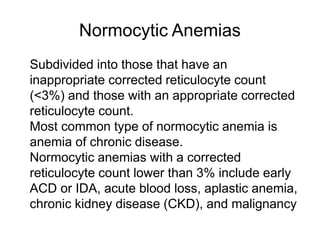
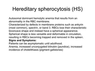
X-linked sideroblastic anemia results from a defect in the δ-ALA synthase gene causing defective enzyme production and laboratory findings similar to acquired sideroblastic anemia. It is treated with pyridoxine and blood transfusions if severe. β-thalassemia results from mutations reducing β-globin chain production, causing imbalanced globin ratios and hemolytic anemia. Trait forms show mild anemia while major requires transfusions. Normocytic anemias are categorized by corrected reticulocyte count; common causes include anemia of chronic disease, blood loss, aplastic anemia, and malignancy. Hemolytic anemias show increased reticulocytes and

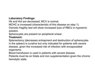
![Signs and Symptoms
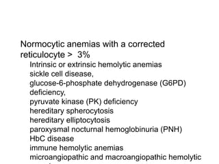
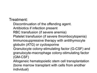
Pain crises occur beginning at age 1 or 2 years.
Vascular occlusion can lead to organ dysfunction or failure, causing bone infarcts,
avascular necrosis,
Acute chest syndrome (chest pain, shortness of breath [SOB], and pulmonary
infiltrates on chest radiograph), chronic leg ulcers, osteomyelitis (salmonella), and
stroke.
Dactylitis (hand-foot syndrome) or swelling of hands and feet can be seen in infants.
Recurrent splenic infarction and autosplenectomy occur at a young age. The
presence of HowellJolly bodies on a peripheral smear signifies impaired or absent
splenic function. Loss of splenic function makes patients increasingly susceptible to
infection by encapsulated bacteria, and daily antibiotic prophylaxis is required for the
treatment of children.
Aplastic crisis can occur in association with parvovirus B19.
Sequestration crisis results when there is entrapment of sickled RBCs in the spleen,
resulting in rapid splenomegaly.
Renal papillary necrosis can occur when microinfarcts of the kidney result in
microhematuria.](https://image.slidesharecdn.com/hematologyreivew4-230109120022-bb6b1582/85/Hematology-reivew-4-pptx-22-320.jpg)
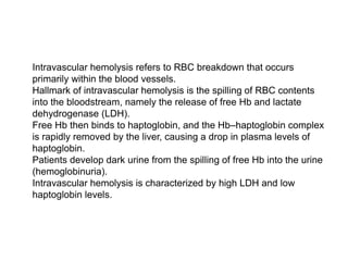
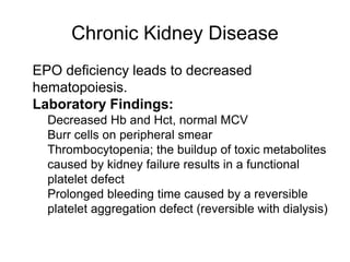
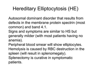
![ Drugs—sulfonamides (trimethoprim-sulfamethoxazole), dapsone,
primaquine, chloroquine, nitrofurantoin
Acidosis (e.g., diabetic ketoacidosis [DKA])
Fava beans (historically, this condition was called favism)
Signs and Symptoms:
Most are asymptomatic
History of neonatal jaundice and cholelithiasis
Episodic signs of anemia (possibly associated with jaundice and
splenomegaly)
Laboratory Findings:
Decreased Hb and Hct, normal MCV
Heinz bodies and/or bite cells on peripheral smear](https://image.slidesharecdn.com/hematologyreivew4-230109120022-bb6b1582/85/Hematology-reivew-4-pptx-28-320.jpg)
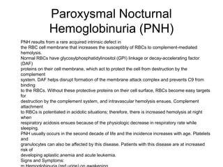
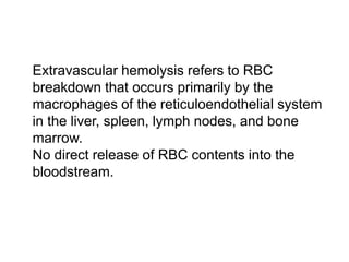
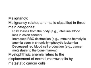
![ Clinical and Laboratory Findings:
m Mild extravascular hemolysis in homozygotes
or HbCC (people with HbC trait [HbAC] are
phenotypically normal)
m Peripheral smear with HbC crystals seen in
RBCs; will also see target cells
m Mild splenomegaly
m Mild anemia
Treatment:
m Usually no treatment is needed, but patients
may supplement with folate to improve anemia.](https://image.slidesharecdn.com/hematologyreivew4-230109120022-bb6b1582/85/Hematology-reivew-4-pptx-37-320.jpg)