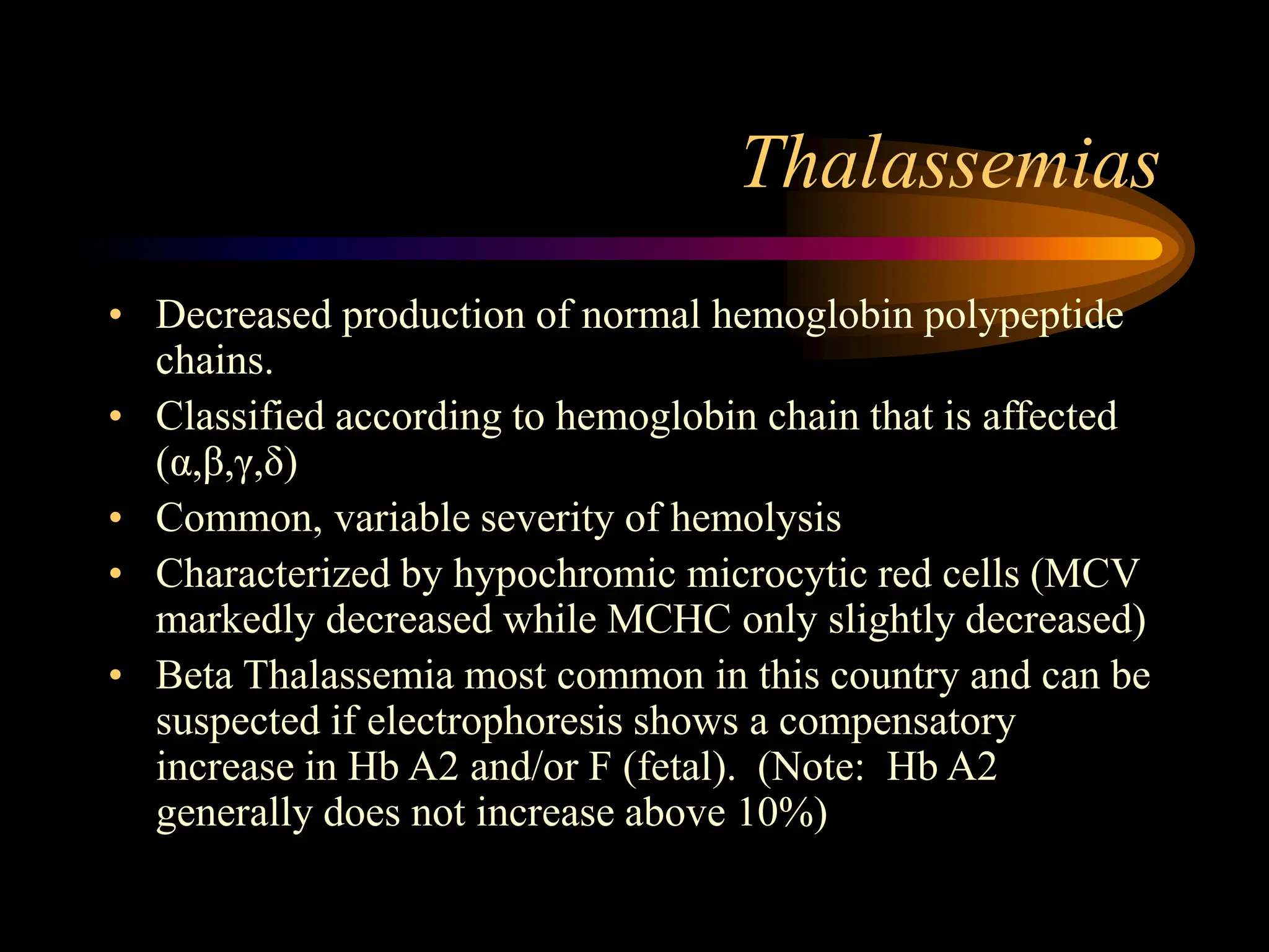
2. Objectives
At the end of this lecture student
should be able to:
1. Define Anaemia
2. Know Cut-off levels of Hb and
Haematocrit for anaemia.
3. Identify the causes of anaemia
4. classify anemia on the basis of morphology
and red blood cell production
5. Know how to Approach an anemia case
6. What is lukaemia?
2
3. 3
Anaemia
• Definition
• → Decreased level of Hb concentration
below a normal cut-off level
A.Cut-off level is specific to population
12. 12
2.Morphological Classification
• Microcytic Anaemia (↓ volume)
A. Iron deficiency anaemia
B. Anaemia of Inflammation
C. Thalassaemias
• Macrocytic Anaemia (↑ volume)
A. Folic Acid deficiency
B. Vit B12 deficiency
3. Normocytic Anaemia
A. ↑ Retics
• Acute blood loss
• Hemolytic anaemias
B. Normal or ↓ Retics
• Anaemia of chronic renal disease
13. 13
Morphological Classification (2)
3. Normocytic Anaemia
A. ↑ Retics
• Acute blood loss
• Hemolytic anaemias
B. Normal or ↓ Retics
• Anaemia of chronic renal disease
14. Anemia Workup - MCV
Iron Deficiency
Anemia of Chronic Disease
Thalassemias
Hemoglobinopathies
Sideroblastic Anemia
Microcytic
Anemia of chronic disease
Early iron deficiency
Hemoglobinopathies
Primary marrow disorders
Combined deficiencies
Increased destruction
Normocytic
Megaloblastic anemias
Liver disease/alcohol
Hemoglobinopathies
Metabolic disorders
Primary marrow disorders
Increased destruction
Macrocytic
Anemia
15. Normochromic, normocytic anemia
with effective erythropoiesis
• INCREASED reticulocyte count
• Acute blood loss
– Very acutely, with hypovolemia, may have
normal blood counts, will become anemic with
volume replenishment
• Hemolytic anemia
– Increased reticulocyte production cannot keep
pace with loss of RBCs peripherally
16. Coomb’s test
• Detects presence of either antibody on RBC
or of antibody in serum
• Helpful in determining if a hemolytic
anemia is immune-mediated
17. Special cases of hemolytic anemia
• Glucose-6 Phosphate Dehydrogenate
deficiency
– More common in African and Mediterranean
populations
– Lack of RBC enzyme makes cells very
sensitive to oxidative stress (infection, certain
drugs)
– Treatment: avoid triggers if possible, especially
inciting drugs
18. Sickle cell disease
• Abnormal hemoglobin causes change in RBC
shape, resulting in constant RBC destruction by
the spleen, functional asplenia, susceptible to
infection
• Arterial occlusion leads to infarcts, pain crises,
acute chest syndrome, stroke, MI
• Keep hydrated, treat pain, take infection seriously
• Also sickle-C and sickle-b-thalassemia
19.
20. Pathophysiology of SCD
In a red blood cell containing mostly Hb S…
…single Hb S
molecules in free in
solution;
allows red cell to be
soft, round, and
deformable
When oxygenated…
+ O2
- O2
- O2
…Hb S molecules
polymerize into
long fibers;
mishapen,
dehydrated and
adherent sickle
cells.
When deoxygenated
+ O2
Sickle Cell Disease: Pharmacologic Treatment
22. Pathophysiology of SCD
Consequences of Hb S polymerization
and RBC sickling
• Red cell injury
• Hemolysis
• RBC dehydration and dense cell formation
• Adhesion of RBC to venule endothelium
• Formation of heterocellular aggregates (WBC, ISC)
• Vasooclusion
• Local hypoxia, increased Hb S polymer formation
• Propagation of vasooclusion in adjacent vasculature
• Dysregulation of vasomotor tone by vasodilator mediators
(NO)
23. Molecular pathology of SCD
The sickle mutation
GAG
Glutamic acid
GTG
Valine
The bs Mutation
The same mutation found in all
bs genes around the world
6th Codon of b-Globin Gene
24. Molecular pathology of SCD
Normal versus sickle beta globin
Sickle Cell Disease: Pharmacologic Treatment
val
glu
1 2 3 4 5 6 7 8 9 10 146
- - - - - - - -
bA
Normal
- - - - - - - -
bS
1 2 3 4 5 6 7 8 9 10 146
Sickle
25. Clinical Pathology of SCD
1. Anemia
2. Vasoocclusion
3. Chronic organ damage
Sickle Cell Disease: Pharmacologic Treatment
26. Clinical Pathology of SCD
1. Anemia
– Chronic intravascular hemolytic anemia
– Acute episodes of severe anemia
Transient red cell aplasia (parvovirus B19)
Acute splenic sequestration
Acute hemolysis (“hyperhemolysis”)
Sickle Cell Disease: Pharmacologic Treatment
28. Beneficial RBC Effects of HU Treatment in SCD-SS
Increase in F-cell numbers and Hb F concentration per F cell
Inhibition of cation depletion and dense-cell formation
Reduction in stress reticulocytes and hemolytic rate
Increased deformability with improved rheology
Inhibition of sickle red cell-endothelium adhesion
Inhibition of sickle erythrocyte adhesion to extracellular
matrix components, including fibronectin,thrombospondin,
and laminin
Hydroxyurea Therapy in Sickle Cell Disease
29. Thalassemias
• Decreased production of normal hemoglobin polypeptide
chains.
• Classified according to hemoglobin chain that is affected
(α,β,γ,δ)
• Common, variable severity of hemolysis
• Characterized by hypochromic microcytic red cells (MCV
markedly decreased while MCHC only slightly decreased)
• Beta Thalassemia most common in this country and can be
suspected if electrophoresis shows a compensatory
increase in Hb A2 and/or F (fetal). (Note: Hb A2
generally does not increase above 10%)
32. Fe++ deficiency anemia
• Most commonly due to chronic bleeding
and erythropoiesis limited by iron stores
that have been depleted
• May be dietary (pica, lack of meat/
vegetables, other)
• Iron balance is very close in menstruating
women, so Fe++ deficiency is not
uncommon with no other source of bleeding
33. Labs
• Iron and ferritin will be low
• TIBC (total iron binding capacity) will be high,
since iron stores are not saturating their binding
sites on transferrin
• Reduced RBC counts (definition of anemia)
• Microcytosis & hypochromia are hallmarks, but
early Fe++ may be normocytic (± hypochromic)
• Usually, MCH and MCHC will both be low
(whereas in macrocytic anemia, the MCH may be
normal while the MCHC is low, because of the
larger cell size)
34. MCV, Retics, Blood
film
Ferritin
Ferritin<
15
Ferritin ≥ 120
TIBC
trial of Fe Rx
anemia
corrected
anemia
not corrected
examine
marrow Fe stores
Fe deficiency excluded
Fe deficiency anemia
Anemia
Ferritin 15-120
High Normal or low
Fe absent Fe present
35. Treatment
• Iron, oral in most cases, parenteral in cases
of malabsorption
• All forms of iron are constipating; the
amount of constipation directly relates to
the amount of elemental iron delivered
– If intolerant of FeSO4 (cheapest), reduce the
dose, rather than switching form
– Start 325 mg QD, increase slowly to TID
• Follow up the cause of the iron deficiency!
36. Macrocytic anemia with ineffective
erythropoiesis
• Low/normal reticulocyte count, macrocytosis
• Most common is folate/B12 deficiency
– Dietary: folate far more common, B12 may occur in
strict vegans
– Pernicious anemia: lack of B12 protection in stomach
and gut
– Poor uptake in terminal ileum (e.g. in Crohn’s disease)
– B12 and folate are essential for cell maturation and
DNA synthesis, erythrocytes end up large, usually
normochromic, since iron is not lacking
• Other: drugs, toxins, myelodysplasia
37. Folate deficiency
• Folate intake is usually dietary, and may be
deficient with low fresh fruit & vegetable
intake
• Folate supplementation of bread prevents
neural tube defects in pregnancy
• PE may include neurological effects if
severe deficiency
38. B12 deficiency
• Less common, usually caused by absorption
problems, rather than dietary deficiency
• B12 needs Intrinsic Factor for protection from
degradation in gut
– Produced by parietal cells of stomach, protects through
gut for uptake at terminal ileum
– Pernicious anemia from immune attack of IF
production
– EtOH-related gastritis can affect IF production, and
liver disease may also contribute to macrocytosis
39. Neurological effects
• Deficiency results in damage to dorsal
columns (sensory) and lateral columns
(motor) of spinal cord
• Decreased vibration sense and position
sense of joints detectable, and may affect
gait, etc.
• May have positive Romberg’s test
• Severe effects may include ataxia and
dementia
40. Labs
• Folate and B12 levels
• Schilling test may be useful to establish
etiology of B12 deficiency
– Assesses radioactive B12 absorption with and
without exogenous IF
• Other tests if pernicious anemia is suspected
– Anti- parietal cell antibodies, anti-IF antibodies
– Secondary causes of poor absorption should be
sought (gastritis, ileal problems, ETOH, etc.)
41. Treatment– supplementation
• Do NOT correct folate levels unless B12 is OK
– Correction of folate deficiency will correct hematologic
abnormalities without correcting neurological
abnormalities
– Check B12 and correct first
• B12 usually 1000 mg I.M. q month
– B12 stores take a long time to deplete; missed doses are
not usually a problem
– Oral supplementation is gaining support; usually
effective in pernicious anemia (1-2 mg PO QD)
• Reticulocyte count should respond in 1 wk
42. Labs
• For all: CBC, reticulocyte count, ±
peripheral smear
• For some:
– B12, folate, hemoglobin electrophoresis
– Bone marrow aspirate to assess possible
defective hematopoiesis
– Other labs to assess other differential diagnoses
(e.g. thyroid function tests, etc.)
43. Tests to define the cause of
hemolysis
• Hemoglobin electrophoresis
• Hemoglobin A2 (beta-thalassemia trait)
• RBC enzymes (G6PD, PK, etc)
• Direct & indirect antiglobulin tests (immune)
• Cold agglutinins
• Osmotic fragility (spherocytosis)
• Acid hemolysis test (PNH)
• Clotting profile (DIC)
NB: These tests do not demonstrate the presence of hemolysis