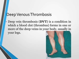
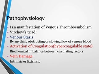
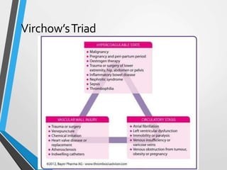
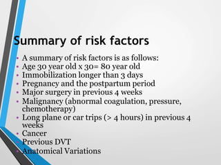
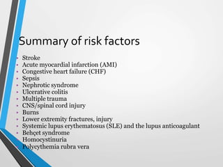
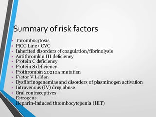
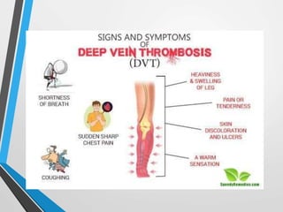
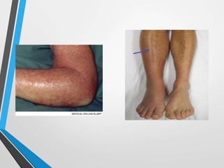
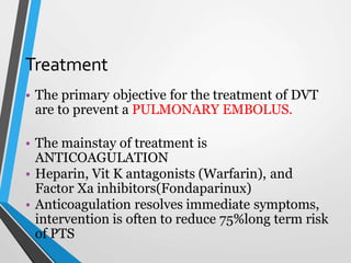
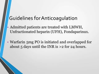
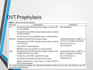
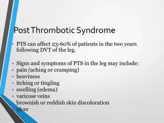
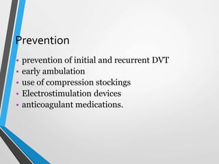
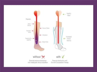
Deep vein thrombosis (DVT) is a blood clot that forms in the deep veins, usually in the legs. Virchow's triad of venous stasis, hypercoagulability, and endothelial injury can lead to thrombus formation. Risk factors include age, immobilization, surgery, cancer, and genetic factors. Patients may experience pain, swelling, warmth, and tenderness. Diagnosis involves a Wells score, D-dimer test, ultrasound or venography. Treatment is anticoagulation with heparin, low molecular weight heparin, fondaparinux, or warfarin to prevent pulmonary embolism. Long-term anticoagulation and compression stockings can help prevent
Deep vein thrombosis (DVT), is the formation of a blood clot in a deep vein, most commonly the legs.[2][a] Symptoms may include pain, swelling, redness, or warmth of the affected area. About half of cases have no symptoms. Complications may include pulmonary embolism, as a result of detachment of a clot which travels to the lungs, and post-thrombotic syndrome.[2][3]
Risk factors include recent surgery, cancer, trauma, lack of movement, obesity, smoking, hormonal birth control, pregnancy and the period following birth, antiphospholipid syndrome, and certain genetic conditions. Genetic factors include deficiencies of antithrombin, protein C, and protein S, and factor V Leiden mutation. The underlying mechanism typically involves some combination of decreased blood flow rate, increased tendency to clot, and injury to the blood vessel wall.
A blood clot in a deep vein, usually in the legs.
This condition is serious because blood clots can loosen and lodge in the lungs.
Leg pain or swelling may occur, but there may be no symptoms.
Treatments include medication and use of compression stockings.
Buerger's disease (thromboangiitis obliterans) is a rare disease of the arteries and veins in the arms and legs. In Buerger's disease, your blood vessels become inflamed, swell and can become blocked with blood clots (thrombi)
Severe hypertension that is a potentially life-threatening condition refers to a hypertensive crisis.
Severe hypertension is further classified into hypertensive emergencies or hypertensive urgencies.
Hypertensive emergency refers to a severe hypertension that is associated with new or progressive end-organ damage. In these clinical situations, blood pressure should be reduced immediately to prevent or minimize organ dysfunction.
Hypertensive urgency refers to severe hypertension without evidence of new or worsening end-organ injury.
A hypertensive emergency is hypertension with acute impairment of one or more
organ systems that can result in irreversible organ damage. Especially:-
Central nervous system
Cardiovascular system
Renal system.
The term hypertensive emergency is primarily used as a specific term for a hypertensive crisis with a diastolic blood pressure greater than or equal to 120mmHg and/or systolic blood pressure greater than or equal to 180mmHg.
Hypertensive emergency differs from hypertensive crisis in that, in the former, there is evidence of acute organ damage.
Lymphoedema is an abnormal swelling of limb due to the collection of excessive amount of high protein fluid secondary to defective lymphatic drainage in the presence of normal capillary filteration.It is very disabiling condition to the patient. In this ppp I have discussed its clinical picture and management in a simple way
MR. MOHAMMAD TALAL AL JOHANY
RESPIRATORY THERAPIST
Meqaat Hospital Madina
POST TEST
https://forms.gle/apSH6sR5mRTwRvjJ6
https://youtube.com/channel/UCUlJw6wef_dhQi3TXNTkn6g
OVID-19 Management experience
What we learned from bedside experience in COVID-19 treatment
Dr. Essam A. Salem, ICU Registrar, Meeqat GENERAL.HOSPITAL, Head OF ICU Unit Meeqat General Hospital
Hassan Mohamed Ali
Associate professor of anesthesia and pain management, Anesthesia department, Cairo University.
MB.B.ch, M Sc, M.D, FCAI, DESA
Meeqat General Hospital, Madinah Munawarah
Unit 8 - Information and Communication Technology (Paper I).pdfThiyagu K
This slides describes the basic concepts of ICT, basics of Email, Emerging Technology and Digital Initiatives in Education. This presentations aligns with the UGC Paper I syllabus.
Francesca Gottschalk - How can education support child empowerment.pptxEduSkills OECD
Francesca Gottschalk from the OECD’s Centre for Educational Research and Innovation presents at the Ask an Expert Webinar: How can education support child empowerment?
Palestine last event orientationfvgnh .pptxRaedMohamed3
An EFL lesson about the current events in Palestine. It is intended to be for intermediate students who wish to increase their listening skills through a short lesson in power point.
Honest Reviews of Tim Han LMA Course Program.pptxtimhan337
Personal development courses are widely available today, with each one promising life-changing outcomes. Tim Han’s Life Mastery Achievers (LMA) Course has drawn a lot of interest. In addition to offering my frank assessment of Success Insider’s LMA Course, this piece examines the course’s effects via a variety of Tim Han LMA course reviews and Success Insider comments.
Instructions for Submissions thorugh G- Classroom.pptxJheel Barad
This presentation provides a briefing on how to upload submissions and documents in Google Classroom. It was prepared as part of an orientation for new Sainik School in-service teacher trainees. As a training officer, my goal is to ensure that you are comfortable and proficient with this essential tool for managing assignments and fostering student engagement.
Acetabularia Information For Class 9 .docxvaibhavrinwa19
Acetabularia acetabulum is a single-celled green alga that in its vegetative state is morphologically differentiated into a basal rhizoid and an axially elongated stalk, which bears whorls of branching hairs. The single diploid nucleus resides in the rhizoid.
The French Revolution, which began in 1789, was a period of radical social and political upheaval in France. It marked the decline of absolute monarchies, the rise of secular and democratic republics, and the eventual rise of Napoleon Bonaparte. This revolutionary period is crucial in understanding the transition from feudalism to modernity in Europe.
For more information, visit-www.vavaclasses.com
How to Make a Field invisible in Odoo 17Celine George
It is possible to hide or invisible some fields in odoo. Commonly using “invisible” attribute in the field definition to invisible the fields. This slide will show how to make a field invisible in odoo 17.
Model Attribute Check Company Auto PropertyCeline George
In Odoo, the multi-company feature allows you to manage multiple companies within a single Odoo database instance. Each company can have its own configurations while still sharing common resources such as products, customers, and suppliers.
Welcome to TechSoup New Member Orientation and Q&A (May 2024).pdfTechSoup
In this webinar you will learn how your organization can access TechSoup's wide variety of product discount and donation programs. From hardware to software, we'll give you a tour of the tools available to help your nonprofit with productivity, collaboration, financial management, donor tracking, security, and more.
The Roman Empire A Historical Colossus.pdfkaushalkr1407
The Roman Empire, a vast and enduring power, stands as one of history's most remarkable civilizations, leaving an indelible imprint on the world. It emerged from the Roman Republic, transitioning into an imperial powerhouse under the leadership of Augustus Caesar in 27 BCE. This transformation marked the beginning of an era defined by unprecedented territorial expansion, architectural marvels, and profound cultural influence.
The empire's roots lie in the city of Rome, founded, according to legend, by Romulus in 753 BCE. Over centuries, Rome evolved from a small settlement to a formidable republic, characterized by a complex political system with elected officials and checks on power. However, internal strife, class conflicts, and military ambitions paved the way for the end of the Republic. Julius Caesar’s dictatorship and subsequent assassination in 44 BCE created a power vacuum, leading to a civil war. Octavian, later Augustus, emerged victorious, heralding the Roman Empire’s birth.
Under Augustus, the empire experienced the Pax Romana, a 200-year period of relative peace and stability. Augustus reformed the military, established efficient administrative systems, and initiated grand construction projects. The empire's borders expanded, encompassing territories from Britain to Egypt and from Spain to the Euphrates. Roman legions, renowned for their discipline and engineering prowess, secured and maintained these vast territories, building roads, fortifications, and cities that facilitated control and integration.
The Roman Empire’s society was hierarchical, with a rigid class system. At the top were the patricians, wealthy elites who held significant political power. Below them were the plebeians, free citizens with limited political influence, and the vast numbers of slaves who formed the backbone of the economy. The family unit was central, governed by the paterfamilias, the male head who held absolute authority.
Culturally, the Romans were eclectic, absorbing and adapting elements from the civilizations they encountered, particularly the Greeks. Roman art, literature, and philosophy reflected this synthesis, creating a rich cultural tapestry. Latin, the Roman language, became the lingua franca of the Western world, influencing numerous modern languages.
Roman architecture and engineering achievements were monumental. They perfected the arch, vault, and dome, constructing enduring structures like the Colosseum, Pantheon, and aqueducts. These engineering marvels not only showcased Roman ingenuity but also served practical purposes, from public entertainment to water supply.
2. DeepVenousThrombosis
• Deep vein thrombosis (DVT) is a condition in
which a blood clot (thrombus) forms in one or
more of the deep veins in your body, usually in
your legs.
3. Blood Flow
• Superficial to deep veins
• 100-150ml blood fills in calf (Peripheral Heart)
and deep veins
• Calf contracts, valves in perforators close and
blood goes up the deep veins
• Coming up next: Pathophysiology
4. Pathophysiology
• Is a manifestation of Venous Thromboembolism
• Virchow’s triad:
• Venous Stasis
By anything obstructing or slowing flow of venous blood
• Activation of Coagulation(hypercoagulable state)
Biochemical imbalance between circulating factors
• Vein Damage
Intrinsic or Extrinsic
5. Development ofThrombosis
• Microscopic thrombus formation and dissolution is
continuous process
• Increased stasis (more microthrombi not washed away
by movement), procoagulant factors, endothelial injury,
favors coagulation more than fibrinolysis, thus a
thrombus
• Coagulation is by Intrinsic or Extrinsic pathway
7. Summary of risk factors
• A summary of risk factors is as follows:
• Age 30 year old x 30= 80 year old
• Immobilization longer than 3 days
• Pregnancy and the postpartum period
• Major surgery in previous 4 weeks
• Malignancy (abnormal coagulation, pressure,
chemotherapy)
• Long plane or car trips (> 4 hours) in previous 4
weeks
• Cancer
• Previous DVT
• Anatomical Variations
9. Summary of risk factors
• Thrombocytosis
• PICC Line> CVC
• Inherited disorders of coagulation/fibrinolysis
• Antithrombin III deficiency
• Protein C deficiency
• Protein S deficiency
• Prothrombin 20210A mutation
• Factor V Leiden
• Dysfibrinogenemias and disorders of plasminogen activation
• Intravenous (IV) drug abuse
• Oral contraceptives
• Estrogens
• Heparin-induced thrombocytopenia (HIT)
10. SIGNS AND SX
• Pain
• Limb Edema
• Tenderness
• Warmth or Erythema
• Can be present or absent, unilateral or bilateral,
mild or severe
• Presentation can be of Pulmonary Embolism,
sudden coughing, haemoptysis, sharp chest pain,
rapid breathing or shortness of breath, light
headedness.
11.
12. Physical Examination
• Homan’s sign
Calf pain on dorsiflexion of foot
Positive in less than 33% of DVT patients and
more than 50% in patients without DVT
Warning: it must be noted that it is of
little diagnostic value and is theoretically
dangerous because of the possibility of
dislodgement of loose clot
13. Physical Examination
• Warmth on area of the clot
• Tense leg
• Red or discolored skin
• The most common abnormal hue is reddish
purple from venous engorgement and
obstruction. Can be cyanotic or white.
• Signs and symptoms depending upon the cause.
16. Low pretest probabilty
• Go for a D Dimer test
• D-dimer levels remain elevated in DVT for about
7 days
• Negative value rules out in low pretest
probability
• Negative result in high pretest probability, has
no value, start anti coagulation nonetheless and
go for ultrasound.
17. Intermediate to High Pretest
Probability of DVT
• Ultrasonography is recommended
• Doppler Ultrasound can depict absent or
abnormal flow even if clot isn’t visible in normal
ultrasound.
18. Venography
• Radiography of a vein after injection of a
radiopaque fluid.
• Conclusive diagnosis historically required
invasive and expensive venography, which is still
considered the criterion standard. Since 1990,
the diagnosis has been obtained noninvasively
by means of sonographic examination.
19. CBC and Coagulation Profile
• Can look for
• Polycythemia
• Leucocytosis
• Thrombocytosis
• Low PT, APTT, hypercoaguable state
20. Further Imaging
• CT Scan and MRI if required
• Suspecting iliac vein or inferior vena caval
thrombosis
21. Who to manage inpatient
• Suspected or proven PE
• Significant CV or pulmonary comorbidity
• Ileofemoral DVT
• Contraindication to anticoagulation
• Disorder of Coagulation
• Pregnancy
• Morbid obesity 150kg+
• Renal Failure
• No compliance, far from hospital, resistance to
therapy, no close follow up.
23. Treatment
• The primary objective for the treatment of DVT
are to prevent a PULMONARY EMBOLUS.
• The mainstay of treatment is
ANTICOAGULATION
• Heparin, Vit K antagonists (Warfarin), and
Factor Xa inhibitors(Fondaparinux)
• Anticoagulation resolves immediate symptoms,
intervention is often to reduce 75%long term risk
of PTS
24. Who not to anticoagulate
• Intracranial Bleeding
• Severe Active Bleeding
• Recent brain, spinal cord, eye surgery
• Pregnancy
• Malignant hypertension
• Relative contraindications include Recent Major
surgery, recent CVA, severe Thrombocytopenia
25. Guidelines forAnticoagulation
• Admitted patients are treated with LMWH,
Unfractionated heparin (UFH), Fondaparinux.
• Warfarin 5mg PO is initiated and overlapped for
about 5 days until the INR is >2 for 24 hours.
26. Unfractionated Heparin
• Standard of care in admitted patients with DVT before
LMWH.
• Prevents extension of thrombus and reduce PE risk
• Heparin must be overlapped for 4-5 days with warfarin
till INR>2
• Because of initial transient hypercoaguable state induced
byWarfarin
27. IV heparin Protocol
• Initial bolus of 80U/kg
• Constant maintenance of 18u/kg/hr
• Half life 60-90 min
• 1ml contains 5000u
• Check the aPTT or heparin activity level 6 hrs after bolus
and adjust infusion rate accordingly
• Continue to check the aPTT or heparin every 6 hrs, until
2 successful values therapeutic
• Monitor aPTT, hematocrit, Platelets every 24 hrs (HIT)
• aPTT is 21-35 sec usually, therapeutic is 1.5 times 50+
28. LMW
H
• Clexane (Enoxaparin)
• Increased Bioavailability, prolonged Half Life.
• Treatment of choice in eligible patients.
• 1mg/kg q12 hours leading to therapeutic peak
0.5-1 iu/mL
• Antagonist?
29. Fondaparinux
• Inhibits factor Xa interrupts blood coagulation
• Generally does not increase PT or PTT
• Dose: <50kg: 5 mg SC once daily
• 50-100 kg: 7.5 Sc once daily
• >100kg 10mg SC once daily
• Patients not responding to Heparin
• Prophylaxis of DVT in patient of HIT until patient
can switch to Warfarin 2.5 mg SC daily
• Antagonist?
30. Warfarin
• Warfarin interferes with hepatic synthesis of vitamin K–
dependent coagulation factors.
• Initiate warfarin on day 1 or 2 of LMWH or
unfractionated heparin therapy and overlap until desired
INR, THEN discontinue heparin
• Start 10 mg PO for 2 days in healthy individuals, 2-5 mg
PO/IV qDay for 2 days
• Check INR after 2 days and adjust dose according to
results
• Typical maintenance dose ranges between 2 and 10
mg/day
• Maintain an INR of 2.0-3.0
31. Treatment duration
• Maintain an INR of 2.0-3.0
• Surgery-provoked DVT or PE: Treatment duration of 3 months
• Transient (reversible) risk factor-induced DVT or PE: Treatment
duration of 3 months
• First unprovoked proximal DVT or PE with low or moderate
bleeding risk: Extended treatment consideration with periodic (ie,
annual) risk-benefit analysis
• First unprovoked proximal DVT or PE with high bleeding risk:
Treatment duration of 3 months
32. Treatment duration
• First unprovoked distal DVT regardless of bleeding risk:
Treatment duration of 3 months
• Second unprovoked DVT or PE with low or moderate
bleeding risk: Extended treatment
• Second unprovoked DVT or PE with high bleeding risk:
Treatment duration of 3 months
• DVT/PE and active cancer: Extended treatment, with
periodic risk-benefit analysis (ACCP recommends
LMWH over vitamin K antagonist therapy)
33. Treatment follow up
• Regular check up on INR, around a week
• Warning signs prompting immediate contact
with doctor, stopping warfarin.
• Bleeding from any site in body.
• it is generally considered dangerous to have
an INR above 4.0. An INR above 5.0 may
require a medical dose of vitamin K to bring
the INR down to a lower level.
34. Endovascular Intervention
• Indications for intervention include the relatively rare phlegmasia
or symptomatic inferior vena cava thrombosis that responds poorly
to anticoagulation alone, or symptomatic iliofemoral or
femoropopliteal DVT in patients with a low risk of bleeding.
• The goals of endovascular therapy include reducing the severity and
duration of lower-extremity symptoms, preventing pulmonary
embolism, diminishing the risk of recurrent venous thrombosis, and
preventing postthrombotic syndrome.
35. Endovascular Intervension Procedures
• Percutaneous transcatheter treatment of
patients with deep venous thrombosis (DVT)
consists of thrombus removal with catheter-
directed thrombolysis, mechanical
thrombectomy, angioplasty, and/or stenting of
venous obstructions.
37. InferiorVenaCava Filters
• Inferior vena cava to trap emboli and minimize venous stasis.
Inferior vena cava filter is a mechanical barrier to the flow of
emboli larger than 4 mm.
• Confirmed acute proximal DVT or acute PE in patient with
contraindication for anticoagulation (this remains the most
common indication for inferior vena cava filter placement)
• Recurrent thromboembolism while on anticoagulation
• Active bleeding complications requiring termination of
anticoagulation therapy
38. • Early ambulation on day 2 after initiation of
outpatient anticoagulant therapy in addition to
effective compression is strongly recommended.
• The regular use of graduated elastic compression
stockings reduced the incidence of PTS by 50%
40. PostThrombotic Syndrome
• PTS can affect 23-60% of patients in the two years
following DVT of the leg.
• Signs and symptoms of PTS in the leg may include:
• pain (aching or cramping)
• heaviness
• itching or tingling
• swelling (edema)
• varicose veins
• brownish or reddish skin discoloration
• ulcer
41. Prevention
• prevention of initial and recurrent DVT
• early ambulation
• use of compression stockings
• Electrostimulation devices
• anticoagulant medications.
42.
43. Treatment
• proper leg elevation
• compression therapy with elastic stockings
• wound care for leg ulcers
• angioplasty and vascular stents in proximal thrombosed
veins by an experienced physician can provide
significant relief of swelling and healing of skin ulcers
• Compression bandages are useful to treat edemas