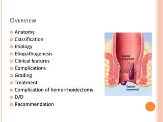
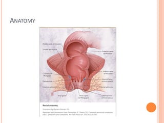
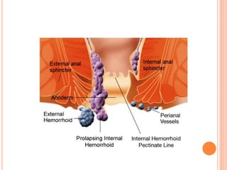
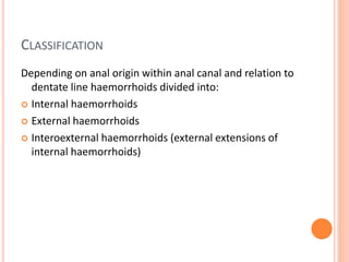
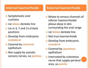
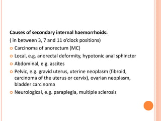
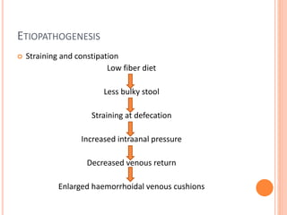
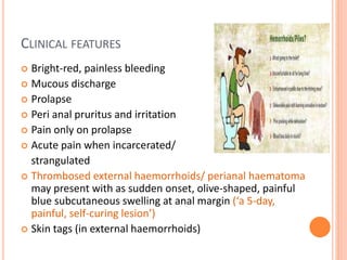
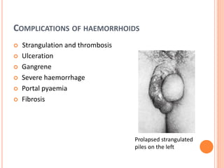
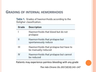
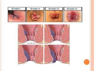
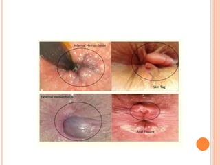
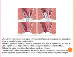
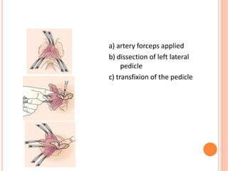
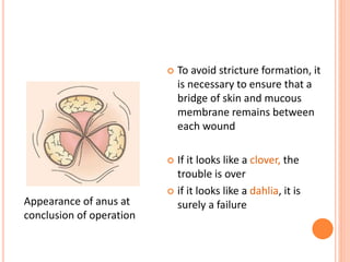
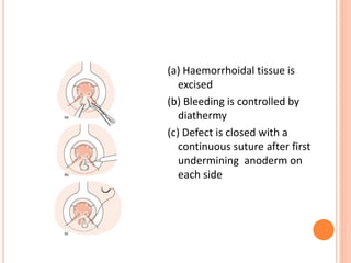
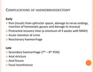
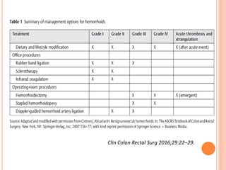
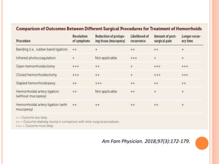
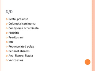
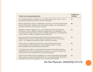
HAEMORRHOIDS
Dr. Somendra Bansal
Dr. Shalu Gupta
SMS Medical College, Jaipur
OVERVIEW
 Anatomy
 Classification
 Etiology
 Etiopathogenesis
 Clinical features
 Complications
 Grading
 Treatment
 Complication of hemorrhoidectomy
 D/D
 Recommendation
ANATOMY
CLASSIFICATION
Depending on anal origin within anal canal and relation to
dentate line haemorrhoids divided into:
 Internal haemorrhoids
 External haemorrhoids
 Interoexternal haemorrhoids (external extensions of
internal haemorrhoids)
 Symptomatic anal
cushions
 Lie above dentate line
 Lie in 3, 7 and 11 o’clock
positions
 Develop from embryonic
endoderm
 Covered by columnar
epithelium
 Not supply by somatic
sensory nerves, so painless
 Relate to venous channels of
inferior haemorrhoidal
plexus deep in skin
surrounding the anal verge
 Lie below dentate line
 Not true haemorrhoids
 Develop from embryonic
ectoderm
 Covered by squamous
epithelium
 Innervated by cutaneous
nerve that supply perianal
area, so painful
Internal haemorrhoids External haemorrhoids
ETIOLOGY
Sedentary life style, Smoking and alcohol
Stress, Family history
Causes of secondary internal haemorrhoids:
( in between 3, 7 and 11 o’clock positions)
 Carcinoma of anorectum (MC)
 Local, e.g. anorectal deformity, hypotonic anal sphincter
 Abdominal, e.g. ascites
 Pelvic, e.g. gravid uterus, uterine neoplasm (fibroid,
carcinoma of the uterus or cervix), ovarian neoplasm,
bladder carcinoma
 Neurological, e.g. paraplegia, multiple sclerosis
ETIOPATHOGENESIS
 Straining and constipation
Low fiber diet
Less bulky stool
Straining at defecation
Increased intraanal pressure
Decreased venous return
Enlarged haemorrhoidal venous cushions
CLINICAL FEATURES
 Bright-red, painless bleeding
 Mucous discharge
 Prolapse
 Peri anal pruritus and irritation
 Pain only on prolapse
 Acute pain when incarcerated/
strangulated
 Thrombosed external haemorrhoids/ perianal haematoma
may present with as sudden onset, olive-shaped, painful
blue subcutaneous swelling at anal margin (‘a 5-day,
painful, self-curing lesion’)
 Skin tags (in external haemorrhoids)
COMPLICATIONS OF HAEMORRHOIDS
 Strangulation and thrombosis
 Ulceration
 Gangrene
 Severe haemorrhage
 Portal pyaemia
 Fibrosis
Prolapsed strangulated
piles on the left
GRADING OF INTERNAL HEMORRHOIDS
Ther Adv Chronic Dis 2017;8(10):141–147
Patients may experience painless bleeding with any grade
 Per rectal examination
 Proctoscopy
 Colonoscopy for exclusion of other causes of rectal
bleeding, especially colorectal malignancy
 CBC
 Coagulation profile
TREATMENT
 Conservative
 Non-surgical
a) Injection sclerotherapy
b) Rubber band ligation
c) Cryotherapy (Lloyd Williams) not often used
d) Infrared photocoagulation (Leicester) not often used
 Surgical
a) Open hemorrhoidectomy (Milligan–Morgan)/ MMH- Gold standard
b) Closed hemorrhoidectomy (Ferguson)
c) Diathermy hemorrhoidectomy
d) Laser hemorrhoidectomy
e) LigaSure hemorrhoidectomy
f) Harmonic scalpel hemorrhoidectomy
g) Semi-closed hemorrhoidectomy
h) Submucosal hemorrhoidectomy (technique of parks)
i) Hemorrhoidal artery ligation
j) Farag procedure
k) Stapled hemorrhoidopexy/ PPH
CONSERVATIVE TREATMENT
 Attempts at normalizing bowel and defaecatory habits:
 only evacuating when natural desire to do so arises
 adopting a defaecatory position to minimize straining
 addition of stool softeners and bulking agents to ease
defaecatory act
 proprietary creams can be inserted into rectum at night and
before defaecation
INJECTION SCLEROTHERAPY
 Aim: to create fibrosis, cause obliteration of vascular
channels and hitch up anorectal mucosa
 Submucosal injection of 5% phenol in arachis oil or
almond oil
 Left lateral position and under direct vision with a
proctoscope, about 5 mL of sclerosant is injected into
apex of pile pedicle
 Procedure is repeated for each pile
 Reassessed after 8 weeks
 If necessary, repeat injections
Correct site (cross) for
injecting a haemorrhoid
 Pain upon injection means that needle is in wrong place
 Too superficial injections: rapid bulging of mucosa, which
turns septic sequelae
 Too deep injections: disastrous consequences, including
pelvic sepsis, prostatitis, impotence and rectovaginal fistula
RUBBER BAND LIGATION
 Efficacious for more bulky piles, but associated with more
discomfort
 Barron’s bander is a commonly available device
 Used to slip tight elastic bands onto base of pedicle of each
haemorrhoid
 Bands cause ischaemic necrosis of piles, which slough off
within 10 days; this may be associated with bleeding
 Three piles may be treated at one session
 Process may be repeated after several weeks if necessary
When an internal hemorrhoid is present in anorectal canal, an anoscope may be used as a
guide to identify hemorrhoidal complex.
A) With a speculum in place, a ligator is positioned over base of hemorrhoid, isolating it.
Some ligators use forceps, whereas others use suction to draw hemorrhoid taut.
B) Once the ligator is positioned at its base, bands are released
C) After the procedure is completed, the constricting bands remain in place until they
eventually fall off (typically because the tissue distal to the constricting bands sloughs)
INDICATIONS FOR HAEMORRHOIDECTOMY
 3rd and 4th degree haemorrhoids
 2nd degree haemorrhoids that have not been cured by
non-operative treatments
 Fibrosed haemorrhoids
 Interoexternal haemorrhoids when the external
haemorrhoid is well defined
 Presence of anorectal conditions requiring surgery
(fistula, fissure, large skin tag)
 Haemorrhoids complicated by strangulation
 Patient preference
OPEN HEMORRHOIDECTOMY
 Milligan–Morgan hemorrhoidectomy (MMH)
 Current gold standard for surgical management
 Hemorrhoidal tissue and vessels involved are excised with
placement of a suture at hemorrhoid pedicle
 But incisions are left open
 Often, because of location, technical difficulties, or extensive
disease with gangrenous hemorrhoidal tissue, open
approach required
 More useful for avoiding subsequent anal stenosis
a) artery forceps applied
b) dissection of left lateral
pedicle
c) transfixion of the pedicle
 To avoid stricture formation, it
is necessary to ensure that a
bridge of skin and mucous
membrane remains between
each wound
 If it looks like a clover, the
trouble is over
 if it looks like a dahlia, it is
surely a failureAppearance of anus at
conclusion of operation
CLOSED HEMORRHOIDECTOMY
 Ferguson hemorrhoidectomy (FH)
 Differs from MMH as the wound is sutured primarily
 After hemorrhoidal pedicle has been mobilized, an
absorbable suture is usually placed at pedicle site
 After hemorrhoidal bundle is excised, mucosal wound and
skin are completely closed with a continuous suture
 MMH may be overall better than FH particularly as regards
complication rate
(a) Haemorrhoidal tissue is
excised
(b) Bleeding is controlled by
diathermy
(c) Defect is closed with a
continuous suture after first
undermining anoderm on
each side
COMPLICATIONS OF HAEMORRHOIDECTOMY
Early
 Pain (results from sphincter spasm, damage to nerve endings,
insertion of hemostatic gauzes and damage to mucosa)
 Protracted recovery time (a minimum of 4 weeks with MMH)
 Acute retention of urine
 Reactionary haemorrhage
Late
 Secondary haemorrhage (7th – 8th POD)
 Anal stricture
 Anal fissure
 Fecal incontinence
CIRCULAR STAPLED HEMORRHOIDOPEXY
 Also known as ‘procedure for prolapse and hemorrhoids
(PPH)’ or stapled anopexy/ mucosectomy/ prolapsectomy
 Introduced by Longo A in 1998
 Employs a circular stapling device, which removes mucosa
and submucosa circumferentially 2-3 cm above dentate line,
anastomosing proximal and distal edges
 Interrupting blood supply to remnant hemorrhoidal tissue
 Less painful and allows quicker recovery than MMH
 High recurrence rate (PPH versus MMH 5.7% vs. 1% at 1 year
and 8.5% vs.1.5% overall)*
*Cochrane Database Syst Rev 2006;(4):CD005393.
DIATHERMY HEMORRHOIDECTOMY
 With diathermy, coagulation occurs at temp. >150 ℃
 Formation of an eschar that seals the bleeding area
 Compared with conventional hemorrhoidectomy:
 Less bleeding
 Shorter operating time
 Lower postoperative analgesic requirement, but with similar post-
operative pain
World J Surg Proced 2014; 4(3): 55-65
LIGASURE HEMORRHOIDECTOMY (LH)
 LigaSure vessel sealing system® (Valleylab) uses a bipolar
electrothermal device without need for sutures, i.e.,
sutureless hemorrhoidectomy
 Aim:
 avoiding painful diathermy burns in the richly innervated anal canal
 allowing better tissue adhesions at the wound site
 decreasing incidence of postoperative hemorrhage
 Shorter operative time
 Postoperative pain and urinary retention
 Day-case surgery
 Earlier return to work
 Additional cost of the disposable device
World J Surg Proced 2014; 4(3): 55-65
HARMONIC SCALPEL HEMORRHOIDECTOMY (HSH)
 Harmonic scalpel® (J & J, Ethicon) is an ultrasonically-
activated instrument
 Vibrates at a rate of 55000 MHz per second
 Able to coagulate small- and medium-sized vessels by
converting electrical energy to a mechanical one
 Less lateral thermal damage
 Benefit of HSH with respect to operative time, blood loss,
postoperative pain, length of hospital stay, and return to
normal activity
 Increased cost
SUBMUCOSAL HEMORRHOIDECTOMY
(TECHNIQUE OF PARKS)
 Designed to reduce postoperative pain and avoid anal and
rectal stenosis
 Indicated for 2nd to 4th degree hemorrhoids
 It includes hemorrhoidectomy with preservation of anal canal
mucosa, reducing surgical wound dimensions and leading to
a shorter healing time
 Mucosa is not included in ligation leads to reduced
postoperative pain
 Surgical time is longer
 Recurrence rate is higher
 Greater risk of bleeding during surgery and postoperatively
World J Surg Proced 2014; 4(3): 55-65
 A Y-shaped incision is made at mucocutaneous junction,
between upper mucosa of the anal canal and anorectal
junction, as an inverted racket incision
 Vascular pedicle is separated from mucosa and sphincter
plane, connecting it afterwards
 Mucosa is then closed with running suture, leaving a small
area open in the perianal region for drainage
DOPPLER-GUIDED HEMORRHOIDAL ARTERY LIGATION
 First described by Morinaga et al in 1995
 Performed under sedation and/or LA
 Involves a proctoscope with a doppler transducer
integrated in probe
 Sequential identification of position and depth of superior
rectal arterial branches (usually 5-7 are found at one level)
 Selectively ligated 2-3 cm above dentate line at two levels
1-1.5 cm apart by absorbable sutures via a lateral ligation
window within scope
 interference with blood supply suppresses bleeding and
volume of hemorrhoids
 Symptomatic relief is usually evident within 6-8 weeks
FARAG PROCEDURE
 Alternative method to ligate hemorrhoidal artery without
doppler guidance
 Piles suture, in which three interrupted sutures are used to
interrupt the blood flow to the prolapsed hemorrhoids
 First suture is passed through mucosa at proximal end of
internal hemorrhoids to occlude superior rectal vessels
 Second suture is passed into distal end of internal
hemorrhoids above dentate line to interrupt connection
between the internal and external hemorrhoidal plexuses
 Third suture between previous two sutures
 Not widely accepted (interruption of the blood flow to
hemorrhoidal cushions cause initial painful congestion)
World J Surg Proced 2014; 4(3): 55-65
SEMI-CLOSED HEMORRHOIDECTOMY
 Involves the pectineal line repair
 Internal hemorrhoid is forced outwards, becoming fully exposed
 For the repair of rectal mucosa
 In upper limit of internal hemorrhoid; 3-4 full-thickness sutures
are made radially, involving mucosa and submucosa
 Craniocaudal length of hemorrhoid to be resected
 Mucosa and submucosa are cut between the ligations
 External part of skin plexus is removed until dentate line with a V-
shaped incision or a racket incision with an external base
 Perfect for voluminous and proximally extended internal
hemorrhoids, whose full dissection would cause a very high
resection of the rectal mucosa
World J Surg Proced 2014; 4(3): 55-65
Clin Colon Rectal Surg 2016;29:22–29.
Am Fam Physician. 2018;97(3):172-179.
D/D
 Rectal prolapse
 Colorectal carcinoma
 Condyloma accuminata
 Proctitis
 Pruritus ani
 IBD
 Pedunculated polyp
 Perianal abscess
 Anal fissure, fistula
 Varicosities
Am Fam Physician. 2018;97(3):172-179.
THANK
YOU

Haemorrhoids

  • 1.
    HAEMORRHOIDS Dr. Somendra Bansal Dr.Shalu Gupta SMS Medical College, Jaipur
  • 2.
    OVERVIEW  Anatomy  Classification Etiology  Etiopathogenesis  Clinical features  Complications  Grading  Treatment  Complication of hemorrhoidectomy  D/D  Recommendation
  • 3.
  • 5.
    CLASSIFICATION Depending on analorigin within anal canal and relation to dentate line haemorrhoids divided into:  Internal haemorrhoids  External haemorrhoids  Interoexternal haemorrhoids (external extensions of internal haemorrhoids)
  • 6.
     Symptomatic anal cushions Lie above dentate line  Lie in 3, 7 and 11 o’clock positions  Develop from embryonic endoderm  Covered by columnar epithelium  Not supply by somatic sensory nerves, so painless  Relate to venous channels of inferior haemorrhoidal plexus deep in skin surrounding the anal verge  Lie below dentate line  Not true haemorrhoids  Develop from embryonic ectoderm  Covered by squamous epithelium  Innervated by cutaneous nerve that supply perianal area, so painful Internal haemorrhoids External haemorrhoids
  • 7.
    ETIOLOGY Sedentary life style,Smoking and alcohol Stress, Family history
  • 8.
    Causes of secondaryinternal haemorrhoids: ( in between 3, 7 and 11 o’clock positions)  Carcinoma of anorectum (MC)  Local, e.g. anorectal deformity, hypotonic anal sphincter  Abdominal, e.g. ascites  Pelvic, e.g. gravid uterus, uterine neoplasm (fibroid, carcinoma of the uterus or cervix), ovarian neoplasm, bladder carcinoma  Neurological, e.g. paraplegia, multiple sclerosis
  • 9.
    ETIOPATHOGENESIS  Straining andconstipation Low fiber diet Less bulky stool Straining at defecation Increased intraanal pressure Decreased venous return Enlarged haemorrhoidal venous cushions
  • 10.
    CLINICAL FEATURES  Bright-red,painless bleeding  Mucous discharge  Prolapse  Peri anal pruritus and irritation  Pain only on prolapse  Acute pain when incarcerated/ strangulated  Thrombosed external haemorrhoids/ perianal haematoma may present with as sudden onset, olive-shaped, painful blue subcutaneous swelling at anal margin (‘a 5-day, painful, self-curing lesion’)  Skin tags (in external haemorrhoids)
  • 11.
    COMPLICATIONS OF HAEMORRHOIDS Strangulation and thrombosis  Ulceration  Gangrene  Severe haemorrhage  Portal pyaemia  Fibrosis Prolapsed strangulated piles on the left
  • 12.
    GRADING OF INTERNALHEMORRHOIDS Ther Adv Chronic Dis 2017;8(10):141–147 Patients may experience painless bleeding with any grade
  • 15.
     Per rectalexamination  Proctoscopy  Colonoscopy for exclusion of other causes of rectal bleeding, especially colorectal malignancy  CBC  Coagulation profile
  • 16.
    TREATMENT  Conservative  Non-surgical a)Injection sclerotherapy b) Rubber band ligation c) Cryotherapy (Lloyd Williams) not often used d) Infrared photocoagulation (Leicester) not often used  Surgical a) Open hemorrhoidectomy (Milligan–Morgan)/ MMH- Gold standard b) Closed hemorrhoidectomy (Ferguson) c) Diathermy hemorrhoidectomy d) Laser hemorrhoidectomy e) LigaSure hemorrhoidectomy f) Harmonic scalpel hemorrhoidectomy g) Semi-closed hemorrhoidectomy h) Submucosal hemorrhoidectomy (technique of parks) i) Hemorrhoidal artery ligation j) Farag procedure k) Stapled hemorrhoidopexy/ PPH
  • 17.
    CONSERVATIVE TREATMENT  Attemptsat normalizing bowel and defaecatory habits:  only evacuating when natural desire to do so arises  adopting a defaecatory position to minimize straining  addition of stool softeners and bulking agents to ease defaecatory act  proprietary creams can be inserted into rectum at night and before defaecation
  • 20.
    INJECTION SCLEROTHERAPY  Aim:to create fibrosis, cause obliteration of vascular channels and hitch up anorectal mucosa  Submucosal injection of 5% phenol in arachis oil or almond oil  Left lateral position and under direct vision with a proctoscope, about 5 mL of sclerosant is injected into apex of pile pedicle  Procedure is repeated for each pile  Reassessed after 8 weeks  If necessary, repeat injections Correct site (cross) for injecting a haemorrhoid
  • 21.
     Pain uponinjection means that needle is in wrong place  Too superficial injections: rapid bulging of mucosa, which turns septic sequelae  Too deep injections: disastrous consequences, including pelvic sepsis, prostatitis, impotence and rectovaginal fistula
  • 22.
    RUBBER BAND LIGATION Efficacious for more bulky piles, but associated with more discomfort  Barron’s bander is a commonly available device  Used to slip tight elastic bands onto base of pedicle of each haemorrhoid  Bands cause ischaemic necrosis of piles, which slough off within 10 days; this may be associated with bleeding  Three piles may be treated at one session  Process may be repeated after several weeks if necessary
  • 23.
    When an internalhemorrhoid is present in anorectal canal, an anoscope may be used as a guide to identify hemorrhoidal complex. A) With a speculum in place, a ligator is positioned over base of hemorrhoid, isolating it. Some ligators use forceps, whereas others use suction to draw hemorrhoid taut. B) Once the ligator is positioned at its base, bands are released C) After the procedure is completed, the constricting bands remain in place until they eventually fall off (typically because the tissue distal to the constricting bands sloughs)
  • 24.
    INDICATIONS FOR HAEMORRHOIDECTOMY 3rd and 4th degree haemorrhoids  2nd degree haemorrhoids that have not been cured by non-operative treatments  Fibrosed haemorrhoids  Interoexternal haemorrhoids when the external haemorrhoid is well defined  Presence of anorectal conditions requiring surgery (fistula, fissure, large skin tag)  Haemorrhoids complicated by strangulation  Patient preference
  • 25.
    OPEN HEMORRHOIDECTOMY  Milligan–Morganhemorrhoidectomy (MMH)  Current gold standard for surgical management  Hemorrhoidal tissue and vessels involved are excised with placement of a suture at hemorrhoid pedicle  But incisions are left open  Often, because of location, technical difficulties, or extensive disease with gangrenous hemorrhoidal tissue, open approach required  More useful for avoiding subsequent anal stenosis
  • 26.
    a) artery forcepsapplied b) dissection of left lateral pedicle c) transfixion of the pedicle
  • 27.
     To avoidstricture formation, it is necessary to ensure that a bridge of skin and mucous membrane remains between each wound  If it looks like a clover, the trouble is over  if it looks like a dahlia, it is surely a failureAppearance of anus at conclusion of operation
  • 28.
    CLOSED HEMORRHOIDECTOMY  Fergusonhemorrhoidectomy (FH)  Differs from MMH as the wound is sutured primarily  After hemorrhoidal pedicle has been mobilized, an absorbable suture is usually placed at pedicle site  After hemorrhoidal bundle is excised, mucosal wound and skin are completely closed with a continuous suture  MMH may be overall better than FH particularly as regards complication rate
  • 29.
    (a) Haemorrhoidal tissueis excised (b) Bleeding is controlled by diathermy (c) Defect is closed with a continuous suture after first undermining anoderm on each side
  • 30.
    COMPLICATIONS OF HAEMORRHOIDECTOMY Early Pain (results from sphincter spasm, damage to nerve endings, insertion of hemostatic gauzes and damage to mucosa)  Protracted recovery time (a minimum of 4 weeks with MMH)  Acute retention of urine  Reactionary haemorrhage Late  Secondary haemorrhage (7th – 8th POD)  Anal stricture  Anal fissure  Fecal incontinence
  • 31.
    CIRCULAR STAPLED HEMORRHOIDOPEXY Also known as ‘procedure for prolapse and hemorrhoids (PPH)’ or stapled anopexy/ mucosectomy/ prolapsectomy  Introduced by Longo A in 1998  Employs a circular stapling device, which removes mucosa and submucosa circumferentially 2-3 cm above dentate line, anastomosing proximal and distal edges  Interrupting blood supply to remnant hemorrhoidal tissue  Less painful and allows quicker recovery than MMH  High recurrence rate (PPH versus MMH 5.7% vs. 1% at 1 year and 8.5% vs.1.5% overall)* *Cochrane Database Syst Rev 2006;(4):CD005393.
  • 33.
    DIATHERMY HEMORRHOIDECTOMY  Withdiathermy, coagulation occurs at temp. >150 ℃  Formation of an eschar that seals the bleeding area  Compared with conventional hemorrhoidectomy:  Less bleeding  Shorter operating time  Lower postoperative analgesic requirement, but with similar post- operative pain World J Surg Proced 2014; 4(3): 55-65
  • 34.
    LIGASURE HEMORRHOIDECTOMY (LH) LigaSure vessel sealing system® (Valleylab) uses a bipolar electrothermal device without need for sutures, i.e., sutureless hemorrhoidectomy  Aim:  avoiding painful diathermy burns in the richly innervated anal canal  allowing better tissue adhesions at the wound site  decreasing incidence of postoperative hemorrhage  Shorter operative time  Postoperative pain and urinary retention  Day-case surgery  Earlier return to work  Additional cost of the disposable device World J Surg Proced 2014; 4(3): 55-65
  • 35.
    HARMONIC SCALPEL HEMORRHOIDECTOMY(HSH)  Harmonic scalpel® (J & J, Ethicon) is an ultrasonically- activated instrument  Vibrates at a rate of 55000 MHz per second  Able to coagulate small- and medium-sized vessels by converting electrical energy to a mechanical one  Less lateral thermal damage  Benefit of HSH with respect to operative time, blood loss, postoperative pain, length of hospital stay, and return to normal activity  Increased cost
  • 36.
    SUBMUCOSAL HEMORRHOIDECTOMY (TECHNIQUE OFPARKS)  Designed to reduce postoperative pain and avoid anal and rectal stenosis  Indicated for 2nd to 4th degree hemorrhoids  It includes hemorrhoidectomy with preservation of anal canal mucosa, reducing surgical wound dimensions and leading to a shorter healing time  Mucosa is not included in ligation leads to reduced postoperative pain  Surgical time is longer  Recurrence rate is higher  Greater risk of bleeding during surgery and postoperatively World J Surg Proced 2014; 4(3): 55-65
  • 37.
     A Y-shapedincision is made at mucocutaneous junction, between upper mucosa of the anal canal and anorectal junction, as an inverted racket incision  Vascular pedicle is separated from mucosa and sphincter plane, connecting it afterwards  Mucosa is then closed with running suture, leaving a small area open in the perianal region for drainage
  • 38.
    DOPPLER-GUIDED HEMORRHOIDAL ARTERYLIGATION  First described by Morinaga et al in 1995  Performed under sedation and/or LA  Involves a proctoscope with a doppler transducer integrated in probe  Sequential identification of position and depth of superior rectal arterial branches (usually 5-7 are found at one level)  Selectively ligated 2-3 cm above dentate line at two levels 1-1.5 cm apart by absorbable sutures via a lateral ligation window within scope  interference with blood supply suppresses bleeding and volume of hemorrhoids  Symptomatic relief is usually evident within 6-8 weeks
  • 39.
    FARAG PROCEDURE  Alternativemethod to ligate hemorrhoidal artery without doppler guidance  Piles suture, in which three interrupted sutures are used to interrupt the blood flow to the prolapsed hemorrhoids  First suture is passed through mucosa at proximal end of internal hemorrhoids to occlude superior rectal vessels  Second suture is passed into distal end of internal hemorrhoids above dentate line to interrupt connection between the internal and external hemorrhoidal plexuses  Third suture between previous two sutures  Not widely accepted (interruption of the blood flow to hemorrhoidal cushions cause initial painful congestion) World J Surg Proced 2014; 4(3): 55-65
  • 40.
    SEMI-CLOSED HEMORRHOIDECTOMY  Involvesthe pectineal line repair  Internal hemorrhoid is forced outwards, becoming fully exposed  For the repair of rectal mucosa  In upper limit of internal hemorrhoid; 3-4 full-thickness sutures are made radially, involving mucosa and submucosa  Craniocaudal length of hemorrhoid to be resected  Mucosa and submucosa are cut between the ligations  External part of skin plexus is removed until dentate line with a V- shaped incision or a racket incision with an external base  Perfect for voluminous and proximally extended internal hemorrhoids, whose full dissection would cause a very high resection of the rectal mucosa World J Surg Proced 2014; 4(3): 55-65
  • 41.
    Clin Colon RectalSurg 2016;29:22–29.
  • 42.
    Am Fam Physician.2018;97(3):172-179.
  • 43.
    D/D  Rectal prolapse Colorectal carcinoma  Condyloma accuminata  Proctitis  Pruritus ani  IBD  Pedunculated polyp  Perianal abscess  Anal fissure, fistula  Varicosities
  • 44.
    Am Fam Physician.2018;97(3):172-179.
  • 45.