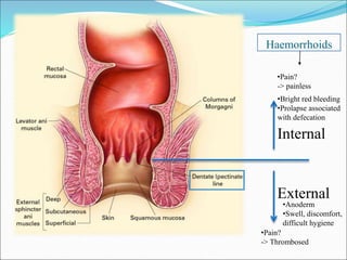
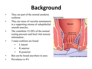
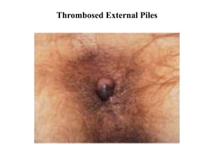
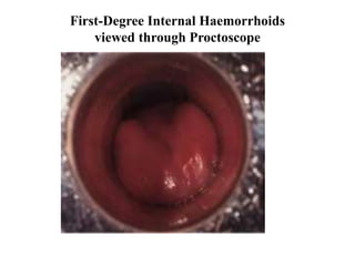
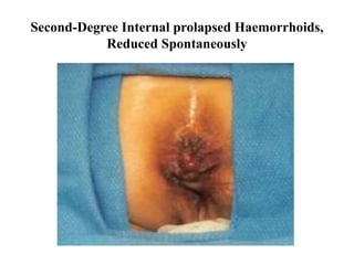
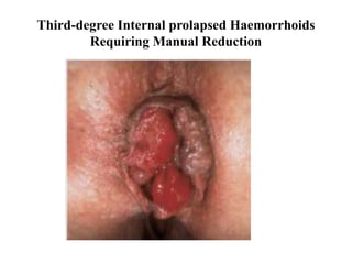
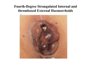
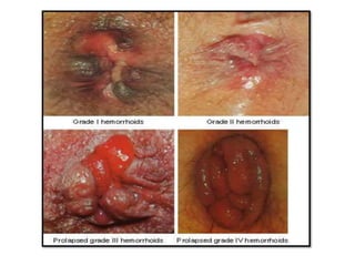
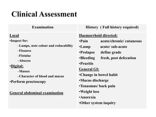
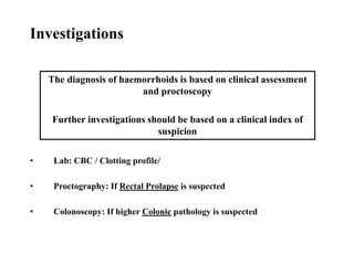
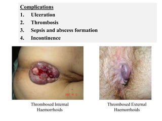
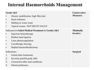
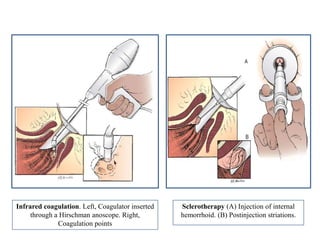
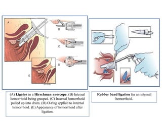
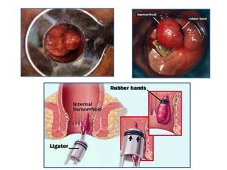
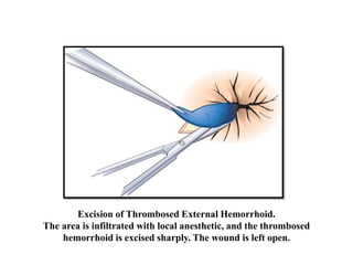
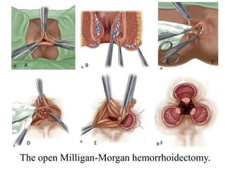
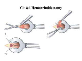
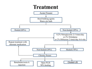
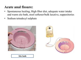
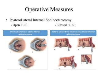
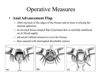
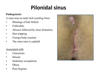
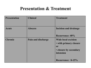
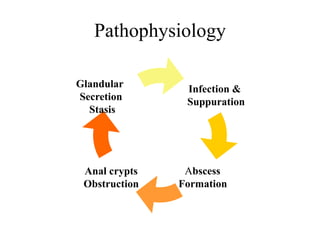
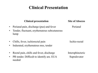
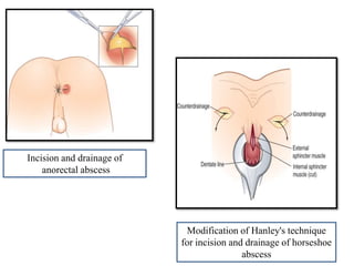
Please find the power point on Hemorrhoids. I tried present it on understandable way and all the contents are reviewed by experts and from very reliable references.
Disclaimer: A lot from this slides were taken also from https://www.slideshare.net/babysurgeon/scrotal-swellings-1 (Dr Selvaraj Balasubramani)
This covers only :
ANATOMY
CAUSES
TORSION OF TESTIS
EPIDIDYMO-ORCHITIS
HYDROCELE
EPIDIDYMAL CYST
VARICOCELE
Types, Investigation, complication and treatment of Incisional herniaimraxid
It is herniation through a weak abdominal scar (scar of previous surgery).
It is common in old age and obese individuals.
Predisposing Factors:
..> Vertical scar, midline scar, lower abdominal scar— may injure the nerves of the abdominal muscles.
...> Scar of major surgeries (biliary, pancreatic).
...> Scar of emergency surgeries (peritonitis, acute abdomen).
For Health Tips: http://MedicoPk.com/
An anorectal abscess is a collection of pus in the anal or rectal region.
It may be caused by infection of an anal fissure, sexually transmitted infections or blocked anal glands.
An anorectal abscess is a collection of pus that builds up in the rectum and anus.
With prompt treatment, client with this condition usually recover very well.
Complications tend occur when treatment is delayed.
This is comprehensive Presentation about IBD, its Classification, major subtypes, eitology, genetics, presentation, diagnosis and treatment.
it Includes Both Crohn's Disease And Ulcerative Colitis in detail
Pathology, Diagnosis, Medical Therapy, Surgical Management of Both the diseases are described
Urinary Tract Fistulas -(VVF) Etiology, Diagnosis, ManagementVikas V
Urinary Tract Fistulas - Etiology, Diagnosis, Management
Surgical and Relevant Anatomy, Classification, eitiology, VVF in Detail, Examination and Diagnosis, Management of VVF - Both Conservative And Surgical Management - Steps of Surgical Management, Post operative Management.
Whipple's procedure - Indications, Steps, ComplicationsVikas V
Whipple's Procedure - Explaining the History of Whipple's Procedure, Indications, Contraindications, Step wise detailed procedure, Complications, Perioperative Management.
The Presentation Includes Detailed Step wise approach to the procedure assisted with Pictorial Representation of The steps
Splenic trauma - Causes, Complications, ManagementVikas V
Splenic Trauma - A detailed Presentation about Splenic Trauma, anatomy of the spleen, Causes of Trauma, Mechanism of Injury, Diagnosis, Management, Surgical management, Steps of Splenectomy, and Complications
Lt hemicolectomy - Surgical Approach, Complications.Vikas V
This is a detailed Presentation of the surgical procedure - Left Hemicolectomy. It deals with the Surgical anatomy, Indications, Surgical Steps, Complications of the Procedure
Hirschsprung Disease - Approach & ManagementVikas V
Hirschsprung Disease. - A developmental Disorder of Intrinsic Component of Enteric Nervous System.
Also known Congenital Megacolon.
This Presentation deals with The eitology, presentation, diagnosis, medical and surgical management & complications of the same.
Cleft Lip and Palate - Presentation.
Cleft Lip and Palate is the 2nd most common Congenital Anomaly after Clubfoot. This presentation goes in depth about the Presentation, eitiology, Genetics, Medical management, Nasoalveolar Moulding, Surgical management of Cleft Lip & Palate
Report Back from SGO 2024: What’s the Latest in Cervical Cancer?bkling
Are you curious about what’s new in cervical cancer research or unsure what the findings mean? Join Dr. Emily Ko, a gynecologic oncologist at Penn Medicine, to learn about the latest updates from the Society of Gynecologic Oncology (SGO) 2024 Annual Meeting on Women’s Cancer. Dr. Ko will discuss what the research presented at the conference means for you and answer your questions about the new developments.
Prix Galien International 2024 Forum ProgramLevi Shapiro
June 20, 2024, Prix Galien International and Jerusalem Ethics Forum in ROME. Detailed agenda including panels:
- ADVANCES IN CARDIOLOGY: A NEW PARADIGM IS COMING
- WOMEN’S HEALTH: FERTILITY PRESERVATION
- WHAT’S NEW IN THE TREATMENT OF INFECTIOUS,
ONCOLOGICAL AND INFLAMMATORY SKIN DISEASES?
- ARTIFICIAL INTELLIGENCE AND ETHICS
- GENE THERAPY
- BEYOND BORDERS: GLOBAL INITIATIVES FOR DEMOCRATIZING LIFE SCIENCE TECHNOLOGIES AND PROMOTING ACCESS TO HEALTHCARE
- ETHICAL CHALLENGES IN LIFE SCIENCES
- Prix Galien International Awards Ceremony
Ozempic: Preoperative Management of Patients on GLP-1 Receptor Agonists Saeid Safari
Preoperative Management of Patients on GLP-1 Receptor Agonists like Ozempic and Semiglutide
ASA GUIDELINE
NYSORA Guideline
2 Case Reports of Gastric Ultrasound
New Directions in Targeted Therapeutic Approaches for Older Adults With Mantl...i3 Health
i3 Health is pleased to make the speaker slides from this activity available for use as a non-accredited self-study or teaching resource.
This slide deck presented by Dr. Kami Maddocks, Professor-Clinical in the Division of Hematology and
Associate Division Director for Ambulatory Operations
The Ohio State University Comprehensive Cancer Center, will provide insight into new directions in targeted therapeutic approaches for older adults with mantle cell lymphoma.
STATEMENT OF NEED
Mantle cell lymphoma (MCL) is a rare, aggressive B-cell non-Hodgkin lymphoma (NHL) accounting for 5% to 7% of all lymphomas. Its prognosis ranges from indolent disease that does not require treatment for years to very aggressive disease, which is associated with poor survival (Silkenstedt et al, 2021). Typically, MCL is diagnosed at advanced stage and in older patients who cannot tolerate intensive therapy (NCCN, 2022). Although recent advances have slightly increased remission rates, recurrence and relapse remain very common, leading to a median overall survival between 3 and 6 years (LLS, 2021). Though there are several effective options, progress is still needed towards establishing an accepted frontline approach for MCL (Castellino et al, 2022). Treatment selection and management of MCL are complicated by the heterogeneity of prognosis, advanced age and comorbidities of patients, and lack of an established standard approach for treatment, making it vital that clinicians be familiar with the latest research and advances in this area. In this activity chaired by Michael Wang, MD, Professor in the Department of Lymphoma & Myeloma at MD Anderson Cancer Center, expert faculty will discuss prognostic factors informing treatment, the promising results of recent trials in new therapeutic approaches, and the implications of treatment resistance in therapeutic selection for MCL.
Target Audience
Hematology/oncology fellows, attending faculty, and other health care professionals involved in the treatment of patients with mantle cell lymphoma (MCL).
Learning Objectives
1.) Identify clinical and biological prognostic factors that can guide treatment decision making for older adults with MCL
2.) Evaluate emerging data on targeted therapeutic approaches for treatment-naive and relapsed/refractory MCL and their applicability to older adults
3.) Assess mechanisms of resistance to targeted therapies for MCL and their implications for treatment selection
Title: Sense of Smell
Presenter: Dr. Faiza, Assistant Professor of Physiology
Qualifications:
MBBS (Best Graduate, AIMC Lahore)
FCPS Physiology
ICMT, CHPE, DHPE (STMU)
MPH (GC University, Faisalabad)
MBA (Virtual University of Pakistan)
Learning Objectives:
Describe the primary categories of smells and the concept of odor blindness.
Explain the structure and location of the olfactory membrane and mucosa, including the types and roles of cells involved in olfaction.
Describe the pathway and mechanisms of olfactory signal transmission from the olfactory receptors to the brain.
Illustrate the biochemical cascade triggered by odorant binding to olfactory receptors, including the role of G-proteins and second messengers in generating an action potential.
Identify different types of olfactory disorders such as anosmia, hyposmia, hyperosmia, and dysosmia, including their potential causes.
Key Topics:
Olfactory Genes:
3% of the human genome accounts for olfactory genes.
400 genes for odorant receptors.
Olfactory Membrane:
Located in the superior part of the nasal cavity.
Medially: Folds downward along the superior septum.
Laterally: Folds over the superior turbinate and upper surface of the middle turbinate.
Total surface area: 5-10 square centimeters.
Olfactory Mucosa:
Olfactory Cells: Bipolar nerve cells derived from the CNS (100 million), with 4-25 olfactory cilia per cell.
Sustentacular Cells: Produce mucus and maintain ionic and molecular environment.
Basal Cells: Replace worn-out olfactory cells with an average lifespan of 1-2 months.
Bowman’s Gland: Secretes mucus.
Stimulation of Olfactory Cells:
Odorant dissolves in mucus and attaches to receptors on olfactory cilia.
Involves a cascade effect through G-proteins and second messengers, leading to depolarization and action potential generation in the olfactory nerve.
Quality of a Good Odorant:
Small (3-20 Carbon atoms), volatile, water-soluble, and lipid-soluble.
Facilitated by odorant-binding proteins in mucus.
Membrane Potential and Action Potential:
Resting membrane potential: -55mV.
Action potential frequency in the olfactory nerve increases with odorant strength.
Adaptation Towards the Sense of Smell:
Rapid adaptation within the first second, with further slow adaptation.
Psychological adaptation greater than receptor adaptation, involving feedback inhibition from the central nervous system.
Primary Sensations of Smell:
Camphoraceous, Musky, Floral, Pepperminty, Ethereal, Pungent, Putrid.
Odor Detection Threshold:
Examples: Hydrogen sulfide (0.0005 ppm), Methyl-mercaptan (0.002 ppm).
Some toxic substances are odorless at lethal concentrations.
Characteristics of Smell:
Odor blindness for single substances due to lack of appropriate receptor protein.
Behavioral and emotional influences of smell.
Transmission of Olfactory Signals:
From olfactory cells to glomeruli in the olfactory bulb, involving lateral inhibition.
Primitive, less old, and new olfactory systems with different path
These lecture slides, by Dr Sidra Arshad, offer a quick overview of physiological basis of a normal electrocardiogram.
Learning objectives:
1. Define an electrocardiogram (ECG) and electrocardiography
2. Describe how dipoles generated by the heart produce the waveforms of the ECG
3. Describe the components of a normal electrocardiogram of a typical bipolar leads (limb II)
4. Differentiate between intervals and segments
5. Enlist some common indications for obtaining an ECG
Study Resources:
1. Chapter 11, Guyton and Hall Textbook of Medical Physiology, 14th edition
2. Chapter 9, Human Physiology - From Cells to Systems, Lauralee Sherwood, 9th edition
3. Chapter 29, Ganong’s Review of Medical Physiology, 26th edition
4. Electrocardiogram, StatPearls - https://www.ncbi.nlm.nih.gov/books/NBK549803/
5. ECG in Medical Practice by ABM Abdullah, 4th edition
6. ECG Basics, http://www.nataliescasebook.com/tag/e-c-g-basics
HOT NEW PRODUCT! BIG SALES FAST SHIPPING NOW FROM CHINA!! EU KU DB BK substit...GL Anaacs
Contact us if you are interested:
Email / Skype : kefaya1771@gmail.com
Threema: PXHY5PDH
New BATCH Ku !!! MUCH IN DEMAND FAST SALE EVERY BATCH HAPPY GOOD EFFECT BIG BATCH !
Contact me on Threema or skype to start big business!!
Hot-sale products:
NEW HOT EUTYLONE WHITE CRYSTAL!!
5cl-adba precursor (semi finished )
5cl-adba raw materials
ADBB precursor (semi finished )
ADBB raw materials
APVP powder
5fadb/4f-adb
Jwh018 / Jwh210
Eutylone crystal
Protonitazene (hydrochloride) CAS: 119276-01-6
Flubrotizolam CAS: 57801-95-3
Metonitazene CAS: 14680-51-4
Payment terms: Western Union,MoneyGram,Bitcoin or USDT.
Deliver Time: Usually 7-15days
Shipping method: FedEx, TNT, DHL,UPS etc.Our deliveries are 100% safe, fast, reliable and discreet.
Samples will be sent for your evaluation!If you are interested in, please contact me, let's talk details.
We specializes in exporting high quality Research chemical, medical intermediate, Pharmaceutical chemicals and so on. Products are exported to USA, Canada, France, Korea, Japan,Russia, Southeast Asia and other countries.
Lung Cancer: Artificial Intelligence, Synergetics, Complex System Analysis, S...Oleg Kshivets
RESULTS: Overall life span (LS) was 2252.1±1742.5 days and cumulative 5-year survival (5YS) reached 73.2%, 10 years – 64.8%, 20 years – 42.5%. 513 LCP lived more than 5 years (LS=3124.6±1525.6 days), 148 LCP – more than 10 years (LS=5054.4±1504.1 days).199 LCP died because of LC (LS=562.7±374.5 days). 5YS of LCP after bi/lobectomies was significantly superior in comparison with LCP after pneumonectomies (78.1% vs.63.7%, P=0.00001 by log-rank test). AT significantly improved 5YS (66.3% vs. 34.8%) (P=0.00000 by log-rank test) only for LCP with N1-2. Cox modeling displayed that 5YS of LCP significantly depended on: phase transition (PT) early-invasive LC in terms of synergetics, PT N0—N12, cell ratio factors (ratio between cancer cells- CC and blood cells subpopulations), G1-3, histology, glucose, AT, blood cell circuit, prothrombin index, heparin tolerance, recalcification time (P=0.000-0.038). Neural networks, genetic algorithm selection and bootstrap simulation revealed relationships between 5YS and PT early-invasive LC (rank=1), PT N0—N12 (rank=2), thrombocytes/CC (3), erythrocytes/CC (4), eosinophils/CC (5), healthy cells/CC (6), lymphocytes/CC (7), segmented neutrophils/CC (8), stick neutrophils/CC (9), monocytes/CC (10); leucocytes/CC (11). Correct prediction of 5YS was 100% by neural networks computing (area under ROC curve=1.0; error=0.0).
CONCLUSIONS: 5YS of LCP after radical procedures significantly depended on: 1) PT early-invasive cancer; 2) PT N0--N12; 3) cell ratio factors; 4) blood cell circuit; 5) biochemical factors; 6) hemostasis system; 7) AT; 8) LC characteristics; 9) LC cell dynamics; 10) surgery type: lobectomy/pneumonectomy; 11) anthropometric data. Optimal diagnosis and treatment strategies for LC are: 1) screening and early detection of LC; 2) availability of experienced thoracic surgeons because of complexity of radical procedures; 3) aggressive en block surgery and adequate lymph node dissection for completeness; 4) precise prediction; 5) adjuvant chemoimmunoradiotherapy for LCP with unfavorable prognosis.
Pulmonary Thromboembolism - etilogy, types, medical- Surgical and nursing man...VarunMahajani
Disruption of blood supply to lung alveoli due to blockage of one or more pulmonary blood vessels is called as Pulmonary thromboembolism. In this presentation we will discuss its causes, types and its management in depth.
Flu Vaccine Alert in Bangalore Karnatakaaddon Scans
As flu season approaches, health officials in Bangalore, Karnataka, are urging residents to get their flu vaccinations. The seasonal flu, while common, can lead to severe health complications, particularly for vulnerable populations such as young children, the elderly, and those with underlying health conditions.
Dr. Vidisha Kumari, a leading epidemiologist in Bangalore, emphasizes the importance of getting vaccinated. "The flu vaccine is our best defense against the influenza virus. It not only protects individuals but also helps prevent the spread of the virus in our communities," he says.
This year, the flu season is expected to coincide with a potential increase in other respiratory illnesses. The Karnataka Health Department has launched an awareness campaign highlighting the significance of flu vaccinations. They have set up multiple vaccination centers across Bangalore, making it convenient for residents to receive their shots.
To encourage widespread vaccination, the government is also collaborating with local schools, workplaces, and community centers to facilitate vaccination drives. Special attention is being given to ensuring that the vaccine is accessible to all, including marginalized communities who may have limited access to healthcare.
Residents are reminded that the flu vaccine is safe and effective. Common side effects are mild and may include soreness at the injection site, mild fever, or muscle aches. These side effects are generally short-lived and far less severe than the flu itself.
Healthcare providers are also stressing the importance of continuing COVID-19 precautions. Wearing masks, practicing good hand hygiene, and maintaining social distancing are still crucial, especially in crowded places.
Protect yourself and your loved ones by getting vaccinated. Together, we can help keep Bangalore healthy and safe this flu season. For more information on vaccination centers and schedules, residents can visit the Karnataka Health Department’s official website or follow their social media pages.
Stay informed, stay safe, and get your flu shot today!
Couples presenting to the infertility clinic- Do they really have infertility...Sujoy Dasgupta
Dr Sujoy Dasgupta presented the study on "Couples presenting to the infertility clinic- Do they really have infertility? – The unexplored stories of non-consummation" in the 13th Congress of the Asia Pacific Initiative on Reproduction (ASPIRE 2024) at Manila on 24 May, 2024.
3. Anorectal Anatomy
Anal verge
Anal canal
Arterial Supply
Inferior rectal A
middle rectal A
Venous drainage
Inferior Rectal V
Middle Rectal V
3 Hemorrhoidal
Complexes
L - Lateral
R - Antero-lateral
R - Posterolateral
Lymphatic drainage
Above dentate: Inf. Mesenteric
Below dentate: Internal Iliac
Nerve Supply
Sympathetic: Superior
hypogastric plexus
Parasympathetic:
S234 (nerviergentis)
Pudendal Nerve:
Motor and Sensory
4. Haemorrhoids
• (Greek: haima = blood, rhoos = flowing;
synonym: piles, Latin: pila = a ball)
• Definition
– Hemorrhoids are normal vascular cushions suspended in the
submucosal layer of the anal canal by longitudinal connective tissue
and muscle fibers.
– Internal haemorrhoids are symptomatic anal cushions and
characteristically lie in the 3, 7 and 11 o’clock positions
– External haemorrhoids relate to venous channels of the inferior
haemorrhoidal plexus deep in the skin surrounding the anal verge
and are not true haemorrhoids
6. Background
• They are part of the normal anoderm
cushions
• They are areas of vascular anastamosis
in a supporting stroma of subepithelial
smooth muscles.
• The contribute 15-20% of the normal
resting pressure and feed vital sensory
information .
• 3 main cushions are found
• L lateral
• R anterior
• R posterior
• But can be found anywhere in anus
• Prevalence is 4%
7. 3 main processes:
1. Increased venous pressure
2. Weakness in supporting fibromuscular stroma
3. Increased internal sphincter tone
Pathogenesis
Abnormal haemorrhoids are dilated cushions of arteriovenous
plexus with stretched suspensory fibromuscular stroma with
prolapsed rectal mucosa
8. Risk factors
Pathological Habitual
1. Chronic diarrhea (IBD)
2. Colon malignancy
3. Portal hypertension
4. Spinal cord injury
5. Rectal surgery
6. Episiotomy
7. Anal intercourse
1. Constipation and straining
2. Low fibre high fat/spicy diet
3. Prolonged sitting in toilet
4. Pregnancy
5. Aging
6. Obesity
7. Office work
8. Family tendency
9. Classification
Degree of prolapse through anus Origin in relation to Dentate line
•1st: Bleed but No Prolapse, Bulge
into Lumen,
•2nd: Spontaneous Reduction
•3rd: Manual Reduction
•4th: Not Reducable
1. Internal: Above DL
2. External: Below DL
3. Mixed
16. Clinical Assessment
Examination History ( Full history required)
Local
•Inspect for:
–Lumps, note colour and reducability
–Fissures
–Fistulae
–Abscess
•Digital:
–Masses
–Character of blood and mucus
•Perform proctoscopy
General abdominal examination
Haemorrhoid directed:
•Pain acute/chronic/ cutaneous
•Lump acute/ sub-acute
•Prolapse define grade
•Bleeding fresh, post defecation
•Pruritis
General GI:
•Change in bowel habit
•Mucus discharge
•Tenasmus/ back pain
•Weight loss
•Anorexia
•Other system inquiry
17. • Lab: CBC / Clotting profile/
• Proctography: If Rectal Prolapse is suspected
• Colonoscopy: If higher Colonic pathology is suspected
The diagnosis of haemorrhoids is based on clinical assessment
and proctoscopy
Further investigations should be based on a clinical index of
suspicion
Investigations
19. Internal Haemorrhoids Management
Grade 1&2
• Dietary modification: high fibre diet
• Stool softeners
• Bathing in warm water
• Topical creams NOT MUCH VALUE
Conservative
Measures
Indicated in Failed Medical Treatment & Grades 3&4
• Injection Sclerotherapy
• Rubber band ligation
• Laser photocoagulation
• Cryotherapy freezing
• Stapled haemorrhoidectomy
Minimally
invasive
Indications:
1. Failed other treatments
2. Severely painful grade 3&4
3. Concurrent other anal conditions
4. Patient preference
Surgical
20. Sclerotherapy (A) Injection of internal
hemorrhoid. (B) Postinjection striations.
Infrared coagulation. Left, Coagulator inserted
through a Hirschman anoscope. Right,
Coagulation points
21. (A) Ligator in a Hirschman anoscope. (B) Internal
hemorrhoid being grasped. (C) Internal hemorrhoid
pulled up into drum. (D) O-ring applied to internal
hemorrhoid. (E) Appearance of hemorrhoid after
ligation.
Rubber band ligation for an internal
hemorrhoid.
22.
23. Excision of Thrombosed External Hemorrhoid.
The area is infiltrated with local anesthetic, and the thrombosed
hemorrhoid is excised sharply. The wound is left open.
26. Grade 4 Hemorrhoid before Reduction Placement of Stapling Device Obturator
Stapling Device
27. Anal Fissure
Definition : An anal fissure (synonym: fissure-in-ano) is a
longitudinal split in the anoderm of the distal anal
canal,which extends from the anal verge proximally
towards, but not beyond, the dentate line.
28. Anal Fissure
• Young & middle aged adults
• Male = Female
• Location – posterior midline (most common)
Anterior midline fissures – more common in females
• In any event, length of each fissure is remarkably constant,
extending from the dentate line to the anal verge and
corresponding roughly to the lower half of the internal
sphincter
29. Pathology
• Acute fissures – heal promptly with conservative
treatment
• Secondary changes if present, it does not heal
readily
– Sentinel pile
– Hypertrophied anal papilla
– Long standing
• Fibrous induration in lateral edges of fissure
• Fibrosis at the base of ulcer (internal sphincter)
– At any stage
• Frank suppuration – intersphincteric / perianal abscess
30. Etiology
• Initiation – trauma
• Why midline posterior fissures are more common?
• Dietary factors
– Decreased risk – raw foods, vegetables, whole grain bread
– Increased risk – white bread sausages etc.
• Secondary fissure
– Crohn’s disease
– Previous anal surgery, especially hemorrhoidectomy
– Fistula-in-ano surgery
– Anterior fissure in females resulting from childbirth
– Long standing loose stools with chronic laxative abuse
31. Pathophysiology
• Initiation – trauma
• Perpetuation of fissure – abnormality of internal anal sphincter
• Higher resting pressure within the internal anal sphincter in pts
with fissures than in normal control
• Rectal distension reflex relaxation of internal anal sphincter
overshoot contractions in these patients sphincter spasm
and pain
• Elevated sphincter pressures cause ischemia of the anal lining
resulting in pain and failure to heal
• Posterior commissure perfused more poorly than the other
portion of the anal canal
32. Clinical Features
• Pain and spasm
– Sharp, agonizing during defecation, recurrent, worsens
constipation.
• Bleeding
– In small amounts,
– approximately 70% of patients note bright red blood on the
toilet paper or stool
• Discharge
– Irritation and pruritis ani due to malodorous discharge of
the pus
• Constipation
• Painless non-healing fissure with occasional bleed – may be a
progenitor of IBD
33. Diagnosis
• Inspection – Acute
fissure is seen as Linear
tear
– most important
• Palpation
• Proctoscopy
34. Treatment
Acute Fissure
Stool-bulking agents
Warm sitz bath
Healed (80%) Non-healed (20%)
0.2% Nitroglycerin 2-3 times/day
or 2% Nifedipine
or 2% Diltiazem 2 times/day 4-6 wks
Repeat treatment with
alternate medication
Non-healed (30%) Healed (70%)
Chronic fissure
Botulinum toxin A
injection
Open PLIS
98% healing
Closed LIS
35. Acute anal fissure:
• Spontaneous healing, High fiber diet, adequate water intake
and warm sitz bath, stool softener/bulk laxative, suppositories
• Sodium tetradecyl sulphate
Sitz bath
36. • By enhancing internal anal sphincter (IAS) relaxation via
– nitric oxide donation
– intracellular Ca2+ depletion
– muscarinic receptor stimulation
– adrenergic inhibition
• This improves blood supply at the site of the fissure that would
promote healing of anal fissures
• Nitric oxide donors and calcium channel blockers, agents that directly
reduce resting anal pressure, has now largely replaced traditional
surgical methods as first-line treatment for chronic anal fissure
Pharmacological Management
38. • Anal Advancement Flap
– After excision of the edges of the fissure and its base overlying the
internal sphincter,
– an inverted house-shaped flap of perianal skin is carefully mobilised
on its blood supply
– advanced without tension to cover the fissure,
– then sutured with interrupted absorbable sutures
Operative Measures
39. Pharmacological Sphincterotomy
• Pharmacological manipulation of anal sphincter tone as an
alternative modality to surgery for the treatment of anal fissure
• Shares the same goal as lateral sphincterotomy without its
possible long-term side effects
• Pharmacological agents lower anal canal resting pressure
producing chemical sphincterotomy without causing permanent
damage to the anal sphincter mechanism
40. • Botulinum Toxin A
• L-arginine
• Gonyautoxin
• Topical sildenafil
Other Agents
41. Pilonidal sinus
Pathogenesis:
A sinus tract at natal cleft resulting from:
• Blockage of hair follicle
• Folliculitis
• Abscess followed by sinus formation.
• Hair trapping
• Foreign body reaction
• The sinus tract is cephald
Associated with:
• Caucasians
• Hirsute
• Sedentary occupations
• Obese
• Poor hygeine
42. Presentation & Treatment
Presentation Clinical Treatment
Acute Abscess Incision and drainage
Recurrence: 40%
Chronic Pain and discharge Wide local excision
• with primary closure
or
• closure by secondary
intension
Recurrence: 8-15%
43. Perianal Abscess
• Infection originates in the intersphincteric plane, most likely in
one of the anal glands.
• This may result in
– simple intersphincteric abscess
– extend vertically either upward
– downwards horizontally
– circumferentially resulting in varied clinical presentations.
44.
45. • 4-10 glands at dentate line.
• Infection of the cryptglandular epithelium resulting from obstruction of the
glands.
• Ascending infection into the intersphincteric space and other potential
spaces.
• Bacteria implicated: E.Coli., Enterococci, bacteroides
• Other causes:
– Crohn
– TB
– Carcinoma, Lymphoma and Leukaemia
– Trauma
– Inflammatory pelvic conditions (appendicitis)
Aetiology & Pathogenesis:
48. Treatment
• Abscesses should be drained when diagnosed.
• Simple and superficial abscesses can most often be drained under
local anesthesia
• Patients who manifest systemic symptoms, immunocompromised
and those with complex, complicated abscesses are best treated in
a hospital setting.
• An intersphincteric abscess is drained by dividing the internal
sphincter at the level of the abscess
49. Modification of Hanley's technique
for incision and drainage of horseshoe
abscess
Incision and drainage of
anorectal abscess
50. Fistula In Ano
• Definition :
– A fistula-in-ano, or anal fistula, is a chronic abnormal
communication, usually lined to some degree by
granulation tissue, which runs outwards from the
anorectal lumen (the internal opening) to an external
opening on the skin of the perineum or buttock (or rarely,
in women, to the vagina).
51. Fistula in Ano
• In anorectal abscess 50% develop a persistent fistula in ano.
• The fistula usually originates in the infected crypt (internal
opening) and tracks to the external opening, usually the site of
prior drainage.
The course of the fistula can often be predicted by the anatomy of
the previous abscess.
• Majority of fistulas are cryptoglandular in origin, trauma, Crohn's
disease, malignancy, radiation, or unusual infections (tuberculosis,
actinomycosis, and chlamydia) may also produce fistulas.
• A complex, recurrent, or non healing fistula should raise the
suspicion of one of these diagnoses.
52. Diagnosis
• Patients present with persistent drainage from the internal and/or
external openings.
• An indurated tract is often palpable.
• Goodsall's rule can be used as a guide in determining the
location of the internal opening
• Fistulas with an external opening anteriorly connect to the
internal opening by a short, radial tract.
• Fistulas with an external opening posteriorly track in a
curvilinear fashion to the posterior midline.
Exceptions: Anterior external opening is greater than 3 cm from the
anal margin. Such fistulas usually track to the posterior midline.
53. Goodsall's rule to determine location of internal opening
Retrograde probing of an anal canal
sometimes reveals the Internal
orifice of the fistula
Goodsall's rule
54. • Fistulas are categorized based upon their relationship to the anal sphincter
complex and treatment options are based upon these classifications:
– Intersphincteric fistula tracks through the distal internal
sphincter and intersphincteric space to an external opening near
the anal verge.
– Transsphincteric fistula often results from an ischiorectal
abscess and extends through both the internal and external
sphincters
– Suprasphincteric fistula originates in the intersphincteric plane
and tracks up and around the entire external sphincter
– Extrasphincteric fistula originates in the rectal wall and tracks
around both sphincters to exit laterally, usually in the ischiorectal
fossa
57. Special investigations
• Manometry :
– functional anal sphincter length,
– resting tone and voluntary squeeze
• Endoanal Ultrasound :
– sphincter integrity
– EUS with hydrogen peroxide, can also be used to delineate fistulae
• MRI :
– Gold Standard
– demonstrate secondary extensions
• Fistulography and computed tomography(CT)
– if Extrasphincteric fistula is suspected.
58. Treatment
• Goal of treatment of fistula in ano
– Eradication of sepsis
– with preservation of continence
• The external opening is usually visible as a red elevation of
granulation tissue with or without concurrent drainage.
• The internal opening may be more difficult to identify.
• Injection of hydrogen peroxide or dilute methylene blue may
be helpful
59. Fistulotomy
• John of Arderne(600 years ago) - fistulous track must be laid open
from its termination to its source.
• Involves division of all those structures lying between the external
and internal openings.
• Used for :
– Intersphincteric fistulae & Trans-sphincteric fistulae
– involving less than 30% of the voluntary musculature
• Granulation tissue is curetted and sent for HPE
Fistulectomy
• This technique involves coring out of the fistula, usually by
diathermy cautery
• It allows better definition of fistula anatomy than fistulotomy
especially the level at which the track crosses the sphincters and the
presence of secondary extensions
60. Fistulotomy. (a) A grooved probe is passed from the external to internal openings. (b) the track
laid open over the probe . (c)The track is curetted to remove granulation tissue, (d) the edges of
the wound are trimmed and the wound may then be marsupialised .
61. Setons (Latin: seta = bristle)
• Loose setons are tied such that there is no tension upon the
encircled tissue; there is no intent to cut the tissue.
• Cutting setons aim to achieve the high fistula eradication rates
associated with fistulotomy, but without the degree of
functional impairment endowed by division of the sphincters
at a single stage
• A variety of seton material has been used, either elastic and
‘self-cutting’ or non-elastic and tightened at intervals, with the
sphincter being divided at varying speeds
62. • High transsphincteric and suprasphincteric fistulas are treated by initial
placement of a seton.
• Extrasphincteric fistulas are rare, and treatment depends upon both the anatomy
of the fistula and its etiology.
• Complex and/or nonhealing fistulas may result from Crohn's disease,
malignancy, radiation proctitis, or unusual infection.
• Proctoscopy should be performed in all cases of complex and/or nonhealing
fistulas to assess the health of the rectal mucosa.
• Biopsies of the fistula tract should be taken to rule out malignancy.
Treatment (contd)
63. • Ligation of intersphincteric fistulous tract (LIFT)
– 2007 by Rojanasakul
– Approach involves Obliteration of the fistula by Ligation
– Perineal incision is made over Intersphincteric groove
– Portion of the intersphincteric fistula tract is excised
– Defect in the internal and external sphincter is suture
ligated with absorbable suture.
– Intersphincteric space - reapproximated with interrupted
absorbable suture
– Skin incision is closed with interrupted chromic.
LIFT PROCEDURE
64. Rectal Prolapse
• Also termed ‘Rectal
procidentia’
• Protrusion of the rectum
beyond the anus
• 6:1 female to male
predominance
• Peak incidence is in the 6th -
7th decades of life
65. Classification of Rectal Prolapse
• Mucosal Prolapse: mucous membrane and submucosa of the
rectum protrude outside the anus for approximately 1–4 cm
• Full thickness Prolapse: prolapse with all layers
• Grade 1: occult prolapse
• Grade 2: prolapse to but not through anus
• Grade 3: any protrusion through anus
66. Risk factors
• Chronic constipation ; Chronic diarrhea
• Female sex - torn perineum ; Male -Urethral Obstruction
• often associated with third-degree haemorrhoids,
mucohaemorrhoidal prolapse
• A/w : cystic fibrosis, neurological disorder, Hirschsprung’s
disease, rectal polyps and maldevelopment of the pelvis.
• Following Sx for Fistula-in-ano
67. Anatomic abnormalities seen in patients with
rectal prolapse
• Deep rectovaginal or rectovesical pouch
• Lax pelvic floor musculature
• Failure of normal relaxation of the external sphincter
• Redundant sigmoids
• Pudendal nerve injury
68. Presentation
Primary complaint -Rectal prolapse(in patient terms -coming out)
May mistake it as haemorrhoids
• Tenesmus
• Bleeding
• Mucus discharge
• Constipation
• Fecal incontinence
• Sensation of incomplete evacuation
• Rectal prolapse can be incarcerated and represent a surgical
emergency.
69. Complications of prolapse
• Ulceration
• Strangulation
• Urinary and fecal incontinence
• Spontaneous rupture with evisceration
70. • Colonoscopy -
• Only if Indicated
• Rule out additional pathology, such as a neoplasm which may be
causing the prolapse
• Anorectal manometry and pudendal nerve terminal motor
latency (PNTML) should be considered in patients with fecal
incontinence
• Patients with constipation should undergo colonic transit
studies
• Dynamic pelvic floor MRI
• Endorectal ultrasound
• Cinedefecography
Investigations
71. Treatment for Mucosal Prolapse
• IN INFANTS AND YOUNG CHILDREN
– Digital repositioning. parents taught to replace the
protrusion; underlying causes are addressed.
– Submucosal injection or banding
• 5% phenol in almond oil or rubber band ligation
• IN ADULTS
– Local treatments Submucosal injection of 5% phenol in
almond oil or rubber band ligation
– Excision of the prolapsed mucosa.
• Endoluminal stapling technique or
• Internal Delorme’s procedure
72. • Surgery :
– PERINEAL APPROACH
• Thiersch operation -
– steel wire, or silastic or nylon tape, placed around anal canal
• Delorme’s operation -
– rectal mucosa is stripped circumferentially
– underlying muscle is plicated with a series of sutures
– concertinaed towards the anal canal
• Altemeier’s procedure -
– full-thickness resection
– hand-sewn or stapled anastomosis
Treatment for Complete Prolapse
75. ABDOMINALAPPROACH
• Principle : fix the rectum in its normal anatomical position
Ripstein procedure:
• Mobilization : dissection between mesorectum and the
presacral fascia
• Anterior mobilization : level of vagina or seminal vesicles
• Polypropylene Mesh : placed around anterior wall of the
rectum and posteriorly between sacrum and rectum
• Done at the level of the peritoneal reflection
• Low recurrence rates: 0-9.6%
• High rate of complications
77. Wells’ posterior Ivalon rectopexy
• First described in 1959
• Low recurrence rates: 3.0-6.0%
• Morbidity rate of up to 19%
• Complications: mesh erosion resulting in fistula formation
Suture rectopexy
• In 1959, Cutait proposed suture rectopexy without the
implantation of mesh.
78. Suture rectopexy with resection
• First described by Frykman in 1955
• Combined resection with rectopexy
• Recommended for rectal prolapse patients with a long, redundant
sigmoid colon
• It has decreased rates of post-operative constipation
• For patients with a long, redundant sigmoid and significant pre-
op constipation, it is the procedure of choice
• Recurrence rates of 0-5%
• Additional theoretical advantage of prevention of sigmoid volvulus
• Complication rates shown to be similar to rectopexy alone
79. Laparoscopy
• Similar recurrence rates and functional
outcomes compared to similar open procedures
• Longer OR times but shorter hospital stays
• Cost analysis shows decreased costs due to
shorter hospital stays
80. (a) Laparoscopic ventral mesh rectopexy: a
prostheticmesh is sutured to the front of
the lower rectum and used to resuspend the
rectum by securing the proximal end of the
mesh to the sacral promontory.
(b)Intraoperative image of a robotic ventral
mesh rectopexy showing suturing of the
mesh to the anterior rectum after dissection
of the rectovaginal septum.
81. Conclusion
• Rectal prolapse is a complicated disease process due to a
combination of factors
• Thorough pre-operative workup is required to determine the
appropriate procedure
• Recent evidence supports that perineal approaches may offer
patients acceptable outcomes not clearly inferior to abdominal
approaches
Editor's Notes
External hemorrhoids are covered with anoderm and are distal to the dentate line; they may swell, causing discomfort and difficult hygiene, but cause severe pain only if actually thrombosed
The physical examination should include inspection during straining, preferably on a commode; digital rectal examination; and anoscopy
Excision of thrombosed external hemorrhoid. The area is infiltrated with local anesthetic, and the thrombosed hemorrhoid is excised sharply. The wound is left open. (by permission of Mayo Foundation.)
Closed hemorrhoidectomy. A, Hemorrhoidal tissues are sharply excised starting just beyond the external component and working proximally, finishing with resection of the internal component. B, The sphincter muscles are preserved by dissecting only the tissues superficial to them. C, The pedicle is transfixed and the defect closed with a running absorbable suture. (by permission of Mayo
Figure 51-10 A, Grade 4 hemorrhoid before reduction. B, Placement of stapling device obturator.
Figure 51-11 Stapling device with circumferential excision of anal canal and hemorrhoid mucosa.
Initially, stapled hemorrhoidopexy was performed with a large standard end-to-end anastomosis (EEA) stapler. Recently, however, a dedicated stapling device specifically designed for this operation was introduced into clinical practice. The stapled hemorrhoidopexy consists of five steps:
Reduce the prolapsed tissue
Gently dilate the anal canal to allow it to accept the instrument.
Place a purse-string suture
Place and fire the stapler
Control any bleeding from the staple line