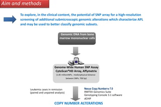
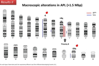
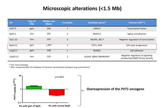
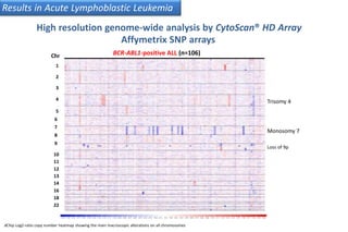
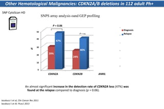
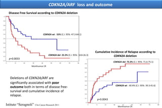
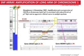
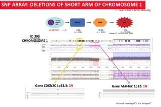
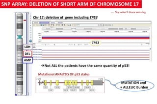
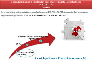
Use of Affymetrix Arrays
(Gene Chip® Human Transcriptome 2.0 Array
HTA and Cytoscan® HD Array)
in hematological malignancy studies
Giovanni Martinelli, MD
Institute of Hematology and Medical Oncology “L.e A. Seragnoli”
University of Bologna, Italy
Finding and Implementing the right clinically actionable cancer biomarker: 360° Tumor profiling
Disclosure
-
-
-
-
Grant and/or MTA from: Novartis, Merck SD, Astrazeneca, Roche, Takeda, J&J,
Mundipharma, Lilly, Pfizer, Cepheid, Onconova, Celgene, Amgen
Consultant/advisor of Celgene, Novartis, Merck SD, Astrazeneca, Roche, Takeda,
JeJ, Mundipharma, Pfizer, BMS, Ariad, Personal Genomics, Quiagen, IL,
Cepheid, Adienne P.
I have no grant or conflict from Affymetrix.
“The statements in this presentation are those of the Author and not of Affymetrix”
Background
• For istace, the genetic hallmark of APL is the t(15;17) (detected by karyotype)
resulting in the fusion of the promyelocytic leukemia (PML) gene and retinoic acid
receptor α (RARα) gene (PML-RARα) (detected by RT-PCR).
• PML-RARα is necessary but not sufficient for the development of APL  Are
additional cooperating genetic events also required for its pathogenesis?
• The development of single nucleotide polymorphism (SNP) Cyto ScanHD®-arrays
now allows to perform genome-wide screens for submicroscopic genomic
alterations with unprecedented informativity and to map all the genes involved in
these alterations.
Nexus Copy NumberTM 7.0
PARTEK Genomics Suite
Genotyping Console 3.1 software
Aim and methods
To explore, in the clinical content, the potential of SNP array for a high-resolution
screening of additional submicroscopic genomic alterations which characterize APL
and may be used to better classify genomic subsets.
Genomic DNA from bone
marrow mononuclear cells
Genome-Wide Human SNP Assay
CytoScan®HD Array, Affymetrix
(1.85 millionSNPs; medianphysical distance
between SNPs: 700 bp)
Leukemia cases in remission
(paired and unpaired analysis)
dCHIP
COPY NUMBER ALTERATIONS
SNP ARRAY: by the new potent CytoScan® HD* Array Affymetrix
CytoScan®HD ARRAY
2.67 milions di markers
750,000 SNP probes
1.9 milioni non-polymorphic probes
•100% Sanger cancer gene coverage
•100% ICCG constitutional gene coverage
•12,000 OMIM® genes
•36,000 RefSeq genes
FDA version approved for
diagnostic use
*for research use only
Patients Variable
Patients (number)
Median age, yrs (range)
Male/Female
De novo AML
M0
M1
M2
M3
M4
M5
Bi-lin
Secondary AML
Cytogenetics
Normal
Complex*
t(15;17)
t(8;21)
inv(3)
inv(16)
other**
+8
NA***
n
105
50 (18-84)
64/41
86 (82%)
6 (7%)
11 (13%)
11 (13%)
28 (33%)
14 (16%)
15 (17%)
1 (1%)
19 (28%)
35 (33%)
8 (8%)
28 (27%)
3 (3%)
3 (3%)
1 (1%)
22 (21%)
2 (2%)
3 (3%)
* Presence of at least 3 chr abnormalities in the absence of t(8;21), inv(16)/t(16;16), t(15;17),and t(11q23);** rare traslocations; *** NA: not available
Principal Component Analysis (PCA)
Results I
PCA showed an evident separation between APL and other AML subtypes →
a peculiar genomeprofile characterizeAPL patients
APL
Karyotype
• NA
• Normal
• Complex
• inv(3)
• inv(16)
• t(15;17)
• t(8;21)
• other
From PartekGenomicsSuite
Identification of multiple copy number alterations
 A wide spectrum of different genetic lesions (gains/losses) involving complete
chromosome arms or submicroscopic genomic intervals were identified in all cases.
 No significant difference in the average number of alterations was detected among
different karyotpye subgroups, except for complex karyotype group.
Karyotype
Normal
Average CNAs* (n)
14
Range
3-44
Complex
t(15;17)
t(8;21)
Trisomy 8
Others
55
8
12
5.5
9
35-92
1-24
6-16
5-6
6-18
p < 0.01
* CNAs: Copy Number Alterations; they includingboth deletions and amplifications
Alteration= region which has a copy number state lower or higher than 2.
At least 5 probesets have been considered.
Results II
Macroscopic alterations in APL (>1.5 Mbp)
loss
gain
-6
For each type of aberration,each line representsa different case (from AffymetrixGenotyping Console v3.1).
+8q
Trisomy 8
-20q
Microscopic alterations (<1.5 Mb)
Chr
1q23.3
6q25.1
7q11.23
8q24.21
11q23.1
12q24.12
Type of
CNA
gain
loss
loss
gain
gain
loss
Median size
(Kbps)
258
202
224
1,907
238
226
# samples
1
2
3
3
1
1
Candidate genes*
LMX1A
AKAP12
MLXIPL, BCL7
PVT1, MYC
NCAM1
ALDH2, BRAP, MAPKAPK5
Function (GO**)
Transcription factor
Signal transduction
Negative regulation of transcription
Cell cycle progression
Cell adhesion
Negative regulation of signaling
tansduction/MAP Kinase activity
Pts with gain of 8q24 Pts with normal 8q24
**GO: Gene Ontology
* After comparisonwith the Database of Genomic Variants(http://projects.tcag.ca/variation/)
p<0.0001
Overexpression of the PVT1 oncogene
Results V
CytoScan® HD Array Affymetrix SNP arrays allow classification of
Acute Promyelocitic Leukemia genomic subgroups
1. No additional chromosomal
abnormalities and low burdenof
CNAs
3. Additional chromosomal
abnormalities and high number of
CNAs
vs
2. Additional chromosomal
abnormalities and low burden of CNAs
Results V
Copy number alterations (CNAs) worsen outcome
ACA: Additional chromosomal abnormalities
ACA+ CNAs > 10 (GroupIII)
(Group I + Group II)
Event Free Survival Time (months)
Stratification according to additional chromosomal abnormalities and a high number of CNAs is
associated with a highly significant shorterevent-freesurvival
Chr
1
2
3
4
5
6
7
8
9
BCR-ABL1-positive ALL (n=106)
Trisomy 4
Monosomy 7
Loss of 9p
10
11
12
13
14
16
18
22
dChip Log2-ratio copy number heatmap showing the main macroscopic alterations on all chromosomes
Results in Acute Lymphoblastic Leukemia
High resolution genome-wide analysis by CytoScan® HD Array
Affymetrix SNP arrays
10
0
20
30
50
40
CDKN2A CDKN2B ANRIL
Diagnosis
Relapse
29%
47%
24%
29%
40%
43%
%
P = ns
An almost significant increase in the detection rate of CDKN2A loss (47%) was
found at the relapse compared to diagnosis (p = 0.06).
Iacobucci I et al, Clin Cancer Res 2011
Iacobucci I et Al. PLos1 2013
Other Hematological Malignancies: CDKN2A/B deletions in 112 adult Ph+
ALL patients
SNP CytoScan HD
SNPS array analysis oand GEP profiling
P = 0.06
p=0.0033
CDKN2A del: 22.2% (C.I. 95%: 18.8-26.3)
CDKN2A/ARF loss and outcome
Disease Free Survival according to CDKN2A deletion
CDKN2A wt : 55% (C.I. 95%: 47.3-64.1)
Cumulative Incidence of Relapse according to
CDKN2A deletion
p=0.0043
CDKN2A wt : 40.4% (C.I. 95%: 39.3-41.6)
Monthssince CR
CDKN2A del: 73.3% (C.I. 95%: 71.6-75.1)
Monthssince CR
Deletions of CDKN2A/ARF are
significantly associated with poor
outcome both in terms of disease free-
survival and cumulative incidence of
relapse.
Istituto “Seragnoli” Clin Cancer Research 2011
SNP ARRAY: AMPLIFICATION OF LONG ARM OF CHROMOSOME 1
… See what’s been missing
Shaughnessy J, Hematology 2005: Amplification and overexpression of
CKS1B at chromosome band 1q21 is associated with reduced levels of
p27 Kip1 and an aggressive clinical course in multiple myeloma
ID 343
CHROMOSOME 1
Gene MDM4 1q32.1: 3N
Istitutodi Ematologia“L. e A. Seràgnoli”
Gene CKS1B 1q21.3: 2N
Gene CDKN2C 1p32.3: 2N Gene FAM46C 1p12: 1N
Istitutodi Ematologia“L. e A. Seràgnoli”
SNP ARRAY: DELETIONS OF SHORT ARM OF CHROMOSOME 1
… See what’s been missing
Leone PE, Clin Canc Res 2008
ID 293
CHROMOSOME 1
SNP ARRAY: DELETION OF SHORT ARM OF CHROMOSOME 17
Chr 17: deletion of gene including TP53
Not ALL the patients have the same quantity of p53!
LOH
DEL
AMP
TP53
MUTATION and
+ ALLELIC Burden
Mutational ANALYSIS OF p53 status
… See what’s been missing
Phase 1b Study Of The MDM2 Antagonist RG7112, the ri-activator of
P53 function, in Combination With 2 Doses/Schedules Of Cytarabine
Karen Yee1, Giovanni Martinelli2, Sarit Assouline3, Margaret Kasner4, Norbert Vey5, Kevin R.
Kelly6, Mark W. Drummond7, Michael Dennis8, Karen Seiter9, Steven Blotner10,
Lori Jukofsky11, Steven Middleton12, Jianguo Zhi12, Gong Chen13, Hua Zhong13 and Gwen
Nichols12
1Princess Margaret Hospital, Toronto, ON, Canada; 2Department of Hematology and Oncological Sciences “L. e A. Seràgnoli”,
University of Bologna, Bologna, Italy; 3Jewish General Hospital, Montreal, QC, Canada; 4Kimmel Cancer Center, Thomas
Jefferson University, Philadelphia, PA; 5Hematology department, Institut Paoli Calmettes, Marseille, France;6University of Texas
Health Science Center at San Antonio, CTRC Institute for Drug Development, San Antonio, TX; 7Beatson West of Scotland
Cancer Centre, Glasgow, United Kingdom; 8Haematology, Christie NHS Foundation Trust, Manchester, United Kingdom; 9New
York Medical College, Valhalla, NY; 10Biostatistics, Hoffmann-LaRoche, Inc, Nutley, NJ;11Hoffmann-LaRoche, Nutley,
NJ; 12Research and Early Development, Hoffmann-LaRoche, Inc, Nutley, NJ;13Bioinformatics, Hoffmann-La Roche, Nutley, NJ
24
p53 symmetric division
The mdm2-mdm4 inhibition restore P53 activation, MYC silencing and p21
down regulation in leukemia
RG7112
oncogene
MDM2
γH2AX
p21 p21-/-
induction of apoptosis
cell cycle stop
asymmetric divisions
dna repair stop
reduced self renewal
The p21 down regulation resembles the mouse model p21-/-
CHROMOTHRIPSIS is frequent associated to p53 structural alteration
Istitutodi Ematologia“L. e A. Seràgnoli”
Tubio J, Nature 2011
Single CATASTROFIC EVENT with breakage
of multiple sites of the region of the chromosome
• Rearrangements
• Deletions
CHROMOTHRIPSIS by SNP ARRAY in MM: CROMOSOMA 16
Marina Martelloet Al. UnpublishedIstitutodi
Ematologia“L. e A. Seràgnoli”
FRAGILE SITE
FRA16D
 Analysis of genes involved in chromosomal translocations
 Comparison between AML and ALL subtypes: aneuploidy vs.
euploidy
2. How does this approach enable the better identification and validation of
actionable biomarkers, translating these into clinical utility and personalized
therapy
Use of Human Transcriptome ®Array
>285,000 full-length transcripts covered:
n >245,000 coding transcripts
n >40,000 non-coding transcripts
n >339,000 probe sets covering exon-exon junctions
Probes designed to maximize exon coverage enable you to
measure all transcript isoforms
Confidence in your results:
n Reproducible: Intra-lot correlation coefficient ≥0.99
n <6.5% coefficient of variation observed for all tissues tested
Minimum total RNA required: 50 ng
GeneChip®Human Transcriptome Array 2.0
Use ofAffymetrixArrays (Human TranscriptomeArray -HTAand Cytoscan HDArray)
in hematological malignancy studies
Gene expression analysis
Benefits of GeneChip Human TranscriptomeArray 2.0:
ex1 ex2 ex3 ex4 ex5 ex6 ex7 ex8
5’ 3’
The extent of IKZF1 deletions correlated with the expression of dominant-negative or
untranslated Ikaros isoforms
 Δ4-7 deletion (65%)
Deletion
 Δ2-7 deletion (30%)
ex1 ex8
5’ 3’
ex2 ex3 ex4 ex5 ex6 ex7
Dominant-negative
Ik6 isoform
Untranslated isoform
Deletion
Iacobucci I et al, Blood 2009
Additional discovery sequencing essential to identify the full range of
Up to 15% of childhoodALL, 26% of young adults
Patients may be detected by
-
-
phosphosignaling analysis / expression profiling
next generation sequencing
Diverse range of alterations converge onABL1/JAK signaling
kinase-activating alterations
Kathryn G. Roberts, Cancer Cell 2012
C.Mullighan, Hematology 2012
IKZF1 Deletions Are Associated With High Rate Of Cumulative Incidence
of Relapse and with Short Disease Free Survival
Martinelli G, Iacobucci I, et al JCO 2009
Ph+ ALL
Characterization of the molecular basis of acute lymphoblastic leukemia
BCR-ABL-like
in adults
The primary objective of the study is to genetically characterize BCR-ABL1-like ALL, to determine their frequency and
prognosis in adult patients and to find NEW BIOMARKERS FOR TARGET THERAPY
GeneChip®Human TranscriptomeArray 2.0
Acute Myeloid Leukemia: Samples characteristics
Normal karyotype
One-two abnormalities
Monosomal karyotype
Complex karyotype
27/50
6/50
5/50
4/50
Other abnormalities 8/50
Rare
translocations
Sample
# 20
FISH
t(6;17)(p21;q11)
RNAseq
STK38 (chr 6p21) – PSMD11 (chr 17q11)
RPL7L1 (chr 6p21) – BC062794 (chr 17q12)
ZEB2 (chr 2q22) – BCL11B (chr 14q)
FAM128A/B (chr 2q21) – CDC42BPB (chr 14q32)
ANO3 (chr 11p14) – CORO1C (chr 12q24)
# 59810
# 21
t(2;14)
t(11;12)
t(3;12)(p22;q24)
monosomal
karyotype
AL049692.1 (chr 11p13) – CNOT2 (chr 12q15)
HINFP (chr 11q23) – RSRC2 (chr 12q24)
NUMA1 (chr 11q13) – SLC35E3 (chr 12q15)
FEZ1 (chr 11q24) – TAOK3 (chr 12q24)
WT1 (chr 11p13) – CNOT2 (chr 12q15)
LIPH (chr 3q27) – PCBP2 (chr 12q13)
NICN1 (chr 3p21) – SPATS2 (chr 12q13)
Simonetti G.
Reference group Cytogenetics abnormalities - no.
Fold difference
(RNAseq sample/ref samples)
1.28 1.32
0.93
0.8
0.4
0.0
1.2
1.63
1.6
2.0
STK38 BC062794 RPL7L1 PSMD11
Increased expression of PSMD11 in the STK38-PSMD11 fusion
GENES
•
•
PSMD11 is upregulated compared to the average of the control group.
PSMD11 is a component of the lid subcomplex of the 26S proteasome, involved in the ATP-
dependent degradation of ubiquitinated proteins. It is required for proteasome assembly.
Simonetti G.
6p21
17q11
Fold difference
(RNAseq sample/ref samples)
0.87
1.00 1.05 1.06 1.18 1.30 1.36 1.48 1.52 1.63 1.75 1.79
1.99
3.91
2.0
1.5
1.0
4.0
3.5
3.0
2.5
0.5
0.0
GENES
Simonetti G. et Al Unpublished Personal comunication .
Increased expression of ZEB2, BCL11B, NUMA1 and HINFP upon gene fusions
14q32
4.5
2q22
2.36
Conclusions
• Despite the overall number of lesions across the patient cohort, novel
regions of micro and macro genetic alteration were identified in de
novo AML-ALL-MM patients by Gene CHIP Human Transcriptome® 2.0
array and by Cyto ScanHD®-arrays :
• Molecular karyotyping is easy obtained,
• Copy neutral loss of heterozygousity (CN-LOH) are friendly
analysed
• Chromothripsis could be identified
• New expressed biomarkers (Bcr-ABL like) for target leukemia
therapy
The identification of additional cytogenetic abnormalities and a high
number of microscopic genetic alterations allows to define subgroups
with worse prognosis, and rapidly stratify these patients to
individualized, personalized leukemia therapy.
Guadagnuolo, Stefania Paolini, Sarah Parisi, Cristina Clissa, Ilaria Iacobucci, Anna
Acknowledgments
Institute of Hematology “L. and A. Seràgnoli”, Bologna
Cristina Papayannidis, Chiara Sartor, Maria Chiara Abbenante, Emanuela Ottaviani, Viviana
Ferrari….,………………………………………
……………………………………..
…………………………………….
Supported by: FP7, European LeukemiaNet, AIL, AIRC, FIRB 2006, Fondazione del Monte di Bologna e
Ravenna
Acknowledgments
•
•
•
•
•
•
•
•
•
•
•
Emanuela Ottaviani
Viviana Guadagnuolo
Stefania Paolini
Cristina Papayannidis
Anna Ferrari
Giorgia Simonetti
Valentina Robustelli
Carolina Terragna
Marina Martello
Ilaria Iacobucci
Michele Baccarani
Institute “L. e A. Seràgnoli” Department of Biopathology,
University Tor Vergata, Rome, Italy
• Francesco Lo Coco
• Hasan Syed Khizer
Department of Clinical and Biological
Sciences, University at Orbassano
• Giuseppe Saglio
• Daniela Cilloni

Use of Affymetrix Arrays (GeneChip® Human Transcriptome 2.0 Array and Cytoscan® HD Array) in haematological malignancy studies

  • 1.
    Use of AffymetrixArrays (Gene Chip® Human Transcriptome 2.0 Array HTA and Cytoscan® HD Array) in hematological malignancy studies Giovanni Martinelli, MD Institute of Hematology and Medical Oncology “L.e A. Seragnoli” University of Bologna, Italy Finding and Implementing the right clinically actionable cancer biomarker: 360° Tumor profiling
  • 2.
    Disclosure - - - - Grant and/or MTAfrom: Novartis, Merck SD, Astrazeneca, Roche, Takeda, J&J, Mundipharma, Lilly, Pfizer, Cepheid, Onconova, Celgene, Amgen Consultant/advisor of Celgene, Novartis, Merck SD, Astrazeneca, Roche, Takeda, JeJ, Mundipharma, Pfizer, BMS, Ariad, Personal Genomics, Quiagen, IL, Cepheid, Adienne P. I have no grant or conflict from Affymetrix. “The statements in this presentation are those of the Author and not of Affymetrix”
  • 3.
    Background • For istace,the genetic hallmark of APL is the t(15;17) (detected by karyotype) resulting in the fusion of the promyelocytic leukemia (PML) gene and retinoic acid receptor α (RARα) gene (PML-RARα) (detected by RT-PCR). • PML-RARα is necessary but not sufficient for the development of APL  Are additional cooperating genetic events also required for its pathogenesis? • The development of single nucleotide polymorphism (SNP) Cyto ScanHD®-arrays now allows to perform genome-wide screens for submicroscopic genomic alterations with unprecedented informativity and to map all the genes involved in these alterations.
  • 4.
    Nexus Copy NumberTM7.0 PARTEK Genomics Suite Genotyping Console 3.1 software Aim and methods To explore, in the clinical content, the potential of SNP array for a high-resolution screening of additional submicroscopic genomic alterations which characterize APL and may be used to better classify genomic subsets. Genomic DNA from bone marrow mononuclear cells Genome-Wide Human SNP Assay CytoScan®HD Array, Affymetrix (1.85 millionSNPs; medianphysical distance between SNPs: 700 bp) Leukemia cases in remission (paired and unpaired analysis) dCHIP COPY NUMBER ALTERATIONS
  • 5.
    SNP ARRAY: bythe new potent CytoScan® HD* Array Affymetrix CytoScan®HD ARRAY 2.67 milions di markers 750,000 SNP probes 1.9 milioni non-polymorphic probes •100% Sanger cancer gene coverage •100% ICCG constitutional gene coverage •12,000 OMIM® genes •36,000 RefSeq genes FDA version approved for diagnostic use *for research use only
  • 6.
    Patients Variable Patients (number) Medianage, yrs (range) Male/Female De novo AML M0 M1 M2 M3 M4 M5 Bi-lin Secondary AML Cytogenetics Normal Complex* t(15;17) t(8;21) inv(3) inv(16) other** +8 NA*** n 105 50 (18-84) 64/41 86 (82%) 6 (7%) 11 (13%) 11 (13%) 28 (33%) 14 (16%) 15 (17%) 1 (1%) 19 (28%) 35 (33%) 8 (8%) 28 (27%) 3 (3%) 3 (3%) 1 (1%) 22 (21%) 2 (2%) 3 (3%) * Presence of at least 3 chr abnormalities in the absence of t(8;21), inv(16)/t(16;16), t(15;17),and t(11q23);** rare traslocations; *** NA: not available
  • 7.
    Principal Component Analysis(PCA) Results I PCA showed an evident separation between APL and other AML subtypes → a peculiar genomeprofile characterizeAPL patients APL Karyotype • NA • Normal • Complex • inv(3) • inv(16) • t(15;17) • t(8;21) • other From PartekGenomicsSuite
  • 8.
    Identification of multiplecopy number alterations  A wide spectrum of different genetic lesions (gains/losses) involving complete chromosome arms or submicroscopic genomic intervals were identified in all cases.  No significant difference in the average number of alterations was detected among different karyotpye subgroups, except for complex karyotype group. Karyotype Normal Average CNAs* (n) 14 Range 3-44 Complex t(15;17) t(8;21) Trisomy 8 Others 55 8 12 5.5 9 35-92 1-24 6-16 5-6 6-18 p < 0.01 * CNAs: Copy Number Alterations; they includingboth deletions and amplifications Alteration= region which has a copy number state lower or higher than 2. At least 5 probesets have been considered.
  • 9.
    Results II Macroscopic alterationsin APL (>1.5 Mbp) loss gain -6 For each type of aberration,each line representsa different case (from AffymetrixGenotyping Console v3.1). +8q Trisomy 8 -20q
  • 10.
    Microscopic alterations (<1.5Mb) Chr 1q23.3 6q25.1 7q11.23 8q24.21 11q23.1 12q24.12 Type of CNA gain loss loss gain gain loss Median size (Kbps) 258 202 224 1,907 238 226 # samples 1 2 3 3 1 1 Candidate genes* LMX1A AKAP12 MLXIPL, BCL7 PVT1, MYC NCAM1 ALDH2, BRAP, MAPKAPK5 Function (GO**) Transcription factor Signal transduction Negative regulation of transcription Cell cycle progression Cell adhesion Negative regulation of signaling tansduction/MAP Kinase activity Pts with gain of 8q24 Pts with normal 8q24 **GO: Gene Ontology * After comparisonwith the Database of Genomic Variants(http://projects.tcag.ca/variation/) p<0.0001 Overexpression of the PVT1 oncogene
  • 11.
    Results V CytoScan® HDArray Affymetrix SNP arrays allow classification of Acute Promyelocitic Leukemia genomic subgroups 1. No additional chromosomal abnormalities and low burdenof CNAs 3. Additional chromosomal abnormalities and high number of CNAs vs 2. Additional chromosomal abnormalities and low burden of CNAs
  • 12.
    Results V Copy numberalterations (CNAs) worsen outcome ACA: Additional chromosomal abnormalities ACA+ CNAs > 10 (GroupIII) (Group I + Group II) Event Free Survival Time (months) Stratification according to additional chromosomal abnormalities and a high number of CNAs is associated with a highly significant shorterevent-freesurvival
  • 13.
    Chr 1 2 3 4 5 6 7 8 9 BCR-ABL1-positive ALL (n=106) Trisomy4 Monosomy 7 Loss of 9p 10 11 12 13 14 16 18 22 dChip Log2-ratio copy number heatmap showing the main macroscopic alterations on all chromosomes Results in Acute Lymphoblastic Leukemia High resolution genome-wide analysis by CytoScan® HD Array Affymetrix SNP arrays
  • 14.
    10 0 20 30 50 40 CDKN2A CDKN2B ANRIL Diagnosis Relapse 29% 47% 24% 29% 40% 43% % P= ns An almost significant increase in the detection rate of CDKN2A loss (47%) was found at the relapse compared to diagnosis (p = 0.06). Iacobucci I et al, Clin Cancer Res 2011 Iacobucci I et Al. PLos1 2013 Other Hematological Malignancies: CDKN2A/B deletions in 112 adult Ph+ ALL patients SNP CytoScan HD SNPS array analysis oand GEP profiling P = 0.06
  • 15.
    p=0.0033 CDKN2A del: 22.2%(C.I. 95%: 18.8-26.3) CDKN2A/ARF loss and outcome Disease Free Survival according to CDKN2A deletion CDKN2A wt : 55% (C.I. 95%: 47.3-64.1) Cumulative Incidence of Relapse according to CDKN2A deletion p=0.0043 CDKN2A wt : 40.4% (C.I. 95%: 39.3-41.6) Monthssince CR CDKN2A del: 73.3% (C.I. 95%: 71.6-75.1) Monthssince CR Deletions of CDKN2A/ARF are significantly associated with poor outcome both in terms of disease free- survival and cumulative incidence of relapse. Istituto “Seragnoli” Clin Cancer Research 2011
  • 16.
    SNP ARRAY: AMPLIFICATIONOF LONG ARM OF CHROMOSOME 1 … See what’s been missing Shaughnessy J, Hematology 2005: Amplification and overexpression of CKS1B at chromosome band 1q21 is associated with reduced levels of p27 Kip1 and an aggressive clinical course in multiple myeloma ID 343 CHROMOSOME 1 Gene MDM4 1q32.1: 3N Istitutodi Ematologia“L. e A. Seràgnoli” Gene CKS1B 1q21.3: 2N
  • 17.
    Gene CDKN2C 1p32.3:2N Gene FAM46C 1p12: 1N Istitutodi Ematologia“L. e A. Seràgnoli” SNP ARRAY: DELETIONS OF SHORT ARM OF CHROMOSOME 1 … See what’s been missing Leone PE, Clin Canc Res 2008 ID 293 CHROMOSOME 1
  • 18.
    SNP ARRAY: DELETIONOF SHORT ARM OF CHROMOSOME 17 Chr 17: deletion of gene including TP53 Not ALL the patients have the same quantity of p53! LOH DEL AMP TP53 MUTATION and + ALLELIC Burden Mutational ANALYSIS OF p53 status … See what’s been missing
  • 19.
    Phase 1b StudyOf The MDM2 Antagonist RG7112, the ri-activator of P53 function, in Combination With 2 Doses/Schedules Of Cytarabine Karen Yee1, Giovanni Martinelli2, Sarit Assouline3, Margaret Kasner4, Norbert Vey5, Kevin R. Kelly6, Mark W. Drummond7, Michael Dennis8, Karen Seiter9, Steven Blotner10, Lori Jukofsky11, Steven Middleton12, Jianguo Zhi12, Gong Chen13, Hua Zhong13 and Gwen Nichols12 1Princess Margaret Hospital, Toronto, ON, Canada; 2Department of Hematology and Oncological Sciences “L. e A. Seràgnoli”, University of Bologna, Bologna, Italy; 3Jewish General Hospital, Montreal, QC, Canada; 4Kimmel Cancer Center, Thomas Jefferson University, Philadelphia, PA; 5Hematology department, Institut Paoli Calmettes, Marseille, France;6University of Texas Health Science Center at San Antonio, CTRC Institute for Drug Development, San Antonio, TX; 7Beatson West of Scotland Cancer Centre, Glasgow, United Kingdom; 8Haematology, Christie NHS Foundation Trust, Manchester, United Kingdom; 9New York Medical College, Valhalla, NY; 10Biostatistics, Hoffmann-LaRoche, Inc, Nutley, NJ;11Hoffmann-LaRoche, Nutley, NJ; 12Research and Early Development, Hoffmann-LaRoche, Inc, Nutley, NJ;13Bioinformatics, Hoffmann-La Roche, Nutley, NJ 24
  • 20.
    p53 symmetric division Themdm2-mdm4 inhibition restore P53 activation, MYC silencing and p21 down regulation in leukemia RG7112 oncogene MDM2 γH2AX p21 p21-/- induction of apoptosis cell cycle stop asymmetric divisions dna repair stop reduced self renewal The p21 down regulation resembles the mouse model p21-/-
  • 21.
    CHROMOTHRIPSIS is frequentassociated to p53 structural alteration Istitutodi Ematologia“L. e A. Seràgnoli” Tubio J, Nature 2011 Single CATASTROFIC EVENT with breakage of multiple sites of the region of the chromosome • Rearrangements • Deletions
  • 22.
    CHROMOTHRIPSIS by SNPARRAY in MM: CROMOSOMA 16 Marina Martelloet Al. UnpublishedIstitutodi Ematologia“L. e A. Seràgnoli” FRAGILE SITE FRA16D
  • 23.
     Analysis ofgenes involved in chromosomal translocations  Comparison between AML and ALL subtypes: aneuploidy vs. euploidy 2. How does this approach enable the better identification and validation of actionable biomarkers, translating these into clinical utility and personalized therapy Use of Human Transcriptome ®Array
  • 24.
    >285,000 full-length transcriptscovered: n >245,000 coding transcripts n >40,000 non-coding transcripts n >339,000 probe sets covering exon-exon junctions Probes designed to maximize exon coverage enable you to measure all transcript isoforms Confidence in your results: n Reproducible: Intra-lot correlation coefficient ≥0.99 n <6.5% coefficient of variation observed for all tissues tested Minimum total RNA required: 50 ng GeneChip®Human Transcriptome Array 2.0 Use ofAffymetrixArrays (Human TranscriptomeArray -HTAand Cytoscan HDArray) in hematological malignancy studies Gene expression analysis Benefits of GeneChip Human TranscriptomeArray 2.0:
  • 25.
    ex1 ex2 ex3ex4 ex5 ex6 ex7 ex8 5’ 3’ The extent of IKZF1 deletions correlated with the expression of dominant-negative or untranslated Ikaros isoforms  Δ4-7 deletion (65%) Deletion  Δ2-7 deletion (30%) ex1 ex8 5’ 3’ ex2 ex3 ex4 ex5 ex6 ex7 Dominant-negative Ik6 isoform Untranslated isoform Deletion Iacobucci I et al, Blood 2009
  • 27.
    Additional discovery sequencingessential to identify the full range of Up to 15% of childhoodALL, 26% of young adults Patients may be detected by - - phosphosignaling analysis / expression profiling next generation sequencing Diverse range of alterations converge onABL1/JAK signaling kinase-activating alterations Kathryn G. Roberts, Cancer Cell 2012 C.Mullighan, Hematology 2012
  • 28.
    IKZF1 Deletions AreAssociated With High Rate Of Cumulative Incidence of Relapse and with Short Disease Free Survival Martinelli G, Iacobucci I, et al JCO 2009 Ph+ ALL
  • 29.
    Characterization of themolecular basis of acute lymphoblastic leukemia BCR-ABL-like in adults The primary objective of the study is to genetically characterize BCR-ABL1-like ALL, to determine their frequency and prognosis in adult patients and to find NEW BIOMARKERS FOR TARGET THERAPY GeneChip®Human TranscriptomeArray 2.0
  • 30.
    Acute Myeloid Leukemia:Samples characteristics Normal karyotype One-two abnormalities Monosomal karyotype Complex karyotype 27/50 6/50 5/50 4/50 Other abnormalities 8/50 Rare translocations Sample # 20 FISH t(6;17)(p21;q11) RNAseq STK38 (chr 6p21) – PSMD11 (chr 17q11) RPL7L1 (chr 6p21) – BC062794 (chr 17q12) ZEB2 (chr 2q22) – BCL11B (chr 14q) FAM128A/B (chr 2q21) – CDC42BPB (chr 14q32) ANO3 (chr 11p14) – CORO1C (chr 12q24) # 59810 # 21 t(2;14) t(11;12) t(3;12)(p22;q24) monosomal karyotype AL049692.1 (chr 11p13) – CNOT2 (chr 12q15) HINFP (chr 11q23) – RSRC2 (chr 12q24) NUMA1 (chr 11q13) – SLC35E3 (chr 12q15) FEZ1 (chr 11q24) – TAOK3 (chr 12q24) WT1 (chr 11p13) – CNOT2 (chr 12q15) LIPH (chr 3q27) – PCBP2 (chr 12q13) NICN1 (chr 3p21) – SPATS2 (chr 12q13) Simonetti G. Reference group Cytogenetics abnormalities - no.
  • 31.
    Fold difference (RNAseq sample/refsamples) 1.28 1.32 0.93 0.8 0.4 0.0 1.2 1.63 1.6 2.0 STK38 BC062794 RPL7L1 PSMD11 Increased expression of PSMD11 in the STK38-PSMD11 fusion GENES • • PSMD11 is upregulated compared to the average of the control group. PSMD11 is a component of the lid subcomplex of the 26S proteasome, involved in the ATP- dependent degradation of ubiquitinated proteins. It is required for proteasome assembly. Simonetti G. 6p21 17q11
  • 32.
    Fold difference (RNAseq sample/refsamples) 0.87 1.00 1.05 1.06 1.18 1.30 1.36 1.48 1.52 1.63 1.75 1.79 1.99 3.91 2.0 1.5 1.0 4.0 3.5 3.0 2.5 0.5 0.0 GENES Simonetti G. et Al Unpublished Personal comunication . Increased expression of ZEB2, BCL11B, NUMA1 and HINFP upon gene fusions 14q32 4.5 2q22 2.36
  • 33.
    Conclusions • Despite theoverall number of lesions across the patient cohort, novel regions of micro and macro genetic alteration were identified in de novo AML-ALL-MM patients by Gene CHIP Human Transcriptome® 2.0 array and by Cyto ScanHD®-arrays : • Molecular karyotyping is easy obtained, • Copy neutral loss of heterozygousity (CN-LOH) are friendly analysed • Chromothripsis could be identified • New expressed biomarkers (Bcr-ABL like) for target leukemia therapy The identification of additional cytogenetic abnormalities and a high number of microscopic genetic alterations allows to define subgroups with worse prognosis, and rapidly stratify these patients to individualized, personalized leukemia therapy.
  • 34.
    Guadagnuolo, Stefania Paolini,Sarah Parisi, Cristina Clissa, Ilaria Iacobucci, Anna Acknowledgments Institute of Hematology “L. and A. Seràgnoli”, Bologna Cristina Papayannidis, Chiara Sartor, Maria Chiara Abbenante, Emanuela Ottaviani, Viviana Ferrari….,……………………………………… …………………………………….. ……………………………………. Supported by: FP7, European LeukemiaNet, AIL, AIRC, FIRB 2006, Fondazione del Monte di Bologna e Ravenna
  • 35.
    Acknowledgments • • • • • • • • • • • Emanuela Ottaviani Viviana Guadagnuolo StefaniaPaolini Cristina Papayannidis Anna Ferrari Giorgia Simonetti Valentina Robustelli Carolina Terragna Marina Martello Ilaria Iacobucci Michele Baccarani Institute “L. e A. Seràgnoli” Department of Biopathology, University Tor Vergata, Rome, Italy • Francesco Lo Coco • Hasan Syed Khizer Department of Clinical and Biological Sciences, University at Orbassano • Giuseppe Saglio • Daniela Cilloni