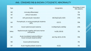
INTEGRATED HAEMATOPATHOLOGY-
NEW APPROACH FOR DIAGNOSIS OF
LEUKAEMIA / LYMPHOMA
Dr Rajesh V Bendre
MD(Path), DNB(Path), DPB
Chief Pathologist & Head Laboratory Services
Jaslok Hospital & Research center
INTRODUCTION
 Revised 2016 Classification for Myeloid (Dr Arber et al) & Lymphoid Neoplasms (Dr Swerdlow et al) -
Reflect a Consensus of opinion of hematopathologists, hematologists, oncologists, and geneticists. It
represents a revision of the prior classification and attempts to incorporate new clinical, prognostic,
morphologic, immunophenotypic and genetic data that have emerged since the last edition (WHO
classification 2008).
 Immense progress in the treatment of blood cancers, but they also represent a future
of cancer research that is genetically driven and increasingly personalized- Modern
Oncology.
 Acute myeloid leukemia (AML) that targets a specific genetic mutation
 Rituximab, a therapy already used to treat a range of cancers of the immune
system, to treat B-cell precursor acute lymphocytic leukemia (BCP-ALL)
 These potent therapies are more effective and potentially safer than standard
chemotherapy because they target specific proteins and mutated gene products while
leaving other cells unharmed.
 The diagnosis & monitoring of leukaemia / lymphoma requires effective integration of
a wide range of diagnostic techniques and expertise. Hence, the need to develop
services that cross traditional boundaries of laboratory specialities is recommended as
guidance.
INTRODUCTION
LEUKAEMIA PANEL REPORTING WORKFLOW
Smear
Examination
• Pathologists review direct smear for morphological assessment and
verify presence/identification of malignant/atypical cells prior flow
cytometry procedures
Cell
Viability
• Cell viability check- 80% as acceptable criteria
• However for Leukemia / Lymphoma studies, though viability is less than
80%, based on the population of interest gated, the Pathologists to take a
call on further processing of sample.
Specimen
Dilution:
• WBC concentration for processing of single markers should be < 25000cells/cumm
Processing
• Lyse –wash staining of cells
Acquisition
• 8 color flow cytometer with BD FACS DIVA &/or Beckman software
Analysis
• Gating & Dot plots, validation of Antibody fluorescence intensity
REPORTING
• Interpretation- Marker Positivity based on Percentage &/or Log shift
Acute Leukaemia case
Type Name Cytogenetics
Percentage of adult
AML patients
M0 minimally differentiated 5%
M1 without maturation 15%
M2 with granulocytic maturation t(8;21)(q22;q22), t(6;9) 25%
M3
Promyelocytic, or acute promyelocytic leukemia
(APL)
t(15;17) 10%
M4 Acute myelomonocytic leukemia inv(16)(p13q22), del(16q) 20%
M4eo
Myelomonocytic together with bone marrow
eosinophilia
inv(16), t(16;16) 5%
M5
Acute monoblastic leukemia (M5a) or
Acute monocytic leukemia (M5b)
del (11q), t(9;11), t(11;19) 10%
M6 Acute erythroid leukemias 5%
M7 Acute megakaryoblastic leukemia t(1;22) 5%
AML- STANDARD FAB & KNOWN CYTOGENETIC ABNORMALITY
 For more than two decades, patients with
myelodysplasia (MDS) or acute myeloid leukemia (AML)
have been classified and treated according to their
cytogenetic characteristics, which represent the most
important prognostic factor of these diseases.
 The first includes patients with the recurrent
unbalanced chromosome aberrations. Loss of whole
chromosome 5 or 7 (-5/-7) or various parts of the long
arms of the two chromosomes (5q-/7q-) or gain of a
whole chromosome 8 (+8) are the most common
unbalanced aberrations.
 The second subgroup comprises patients with recurrent
balanced chromosome aberrations, mainly reciprocal
translocations. These are often observed as sole
abnormalities, in younger patients and in cases
presenting as overt AML.
 The third subgroup includes patients with a normal karyotype.
They present as either MDS with a favorable prognosis or as
AML with an intermediate prognosis
 In addition to chromosomal aberrations, gene mutations and
duplications are frequently observed in AML patients.
Approximately 40-50% of AML patients have a normal karyotype,
but have shown a wide range of clinical outcomes . Therefore, it is
particularly important to identify molecular prognostic factors
that can improve risk stratification in this group of patients. It has
been shown that 85% of AML patients with a normal karyotype
have gene mutations, in particular, NPM1, FLT3 and CEBPA. These
mutations have prognostic significance and are being used in
diagnosis
 Patients with a normal karyotype were previously poorly
characterized with respect to genetic changes. More recently,
particularly in this subgroup of patients with AML, mutations of
the FLT3 gene encoding a receptor tyrosine kinase,of
the CEBPA gene encoding a hematopoietic transcription factor
and of the NPM1 transcription regulating gene have been
observed.
 In leukemogenesis, a cooperation between two general types of
gene mutations have been proposed . Class I mutations result in
constitutive activation of receptor tyrosine kinases or genes
downstream in the RAS–BRAF–MEK–ERK signal-transduction
pathway, thereby stimulating cell cycling and proliferation. Class II
mutations, on the contrary, inactivate hematopoietic transcription
factors and result in loss of differentiation.
AML/ETO:t(8;21)
PML/RARA:t(15;17)
CBFB/MYH1:inv(16)
MLL:t(11q23)
AML with
Chromosomal
abnormalities
AML/ETO
CBFB
C-Kit mutation
Without c-Kit
mutation
Better Risk
With C-Kit
Mutation
Intermediate risk
PML/RARA
Better Risk
+8
Intermediate risk
Complex Karyotype
-5/5q or -7/7q
deletion
MLL
BCR/ABL
Poor Risk
AML without
chromosomal
abnormalities
AML
Molecular Testing
Risk Assessment
AML/ETO:t(8;21)
PML/RARA:t(15;17)
CBFB/MYH1:inv(16)
MLL:t(11q23)
AML with
Chromosomal
abnormalities
AML without
chromosomal
abnormalities
NPM1or CEBPA
mutations in absence
of FLT3-ITD
No NPM1, or CEBPA or
FLT3-ITD mutations
FLT3-ITD mutations
Poor Risk
Intermediate Risk
Better Risk
AML
Molecular
Testing
FLT3
NPM1
Acute Lymphoblastic Leukemia
• Hyperdiploidy (51-65
chromosomes) Trisomies of
chromosomes 4,10 and 17.
• t(12;21)(p13;q22):TEL-AML1
Favorable markers
• t(1;19)(q23;p13.3):E2A-PBX1
Intermediate
markers
• Hypodiploidy (<44
chromosomes)
• t(11q23):MLL rearrangements
• t(9;22)(q34;q11.2)
Poor risk markers
t(9;22)(q34;q11) (BCR/ABL1): Cases of precursor B-
cell ALL with t(9;22) are associated with a poor
prognosis. The t(9;22) is observed in about 2 to 5
percent of children compared with about 30
percent of adults
11q23 (MLL) abnormalities: Translocations
involving the MLL gene, which may involve one of
many partner genes, are associated with a poor
prognosis in precursor B-cell ALL.
t(12;21)(p13;q22) (TEL/AML1): This abnormality
cannot be detected by traditional banded
karyotyping. Cases of precursor B-cell ALL with
t(12;21) are associated with a favorable prognosis.
The t(12;21), which is detectable only by FISH or
polymerase chain reaction (PCR) analysis, is
observed in about 25 percent of children with B-
lineage leukemia, compared with about 3 percent
of adults.
Interphase Cell----Split
in MLL
t(4;11) by centromeric /
Loci specific probe
Acute Leukemia
Myeloproliferative neoplasms (MPNs)
• Chronic myelogenous leukemia (CML)
• Chronic neutrophilic leukemia
• Polycythemia vera (PV)
• Primary myelofibrosis (PMF)
• Essential thrombocythemia (ET)
• Chronic eosinophilic leukemia
CHRONIC MYELOID LEUKEMIA
Chromosomal Analysis for Philadelphia chromosome
FISH for BCR/ABL fusion detection for a faster
turn around time.
BCR/ABL MRD status Monitoring the response
to treatment Real Time RQ-PCR
Monitoring
Tyrosine kinase mutation analysis Resistance to
Imatinib treatment : ABL Kinase Domain
mutations. Relapse
Treatment Response for CML
Definitions of Responses to Treatments
Hematologic Response
Complete Hematologic response
1) Normal PB counts (WBC < 10 and plt < 450)
2) Normal WBC differential
3) No Dz symptoms
4) Normalization of the size of the liver and spleen
Cytogenetic Responses: Ph+ Metaphases
1) complete: 0%
2) partial: 1% - 35%
3) minor: 36% - 65%
4) minimal: 66% - 95%
5) none: 96% - 100%
Molecular Responses: ratio of Bcr-Abl/Abl
Major Molecular Response
3-log10 reduction from initial diagnosis sample (i.e. 25 →0.025)
BCR –ABL IS BY RT-PCR
Every three months RQ-PCR
Every 6 months Cytogenetic
Response by CK/FISH
MMR not achieved Resistance
workup
ABL Kinase domain mutation
analysis
T315I V299L T315A Y253H
E255k/V F359V/C/I
IMATINIB RESISTANCE MUTATIONAL ANALYSIS
Summary of the most appropriate
alternative therapeutic options based on the
BCR-ABL KD mutation status
• T315I - HSCT (hematopoietic stem cell
transplantation )or investigational drugs
• V299L, T315A, and F317L/V/I/C -
Consider nilotinib rather than dasatinib
• Y253H, E255K/V, and F359V/C/I -
Consider dasatinib rather than nilotinib
• Any other mutation - Consider high-dose
imatinib* (No sufficient data on dose
escalation available to indicate if
mutations with lower IC50 values are
sensitive to high-dose imatinib) or
dasatinib or nilotinib
Chronic Myeloproliferative disorders
Tefferiand Vardiman.Leukemia, Sep 20 Epub, 2007
Chronic Myeloproliferative disorders
Polycythaemia Vera (PV)
~50% with Essential Thrombocythaemia (ET) or Idiopathic Myelofibrosis (IMF).
JAK2 V617 F Mutation
Negative
Exon 12 and 13
Mutations
Negative
MPL & Calreticulin
Mutations
Positive
No
further
testing
Positive
No further
testing
PRIMARY EOSINOPHILIA- APPROACH
CLL
Markers of Low risk
Mutated IgVH gene
ZAP-70 negative
CD38 negative
13q deletion
Markers of High Risk
Unmutated IgVH
ZAP-70 positive
CD38 positive
11q deletion (ATM)
Markers of Greatest Risk
17p/p53 deletion
P53 gene mutation
ASSESSMENT OF PROGNOSIS IN CLL
Cytogenetics of Multiple Myeloma- FISH Panel
Test Probe Gene(s)/Unique Sequence Prognosis
Deletion 13q 13q14.3 D13S319 Short event free
survival
Deletion 17p 17p13.1 p53 High risk marker
Ploidy analysis 11q23/15q22/9q34 MLL/PML/ABL Favorable Prognosis
IGH rearrangement 14q32 IGH
If an IGH rearrangement is detected, additional FISH experiments will be performed
to detect t(11;14), t(4;14) or t(14;16)
Translocation: t(11;14) t(11;14)(q13;q32) IGH/CCND1XT Good Prognosis
Translocation: t(4;14) t(4;14)(p16;q32) FGFR3/IGH Poor Prognosis
Translocation: t(14;16) t(14;16)(q32;q23.1) IGH/MAF Poor Prognosis
Lymphomas
NON-HOGDKINS LYMPHOMA
Burkitts Lymphomas c-MYC translocations/réarrangements - t(8;14)/
t(8;22)/ t(2;8)
Follicular Lymphomas t(14;18): IGH/BCL2
Mantle Cell Lymphomas t(11;14) IGH/CCND1
Anaplastic Large cell lymphoma t(2;5):ALK/NPM1
translocations/rearrangement
FISH IN LYMPHOMAS
• A rapid, robust, cheap and relatively easily
applicable technique that can be applied to
formalin fixed and paraffin embedded
tissues and does not need vital, growing
cells or specific culture systems
• A further advantage is a short turnaround
time.
• The major limitation of FISH is that is
targeted to the detection of specific
abnormalities and the genome wide
perspective offered by conventional
cytogenetics is lost.
Follicular lymphoma characterized by the presence of the t(14;18) translocation that juxtaposes the BCL2 locus
on chromosome 18q21 to the immunoglobulin H (IGH) locus on chromosome 14q32, resulting in the
overexpression of the antiapoptotic protein BCL2.
BCL2/IgH t(14;18) Fusion (tissue)
It a common cytogenetic abnormality in human lymphoma and is observed in about 85% of follicular
lymphoma (FL) and up to one-third of diffuse lymphomas (DL).
FOLLICULAR LYMPHOMA
Rearrangements of the proto oncogene C-myc (or MYC) have been consistently found in tumor
cells of patients suffering from Burkitt’s lymphoma.
MyC (8q24) Break, TC (tissue)
BURKITT LYMPHOMA
The most common chromosomal translocation in mantle cell lymphoma is t(11;14), resulting in up-regulation
of cyclin D1.
The CCND1/IGH t(11;14)(q13;q32) specific DNA probe is optimized to detect the reciprocal translocation
t(11;14)
CCND1/IgH t(11;14) Fusion tissue
MANTLE CELL LYMPHOMA
Cyclin D1 IHC – Positive
Diagnosis on IHC is better than On Flow-
cytometry as there is a background
fluorescence
Better technique to confirm MCL is by
FISH t(11,14)
Ki 67 index
Integrated Haematopathology- New Approach
 Integrated Hematopathology- includes
one or more of the following tests:
 Microscopy/Morphology
 Immunohistochemistry (IHC)
 Multi-color Cytometry
 Cytogenetics- Cell enrichment for In
Situ Hybridization/FISH and/or
molecular analysis
 Molecular Genetics - Qualitative &
Quantitative PCR, Sequencing &
Fragment Analysis
Integrated Haematopathology- New Approach
Performance parameters of different genetic techniques used in diagnosis of
leukemia.
Technique
Sample
type
Speed
(days)
Sensitivity
(%)
Resolution
Whole
genome
Detect
balanced
rearrangem
ents
G-band
metaphase
cytogenetics
BM/PB 8-12 1–5 3–5 Mb Yes Yes
FISH BM/PB 1-2 0.5–1 100 kb No Yes
Array CGH BM/PB 3-5 20–30 50 kb Yes No
SNP arrays BM/PB 3-5 20–30 25 kb Yes No
Molecular
(PCR- NGS
based)
BM/PB 5-7 0.001 1 bp No Yes
MOLECULAR TECHNOLOGIES IN HEMATOLOGY
Molecular technique Applications Advantages
Fluorescence in situ
hybridization (FISH)
centromeric enumeration
probes (CEP), chromosome
painting probes, dual-color
dual fusion probes (D-FISH)
and dual-color break-apart
probes
• BCR to ABL gene at 9q34, t(9;22) variant in chronic myeloid
leukemia (CML) and acute promyelocytic leukemia (APL)
• demonstration of t(11;18)(q21;q21) in mucosa-associated
lymphoid tissue (MALT) lymphoma [6]; the detection of clonal
abnormalities (deleted 5q, trisomy 8 and monosomy 7) in
myelodysplastic syndromes (MDS) with minimal dysplasia; 11q23
abnormalities in acute myeloid leukemia (AML) [7]; the
demonstration of hyper- or hypo-diploidy and occult
translocations in acute lymphoid leukemia (ALL), which is hard to
grow in metaphase and is one of the difficulties of cytogenetic
detections [8]; myeloproliferative neoplasms (MPN) with
eosinophilia and interstitial deletion on short arm of chromosome
4; chronic lymphocytic leukemia (CLL) with trisomy 12 and cryptic
t(15;17) variant – ins(17;15) in APL
MOLECULAR TECHNOLOGIES IN HEMATOLOGY
Molecular technique Applications Advantages
Polymerase Chain Reaction
(PCR) & Various derivatives
RT-PCR, Q-PCR, multiplex
PCR, gap-PCR, ARMS and IS-
PCR
Unlike FISH, PCR and its
variants only detect
breakpoints covered by
designated specific primers
• Application of PCR include JAK2 V617F mutations; FLT3, NPM1,
and CEBPA mutations in acute myeloid leukemia with normal
cytogenetics (CN-AML) [11]; and gene rearrangements such as Ig
heavy chain gene rearrangement for B-cell malignancies and T-cell
receptor gamma chain gene rearrangement for T-cell malignancies
DNA Sequencing DNA sequencing is a post-
PCR analysis and a
confirmation method for
mutations.
• Applications include non-deleted α thalassaemia, F8 mutation in
HA, AML with normal karyotype, AML with NPM1 exon 12 mutation,
AML with mutated CEBPA (not common), FLT3 exon 14 tandem
duplication and other mutated genes – KIT, MLL, WT1, NRAS, KRAS;
and enquiries on normal or specific karyotypes
Massively parallel
or so called next generation
sequencing (NGS)
whole cancer genome
sequencing has revealed
novel candidate genes that
may contribute to leukemic
pathogenesis
• T-cell acute lymphoblastic leukemia (T-ALL), which is predominant
in male, the X-linked PHF6 gene was reported to be a tumor
suppressor gene
• AML with cryptic manifestation, driver mutations such as NPM1 and
CEBPA mutations in AML
• Potential markers include TP53 mutations in MDS and NOTCH1,
SF3B1 and BIRC3 mutations in CLL
Do we really need to do everything on every
case?
 PB, BM aspirate and biopsy should be
reviewed together
 Increased utility of karyotype and molecular
genetic testing in myeloid disorders
 Does every case need karyotype and BCR-
ABL1 ?
 Is routine flow cytometry necessary for most
MDS and MPNs?
 When do you perform both
immunohistochemistry and flow cytometry
immunophenotyping?
CONCLUSION
 Pathologists Role as the Gate-Keepers
 Suggest more appropriate tests to get to
the correct diagnosis
 Appropriate use of ancillary and
molecular biological methods require
experience and knowledge of limits of
each technique & thus Avoid unnecessary
tests
 Put all the information together
Integrated haematopathology

Integrated haematopathology

  • 1.
    INTEGRATED HAEMATOPATHOLOGY- NEW APPROACHFOR DIAGNOSIS OF LEUKAEMIA / LYMPHOMA Dr Rajesh V Bendre MD(Path), DNB(Path), DPB Chief Pathologist & Head Laboratory Services Jaslok Hospital & Research center
  • 2.
    INTRODUCTION  Revised 2016Classification for Myeloid (Dr Arber et al) & Lymphoid Neoplasms (Dr Swerdlow et al) - Reflect a Consensus of opinion of hematopathologists, hematologists, oncologists, and geneticists. It represents a revision of the prior classification and attempts to incorporate new clinical, prognostic, morphologic, immunophenotypic and genetic data that have emerged since the last edition (WHO classification 2008).
  • 3.
     Immense progressin the treatment of blood cancers, but they also represent a future of cancer research that is genetically driven and increasingly personalized- Modern Oncology.  Acute myeloid leukemia (AML) that targets a specific genetic mutation  Rituximab, a therapy already used to treat a range of cancers of the immune system, to treat B-cell precursor acute lymphocytic leukemia (BCP-ALL)  These potent therapies are more effective and potentially safer than standard chemotherapy because they target specific proteins and mutated gene products while leaving other cells unharmed.  The diagnosis & monitoring of leukaemia / lymphoma requires effective integration of a wide range of diagnostic techniques and expertise. Hence, the need to develop services that cross traditional boundaries of laboratory specialities is recommended as guidance. INTRODUCTION
  • 4.
    LEUKAEMIA PANEL REPORTINGWORKFLOW Smear Examination • Pathologists review direct smear for morphological assessment and verify presence/identification of malignant/atypical cells prior flow cytometry procedures Cell Viability • Cell viability check- 80% as acceptable criteria • However for Leukemia / Lymphoma studies, though viability is less than 80%, based on the population of interest gated, the Pathologists to take a call on further processing of sample. Specimen Dilution: • WBC concentration for processing of single markers should be < 25000cells/cumm Processing • Lyse –wash staining of cells Acquisition • 8 color flow cytometer with BD FACS DIVA &/or Beckman software Analysis • Gating & Dot plots, validation of Antibody fluorescence intensity REPORTING • Interpretation- Marker Positivity based on Percentage &/or Log shift
  • 5.
  • 6.
    Type Name Cytogenetics Percentageof adult AML patients M0 minimally differentiated 5% M1 without maturation 15% M2 with granulocytic maturation t(8;21)(q22;q22), t(6;9) 25% M3 Promyelocytic, or acute promyelocytic leukemia (APL) t(15;17) 10% M4 Acute myelomonocytic leukemia inv(16)(p13q22), del(16q) 20% M4eo Myelomonocytic together with bone marrow eosinophilia inv(16), t(16;16) 5% M5 Acute monoblastic leukemia (M5a) or Acute monocytic leukemia (M5b) del (11q), t(9;11), t(11;19) 10% M6 Acute erythroid leukemias 5% M7 Acute megakaryoblastic leukemia t(1;22) 5% AML- STANDARD FAB & KNOWN CYTOGENETIC ABNORMALITY
  • 8.
     For morethan two decades, patients with myelodysplasia (MDS) or acute myeloid leukemia (AML) have been classified and treated according to their cytogenetic characteristics, which represent the most important prognostic factor of these diseases.  The first includes patients with the recurrent unbalanced chromosome aberrations. Loss of whole chromosome 5 or 7 (-5/-7) or various parts of the long arms of the two chromosomes (5q-/7q-) or gain of a whole chromosome 8 (+8) are the most common unbalanced aberrations.  The second subgroup comprises patients with recurrent balanced chromosome aberrations, mainly reciprocal translocations. These are often observed as sole abnormalities, in younger patients and in cases presenting as overt AML.  The third subgroup includes patients with a normal karyotype. They present as either MDS with a favorable prognosis or as AML with an intermediate prognosis
  • 9.
     In additionto chromosomal aberrations, gene mutations and duplications are frequently observed in AML patients. Approximately 40-50% of AML patients have a normal karyotype, but have shown a wide range of clinical outcomes . Therefore, it is particularly important to identify molecular prognostic factors that can improve risk stratification in this group of patients. It has been shown that 85% of AML patients with a normal karyotype have gene mutations, in particular, NPM1, FLT3 and CEBPA. These mutations have prognostic significance and are being used in diagnosis  Patients with a normal karyotype were previously poorly characterized with respect to genetic changes. More recently, particularly in this subgroup of patients with AML, mutations of the FLT3 gene encoding a receptor tyrosine kinase,of the CEBPA gene encoding a hematopoietic transcription factor and of the NPM1 transcription regulating gene have been observed.  In leukemogenesis, a cooperation between two general types of gene mutations have been proposed . Class I mutations result in constitutive activation of receptor tyrosine kinases or genes downstream in the RAS–BRAF–MEK–ERK signal-transduction pathway, thereby stimulating cell cycling and proliferation. Class II mutations, on the contrary, inactivate hematopoietic transcription factors and result in loss of differentiation.
  • 11.
    AML/ETO:t(8;21) PML/RARA:t(15;17) CBFB/MYH1:inv(16) MLL:t(11q23) AML with Chromosomal abnormalities AML/ETO CBFB C-Kit mutation Withoutc-Kit mutation Better Risk With C-Kit Mutation Intermediate risk PML/RARA Better Risk +8 Intermediate risk Complex Karyotype -5/5q or -7/7q deletion MLL BCR/ABL Poor Risk AML without chromosomal abnormalities AML Molecular Testing Risk Assessment
  • 12.
    AML/ETO:t(8;21) PML/RARA:t(15;17) CBFB/MYH1:inv(16) MLL:t(11q23) AML with Chromosomal abnormalities AML without chromosomal abnormalities NPM1orCEBPA mutations in absence of FLT3-ITD No NPM1, or CEBPA or FLT3-ITD mutations FLT3-ITD mutations Poor Risk Intermediate Risk Better Risk AML Molecular Testing FLT3 NPM1
  • 13.
    Acute Lymphoblastic Leukemia •Hyperdiploidy (51-65 chromosomes) Trisomies of chromosomes 4,10 and 17. • t(12;21)(p13;q22):TEL-AML1 Favorable markers • t(1;19)(q23;p13.3):E2A-PBX1 Intermediate markers • Hypodiploidy (<44 chromosomes) • t(11q23):MLL rearrangements • t(9;22)(q34;q11.2) Poor risk markers t(9;22)(q34;q11) (BCR/ABL1): Cases of precursor B- cell ALL with t(9;22) are associated with a poor prognosis. The t(9;22) is observed in about 2 to 5 percent of children compared with about 30 percent of adults 11q23 (MLL) abnormalities: Translocations involving the MLL gene, which may involve one of many partner genes, are associated with a poor prognosis in precursor B-cell ALL. t(12;21)(p13;q22) (TEL/AML1): This abnormality cannot be detected by traditional banded karyotyping. Cases of precursor B-cell ALL with t(12;21) are associated with a favorable prognosis. The t(12;21), which is detectable only by FISH or polymerase chain reaction (PCR) analysis, is observed in about 25 percent of children with B- lineage leukemia, compared with about 3 percent of adults.
  • 14.
    Interphase Cell----Split in MLL t(4;11)by centromeric / Loci specific probe Acute Leukemia
  • 15.
    Myeloproliferative neoplasms (MPNs) •Chronic myelogenous leukemia (CML) • Chronic neutrophilic leukemia • Polycythemia vera (PV) • Primary myelofibrosis (PMF) • Essential thrombocythemia (ET) • Chronic eosinophilic leukemia
  • 16.
    CHRONIC MYELOID LEUKEMIA ChromosomalAnalysis for Philadelphia chromosome FISH for BCR/ABL fusion detection for a faster turn around time. BCR/ABL MRD status Monitoring the response to treatment Real Time RQ-PCR Monitoring Tyrosine kinase mutation analysis Resistance to Imatinib treatment : ABL Kinase Domain mutations. Relapse
  • 17.
    Treatment Response forCML Definitions of Responses to Treatments Hematologic Response Complete Hematologic response 1) Normal PB counts (WBC < 10 and plt < 450) 2) Normal WBC differential 3) No Dz symptoms 4) Normalization of the size of the liver and spleen Cytogenetic Responses: Ph+ Metaphases 1) complete: 0% 2) partial: 1% - 35% 3) minor: 36% - 65% 4) minimal: 66% - 95% 5) none: 96% - 100% Molecular Responses: ratio of Bcr-Abl/Abl Major Molecular Response 3-log10 reduction from initial diagnosis sample (i.e. 25 →0.025)
  • 18.
    BCR –ABL ISBY RT-PCR
  • 19.
    Every three monthsRQ-PCR Every 6 months Cytogenetic Response by CK/FISH MMR not achieved Resistance workup ABL Kinase domain mutation analysis T315I V299L T315A Y253H E255k/V F359V/C/I IMATINIB RESISTANCE MUTATIONAL ANALYSIS Summary of the most appropriate alternative therapeutic options based on the BCR-ABL KD mutation status • T315I - HSCT (hematopoietic stem cell transplantation )or investigational drugs • V299L, T315A, and F317L/V/I/C - Consider nilotinib rather than dasatinib • Y253H, E255K/V, and F359V/C/I - Consider dasatinib rather than nilotinib • Any other mutation - Consider high-dose imatinib* (No sufficient data on dose escalation available to indicate if mutations with lower IC50 values are sensitive to high-dose imatinib) or dasatinib or nilotinib
  • 20.
    Chronic Myeloproliferative disorders TefferiandVardiman.Leukemia, Sep 20 Epub, 2007
  • 21.
    Chronic Myeloproliferative disorders PolycythaemiaVera (PV) ~50% with Essential Thrombocythaemia (ET) or Idiopathic Myelofibrosis (IMF). JAK2 V617 F Mutation Negative Exon 12 and 13 Mutations Negative MPL & Calreticulin Mutations Positive No further testing Positive No further testing
  • 22.
  • 23.
    CLL Markers of Lowrisk Mutated IgVH gene ZAP-70 negative CD38 negative 13q deletion Markers of High Risk Unmutated IgVH ZAP-70 positive CD38 positive 11q deletion (ATM) Markers of Greatest Risk 17p/p53 deletion P53 gene mutation ASSESSMENT OF PROGNOSIS IN CLL
  • 24.
    Cytogenetics of MultipleMyeloma- FISH Panel Test Probe Gene(s)/Unique Sequence Prognosis Deletion 13q 13q14.3 D13S319 Short event free survival Deletion 17p 17p13.1 p53 High risk marker Ploidy analysis 11q23/15q22/9q34 MLL/PML/ABL Favorable Prognosis IGH rearrangement 14q32 IGH If an IGH rearrangement is detected, additional FISH experiments will be performed to detect t(11;14), t(4;14) or t(14;16) Translocation: t(11;14) t(11;14)(q13;q32) IGH/CCND1XT Good Prognosis Translocation: t(4;14) t(4;14)(p16;q32) FGFR3/IGH Poor Prognosis Translocation: t(14;16) t(14;16)(q32;q23.1) IGH/MAF Poor Prognosis
  • 25.
    Lymphomas NON-HOGDKINS LYMPHOMA Burkitts Lymphomasc-MYC translocations/réarrangements - t(8;14)/ t(8;22)/ t(2;8) Follicular Lymphomas t(14;18): IGH/BCL2 Mantle Cell Lymphomas t(11;14) IGH/CCND1 Anaplastic Large cell lymphoma t(2;5):ALK/NPM1 translocations/rearrangement
  • 26.
    FISH IN LYMPHOMAS •A rapid, robust, cheap and relatively easily applicable technique that can be applied to formalin fixed and paraffin embedded tissues and does not need vital, growing cells or specific culture systems • A further advantage is a short turnaround time. • The major limitation of FISH is that is targeted to the detection of specific abnormalities and the genome wide perspective offered by conventional cytogenetics is lost.
  • 27.
    Follicular lymphoma characterizedby the presence of the t(14;18) translocation that juxtaposes the BCL2 locus on chromosome 18q21 to the immunoglobulin H (IGH) locus on chromosome 14q32, resulting in the overexpression of the antiapoptotic protein BCL2. BCL2/IgH t(14;18) Fusion (tissue) It a common cytogenetic abnormality in human lymphoma and is observed in about 85% of follicular lymphoma (FL) and up to one-third of diffuse lymphomas (DL). FOLLICULAR LYMPHOMA
  • 28.
    Rearrangements of theproto oncogene C-myc (or MYC) have been consistently found in tumor cells of patients suffering from Burkitt’s lymphoma. MyC (8q24) Break, TC (tissue) BURKITT LYMPHOMA
  • 29.
    The most commonchromosomal translocation in mantle cell lymphoma is t(11;14), resulting in up-regulation of cyclin D1. The CCND1/IGH t(11;14)(q13;q32) specific DNA probe is optimized to detect the reciprocal translocation t(11;14) CCND1/IgH t(11;14) Fusion tissue MANTLE CELL LYMPHOMA
  • 30.
    Cyclin D1 IHC– Positive Diagnosis on IHC is better than On Flow- cytometry as there is a background fluorescence Better technique to confirm MCL is by FISH t(11,14) Ki 67 index
  • 31.
    Integrated Haematopathology- NewApproach  Integrated Hematopathology- includes one or more of the following tests:  Microscopy/Morphology  Immunohistochemistry (IHC)  Multi-color Cytometry  Cytogenetics- Cell enrichment for In Situ Hybridization/FISH and/or molecular analysis  Molecular Genetics - Qualitative & Quantitative PCR, Sequencing & Fragment Analysis
  • 32.
  • 33.
    Performance parameters ofdifferent genetic techniques used in diagnosis of leukemia. Technique Sample type Speed (days) Sensitivity (%) Resolution Whole genome Detect balanced rearrangem ents G-band metaphase cytogenetics BM/PB 8-12 1–5 3–5 Mb Yes Yes FISH BM/PB 1-2 0.5–1 100 kb No Yes Array CGH BM/PB 3-5 20–30 50 kb Yes No SNP arrays BM/PB 3-5 20–30 25 kb Yes No Molecular (PCR- NGS based) BM/PB 5-7 0.001 1 bp No Yes MOLECULAR TECHNOLOGIES IN HEMATOLOGY
  • 34.
    MOLECULAR TECHNOLOGIES INHEMATOLOGY Molecular technique Applications Advantages Fluorescence in situ hybridization (FISH) centromeric enumeration probes (CEP), chromosome painting probes, dual-color dual fusion probes (D-FISH) and dual-color break-apart probes • BCR to ABL gene at 9q34, t(9;22) variant in chronic myeloid leukemia (CML) and acute promyelocytic leukemia (APL) • demonstration of t(11;18)(q21;q21) in mucosa-associated lymphoid tissue (MALT) lymphoma [6]; the detection of clonal abnormalities (deleted 5q, trisomy 8 and monosomy 7) in myelodysplastic syndromes (MDS) with minimal dysplasia; 11q23 abnormalities in acute myeloid leukemia (AML) [7]; the demonstration of hyper- or hypo-diploidy and occult translocations in acute lymphoid leukemia (ALL), which is hard to grow in metaphase and is one of the difficulties of cytogenetic detections [8]; myeloproliferative neoplasms (MPN) with eosinophilia and interstitial deletion on short arm of chromosome 4; chronic lymphocytic leukemia (CLL) with trisomy 12 and cryptic t(15;17) variant – ins(17;15) in APL
  • 35.
    MOLECULAR TECHNOLOGIES INHEMATOLOGY Molecular technique Applications Advantages Polymerase Chain Reaction (PCR) & Various derivatives RT-PCR, Q-PCR, multiplex PCR, gap-PCR, ARMS and IS- PCR Unlike FISH, PCR and its variants only detect breakpoints covered by designated specific primers • Application of PCR include JAK2 V617F mutations; FLT3, NPM1, and CEBPA mutations in acute myeloid leukemia with normal cytogenetics (CN-AML) [11]; and gene rearrangements such as Ig heavy chain gene rearrangement for B-cell malignancies and T-cell receptor gamma chain gene rearrangement for T-cell malignancies DNA Sequencing DNA sequencing is a post- PCR analysis and a confirmation method for mutations. • Applications include non-deleted α thalassaemia, F8 mutation in HA, AML with normal karyotype, AML with NPM1 exon 12 mutation, AML with mutated CEBPA (not common), FLT3 exon 14 tandem duplication and other mutated genes – KIT, MLL, WT1, NRAS, KRAS; and enquiries on normal or specific karyotypes Massively parallel or so called next generation sequencing (NGS) whole cancer genome sequencing has revealed novel candidate genes that may contribute to leukemic pathogenesis • T-cell acute lymphoblastic leukemia (T-ALL), which is predominant in male, the X-linked PHF6 gene was reported to be a tumor suppressor gene • AML with cryptic manifestation, driver mutations such as NPM1 and CEBPA mutations in AML • Potential markers include TP53 mutations in MDS and NOTCH1, SF3B1 and BIRC3 mutations in CLL
  • 36.
    Do we reallyneed to do everything on every case?  PB, BM aspirate and biopsy should be reviewed together  Increased utility of karyotype and molecular genetic testing in myeloid disorders  Does every case need karyotype and BCR- ABL1 ?  Is routine flow cytometry necessary for most MDS and MPNs?  When do you perform both immunohistochemistry and flow cytometry immunophenotyping? CONCLUSION  Pathologists Role as the Gate-Keepers  Suggest more appropriate tests to get to the correct diagnosis  Appropriate use of ancillary and molecular biological methods require experience and knowledge of limits of each technique & thus Avoid unnecessary tests  Put all the information together

Editor's Notes