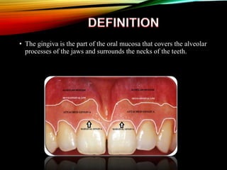
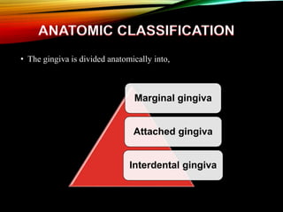
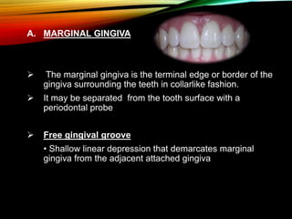
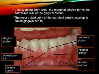
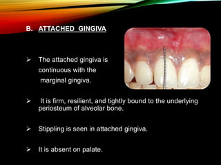
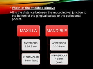
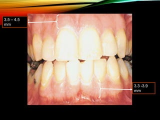
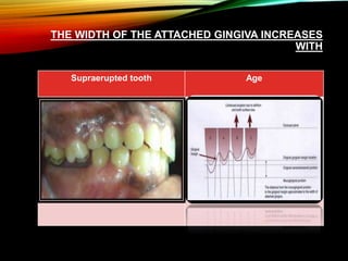
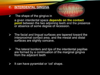
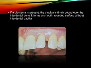
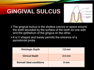
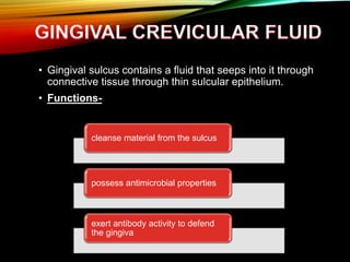
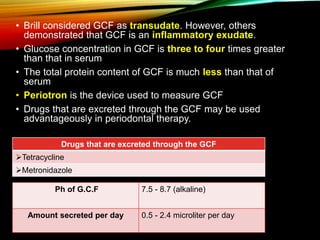
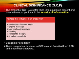
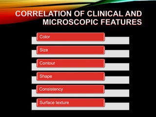
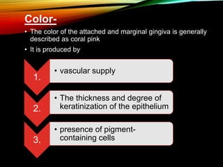
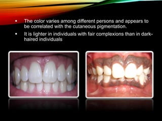
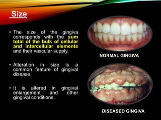
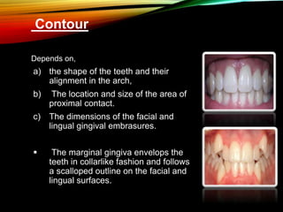
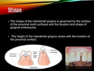
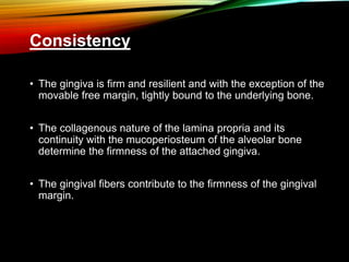
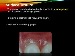
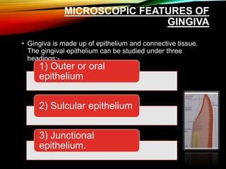
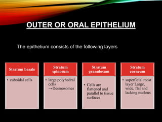
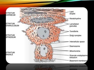
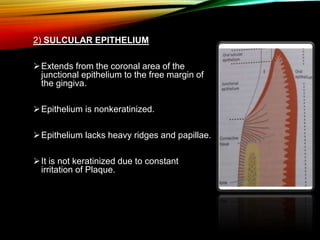
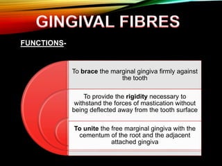
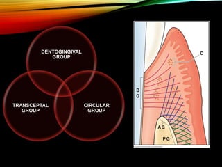
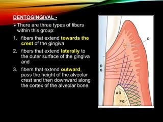
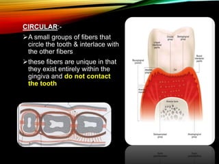
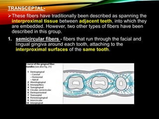
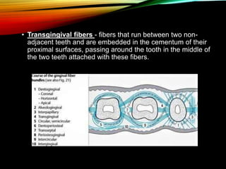
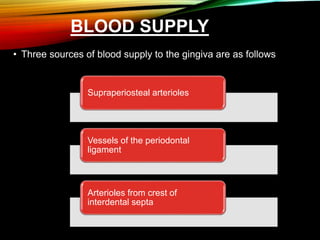
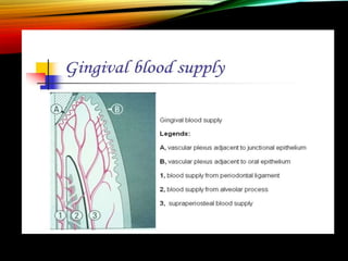
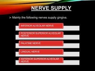
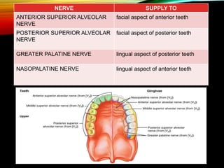
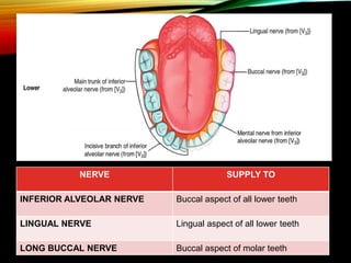
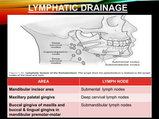
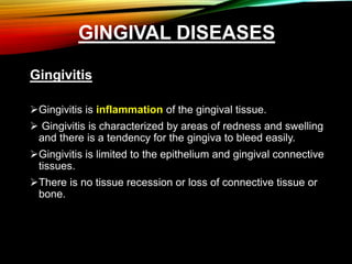
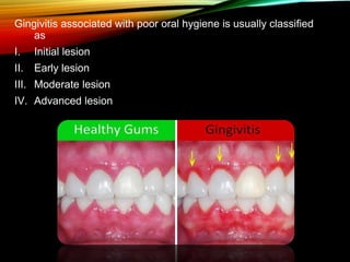
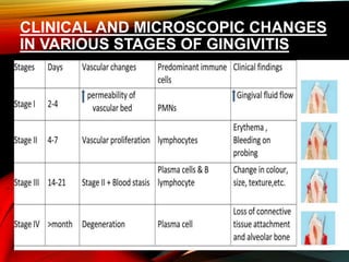
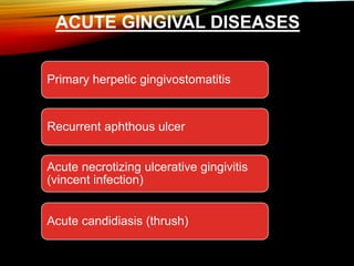
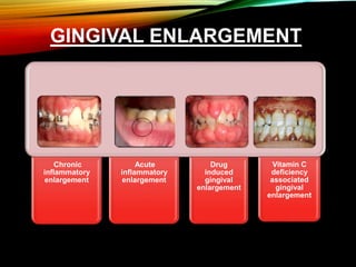
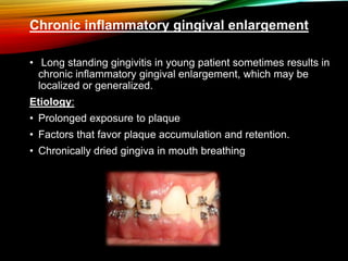
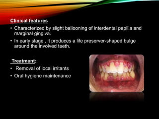
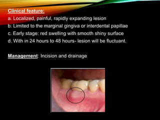
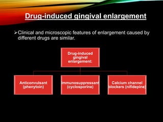
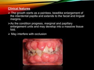
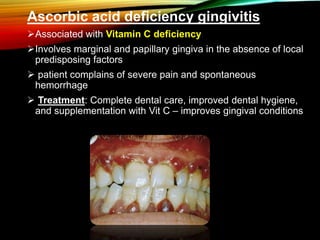
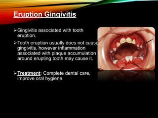
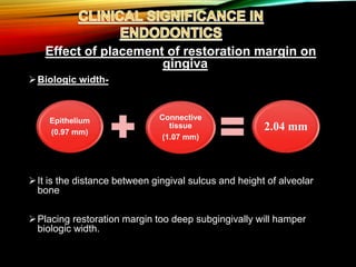
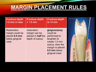
• DEFINITION
• ANATOMIC CLASSIFICATION
• GINGIVAL SULCUS
• GINGIVAL CREVICULAR FLUID
• CLINICAL FEATURES OF GINGIVA
• MICROSCOPIC FEATURES OF GINGIVA
• GINGIVAL FIBRES
• DIFFERENT TYPES OF EPITHELIAL LAYERS
• ARTERIAL SUPPLY
• NERVE SUPPLY
• LYMPHATIC DRAINAGE
• GINGIVAL DISEASES
• CLINICAL SIGNIFICANCE IN ENDODONTICS
• The gingiva is the part of the oral mucosa that covers the alveolar
processes of the jaws and surrounds the necks of the teeth.
• The gingiva is divided anatomically into,
Marginal gingiva
Attached gingiva
Interdental gingiva
A. MARGINAL GINGIVA
 The marginal gingiva is the terminal edge or border of the
gingiva surrounding the teeth in collarlike fashion.
 It may be separated from the tooth surface with a
periodontal probe
 Free gingival groove
• Shallow linear depression that demarcates marginal
gingiva from the adjacent attached gingiva
• Usually about 1mm wide, the marginal gingiva forms the
soft tissue wall of the gingival sulcus
• The most apical point of the marginal gingiva scallop is
called gingival zenith
Interdental
gingiva
Attached
gingiva
Alveolar
mucosa
Marginal
gingiva
Mucogingival
line
Gingival
zenith
B. ATTACHED GINGIVA
 The attached gingiva is
continuous with the
marginal gingiva.
 It is firm, resilient, and tightly bound to the underlying
periosteum of alveolar bone.
 Stippling is seen in attached gingiva.
 It is absent on palate.
• Width of the attached gingiva
It is the distance between the mucogingival junction to
the bottom of the gingival sulcus or the periodontal
pocket.
MAXILLA
ANTERIORS
3.5-4.5 mm
1st PREMOLAR
1.9 mm (least)
MANDIBLE
ANTERIORS
3.3-3.9 mm
1st PREMOLAR
1.8 mm
(least)
3.5 – 4.5
mm
3.3 -3.9
mm
THE WIDTH OF THE ATTACHED GINGIVA INCREASES
WITH
Supraerupted tooth Age
C. INTERDENTAL GINGIVA
 The shape of the gingiva in
a given interdental space depends on the contact
point between the two adjoining teeth and the presence
or absence of some degree of recession.
 The facial and lingual surfaces are tapered toward the
interproximal contact area, and the mesial and distal
surfaces are slightly concave.
 The lateral borders and tips of the interdental papillae
are formed by a continuation of the marginal gingiva
from the adjacent teeth
 It can have pyramidal or ‘col’ shape.
• If a diastema is present, the gingiva is firmly bound over the
interdental bone & forms a smooth, rounded surface without
interdental papilla
The gingival sulcus is the shallow crevice or space around
the tooth bounded by the surface of the tooth on one side
and the epithelium of the gingiva on the other.
It is V shaped and barely permits the entrance of a
periodontal probe
Histologic Depth 1.8 mm
Clinical Depth 2-3 mm
Normal/ ideal conditions 0 mm
• Gingival sulcus contains a fluid that seeps into it through
connective tissue through thin sulcular epithelium.
• Functions-
cleanse material from the sulcus
possess antimicrobial properties
exert antibody activity to defend
the gingiva
Ph of G.C.F 7.5 - 8.7 (alkaline)
Amount secreted per day 0.5 - 2.4 microliter per day
• Brill considered GCF as transudate. However, others
demonstrated that GCF is an inflammatory exudate.
• Glucose concentration in GCF is three to four times greater
than that in serum
• The total protein content of GCF is much less than that of
serum
• Periotron is the device used to measure GCF
• Drugs that are excreted through the GCF may be used
advantageously in periodontal therapy.
Drugs that are excreted through the GCF
Tetracycline
Metronidazole
CLINICAL SIGNIFICANCE (G.C.F)
• The amount of GCF is greater when inflammation is present and
is sometimes proportional to the severity of inflammation.
• Circadian Periodicity.
There is a gradual increase in GCF amount from 6 AM to 10 PM
and a decrease afterward.'
Factors that influence GCF production
mastication of coarse foods
gingival massage
hormonal contraceptives
smoking
periodontal therapy.
circadian periodicity
Color
Size
Contour
Shape
Consistency
Surface texture
Color-
• The color of the attached and marginal gingiva is generally
described as coral pink
• It is produced by
1.
• vascular supply
2.
• The thickness and degree of
keratinization of the epithelium
3.
• presence of pigment-
containing cells
 The color varies among different persons and appears to
be correlated with the cutaneous pigmentation.
 It is lighter in individuals with fair complexions than in dark-
haired individuals
NORMAL GINGIVA
Size
• The size of the gingiva
corresponds with the sum
total of the bulk of cellular
and intercellular elements
and their vascular supply.
• Alteration in size is a
common feature of gingival
disease.
• It is altered in gingival
enlargement and other
gingival conditions.
DISEASED GINGIVA
Contour
Depends on,
a) the shape of the teeth and their
alignment in the arch,
b) The location and size of the area of
proximal contact.
c) The dimensions of the facial and
lingual gingival embrasures.
 The marginal gingiva envelops the
teeth in collarlike fashion and follows
a scalloped outline on the facial and
lingual surfaces.
Shape
• The shape of the interdental gingiva is governed by the contour
of the proximal tooth surfaces and the location and shape of
gingival embrasures.
• The height of the interdental gingiva varies with the location of
the proximal contact.
Consistency
• The gingiva is firm and resilient and with the exception of the
movable free margin, tightly bound to the underlying bone.
• The collagenous nature of the lamina propria and its
continuity with the mucoperiosteum of the alveolar bone
determine the firmness of the attached gingiva.
• The gingival fibers contribute to the firmness of the gingival
margin.
Surface Texture
• The gingiva presents a textured surface similar to an orange peel
and is referred to as being stippled.
• Stippling is best viewed by drying the gingiva
• It is a feature of healthy gingiva.
• only attached gingiva is stippled; the marginal gingiva is
not.
• Reduction or loss of stippling is a common sign of
gingival disease
• When the gingiva is restored to health the stippled
appearance returns.
• Stippling varies with age. It is absent in infancy,
appears in some children at about 5 years of age,
increases until adulthood, and frequently begins to
disappear in old age.
Position
• The position of the gingiva refers to the level at which the
gingival margin is attached to the tooth. (apparent position).
• The level of epithelial attachment to tooth is referred to as
the actual position
CONTINUOUS TOOTH ERUPTION
• Eruption does not cease when teeth meet their functional
antagonists but continues throughout life.
Active eruption Passive eruption
It is the movement of the
teeth in the direction of the
occlusal plane
It is the exposure of the
teeth by apical migration of
gingiva
PASSIVE ERUPTION
MICROSCOPIC FEATURES OF
GINGIVA
• Gingiva is made up of epithelium and connective tissue.
The gingival epithelium can be studied under three
headings:-
1) Outer or oral
epithelium
2) Sulcular epithelium
3) Junctional
epithelium.
OUTER OR ORAL EPITHELIUM
The epithelium consists of the following layers
Stratum basale
• cuboidal cells
Stratum
spinosum
• large polyhedral
cells
→Desmosomes
Stratum
granulosum
• Cells are
flattened and
parallel to tissue
surfaces
Stratum
corneum
• superficial most
layer Large,
wide, flat and
lacking nucleus
2) SULCULAR EPITHELIUM
Extends from the coronal area of the
junctional epithelium to the free margin of
the gingiva.
Epithelium is nonkeratinized.
Epithelium lacks heavy ridges and papillae.
It is not keratinized due to constant
irritation of Plaque.
3) JUNCTIONAL EPITHELIUM
Cells become cuboidal after ameloblasts have finished
formation of enamel.
It forms a collar around the fully erupted tooth
It consist of stratified squamous nonkeratinizing
epithelium.
Length of junctional epithelium 0.25-1.35 mm
FUNCTIONS-
To brace the marginal gingiva firmly against
the tooth
To provide the rigidity necessary to
withstand the forces of mastication without
being deflected away from the tooth surface
To unite the free marginal gingiva with the
cementum of the root and the adjacent
attached gingiva
DENTOGINGIVAL
GROUP
CIRCULAR
GROUP
TRANSCEPTAL
GROUP
DENTOGINGIVAL -
There are three types of fibers
within this group:
1. fibers that extend towards the
crest of the gingiva
2. fibers that extend laterally to
the outer surface of the gingiva
and
3. fibers that extend outward,
pass the height of the alveolar
crest and then downward along
the cortex of the alveolar bone.
CIRCULAR:-
A small groups of fibers that
circle the tooth & interlace with
the other fibers
these fibers are unique in that
they exist entirely within the
gingiva and do not contact
the tooth
TRANSCEPTAL-
These fibers have traditionally been described as spanning the
interproximal tissue between adjacent teeth, into which they
are embedded. However, two other types of fibers have been
described in this group.
1. semicircular fibers - fibers that run through the facial and
lingual gingiva around each tooth, attaching to the
interproximal surfaces of the same tooth.
• Transgingival fibers - fibers that run between two non-
adjacent teeth and are embedded in the cementum of their
proximal surfaces, passing around the tooth in the middle of
the two teeth attached with these fibers.
BASED ON DEGREE OF KERATINIZATION
1) KERATINIZED (75%) :- In which the superficial cells form
scales of keratin & lose their nuclei. A stratum granulosum
is present.
2) PARAKERATINIZED (15%) :- In which the superficial cells
retain pyknotic nuclei & show some signs of being
keratinized. However, the stratum granulosum is generally
absent.
3) NON-KERATINIZED (10%) :- In which the surface cells
are nucleated & show no signs of keratinization.
BLOOD SUPPLY
• Three sources of blood supply to the gingiva are as follows
Supraperiosteal arterioles
Vessels of the periodontal
ligament
Arterioles from crest of
interdental septa
NERVE SUPPLY
 Mainly the following nerves supply gingiva.
INFERIOR ALVEOLAR NERVE
POSTERIOR SUPERIOR ALVEOLAR
NERVE
PALATINE NERVE
LINGUAL NERVE
ANTERIOR SUPERIOR ALVEOLAR
NERVE
NERVE SUPPLY TO
ANTERIOR SUPERIOR ALVEOLAR
NERVE
facial aspect of anterior teeth
POSTERIOR SUPERIOR ALVEOLAR
NERVE
facial aspect of posterior teeth
GREATER PALATINE NERVE lingual aspect of posterior teeth
NASOPALATINE NERVE lingual aspect of anterior teeth
NERVE SUPPLY TO
INFERIOR ALVEOLAR NERVE Buccal aspect of all lower teeth
LINGUAL NERVE Lingual aspect of all lower teeth
LONG BUCCAL NERVE Buccal aspect of molar teeth
LYMPHATIC DRAINAGE
AREA LYMPH NODE
Mandibular incisor area Submental lymph nodes
Maxillary palatal gingiva Deep cervival lymph nodes
Buccal gingiva of maxilla and
buccal & lingual gingiva in
mandibular premolar-molar
Submandibular lymph nodes
GINGIVAL DISEASES
Gingivitis
Gingivitis is inflammation of the gingival tissue.
 Gingivitis is characterized by areas of redness and swelling
and there is a tendency for the gingiva to bleed easily.
Gingivitis is limited to the epithelium and gingival connective
tissues.
There is no tissue recession or loss of connective tissue or
bone.
Gingivitis associated with poor oral hygiene is usually classified
as
I. Initial lesion
II. Early lesion
III. Moderate lesion
IV. Advanced lesion
CLINICAL AND MICROSCOPIC CHANGES
IN VARIOUS STAGES OF GINGIVITIS
ACUTE GINGIVAL DISEASES
Primary herpetic gingivostomatitis
Recurrent aphthous ulcer
Acute necrotizing ulcerative gingivitis
(vincent infection)
Acute candidiasis (thrush)
PRIMARY HERPETIC
GINGIVOSTOMATITIS
Caused by HSV type 1.
Age- Children younger than 6 yrs, but also may be seen in
adolescents and adults.
Primary infection is asymptomatic
Manifestations include blister on the lip so disease commonly
called recurrent herpes labialis.
Location-
hard palate
Attached gingiva
oral mucosa.
Characteristic oral finding:
 Diffuse erythematous involvement of
gingiva.
lip become hemorrhagic
Course is self limited to 7-10 days.
Infants show irritability and refusal to eat
Pain upon swallowing
Extra oral symptoms:
a. Cervical lymphadenopathy
b. Fever ( 101- 105 F)
c. Generalized malaise, irritability
• Treatment-
Symptomatic & supportive.
Application of mild anesthetic such
as dyclonine hydrochloride(0.5%)
Bed rest , soft diet are recommended during the febrile stage
& the person should be kept well hydrated.
Pyrexia - paracetamol suspension and secondary infection
of ulcers may be prevented using chlorhexidine.
In severe case, systemic acyclovir (200 mg 5 times daily for
5 days).
ACUTE NECROTIZING
ULCERATIVE GINGIVITIS
Characterized by sloughing of gingival tissue
 Predisposing factors:
Local: poor oral hygiene, pre-existing gingivitis and
smoking
Systemic: Emotional stress
 Nutritional deficiency – Vit B and C
Clinical features –
Characteristic lesions are punched out, crater like
depression at the crest of interdental papillae
 Surface of gingival craters is covered by pseudomembranous
slough.
 Linear erythma.
Treatment
Perform debridement under local anesthesia.
Remove pseudo membrane.
Patient counselling should include specific oral hygiene
instructions, instruction on proper nutrition, For any signs of
systemic involvement, the recommended antibiotics are:
Amoxicillin 500 mg every 6hr x daily for 7 days
Metronidazole 500 mg twice daily for 7 days
GINGIVAL ENLARGEMENT
Chronic
inflammatory
enlargement
Acute
inflammatory
enlargement
Drug
induced
gingival
enlargement
Vitamin C
deficiency
associated
gingival
enlargement
Chronic inflammatory gingival enlargement
• Long standing gingivitis in young patient sometimes results in
chronic inflammatory gingival enlargement, which may be
localized or generalized.
Etiology:
• Prolonged exposure to plaque
• Factors that favor plaque accumulation and retention.
• Chronically dried gingiva in mouth breathing
Clinical features
• Characterized by slight ballooning of interdental papilla and
marginal gingiva.
• In early stage , it produces a life preserver-shaped bulge
around the involved teeth.
Treatment:
• Removal of local irritants
• Oral hygiene maintenance
Acute inflammatory enlargement
• Gingival abscess Is a localized, painful rapidly expanding
lesion that is usually of sudden onset
• Etiology:
a. Irritation from foreign substance
b. Tooth brush bristle
c. Piece of apple core
d. Lobster shell fragment –embedded
in to gingiva
Clinical feature:
a. Localized, painful, rapidly expanding lesion
b. Limited to the marginal gingiva or interdental papillae
c. Early stage: red swelling with smooth shiny surface
d. With in 24 hours to 48 hours- lesion will be fluctuant.
Management: Incision and drainage
Drug-induced gingival enlargement
Clinical and microscopic features of enlargement caused by
different drugs are similar.
Drug-induced
gingival
enlargement:
Anticonvulsant
(phenytoin)
Immunosuppressant
(cyclosporine)
Calcium channel
blockers (nifidepine)
Clinical features
 The growth starts as a painless, beadlike enlargement of
the interdental papilla and extends to the facial and lingual
margins.
As the condition progress, marginal and papillary
enlargement units and may develop into a massive tissue
fold.
 May interfere with occlusion
Ascorbic acid deficiency gingivitis
Associated with Vitamin C deficiency
Involves marginal and papillary gingiva in the absence of local
predisposing factors
 patient complains of severe pain and spontaneous
hemorrhage
 Treatment: Complete dental care, improved dental hygiene,
and supplementation with Vit C – improves gingival conditions
Eruption Gingivitis
Gingivitis associated with tooth
eruption.
Tooth eruption usually does not cause
gingivitis, however inflammation
associated with plaque accumulation
around erupting tooth may cause it.
Treatment: Complete dental care,
improve oral hygiene.
Effect of placement of restoration margin on
gingiva
Biologic width-
It is the distance between gingival sulcus and height of alveolar
bone
Placing restoration margin too deep subgingivally will hamper
biologic width.
Epithelium
(0.97 mm)
Connective
tissue
(1.07 mm)
2.04 mm
MARGIN PLACEMENT RULES
• ,
If sulcus depth
1.5 mm or less
If sulcus depth
> 1.5 mm
If sulcus depth
is >2 mm
Restoration
margin could be
placed 0.5 mm
below gingival
crest
restoration
margin can be
placed in half the
depth of sulcus
gingivectomy
could be
performed to
lenghten &
create 1.5 mm
sulcus, then the
margin is placed
0.5 mm below
gingival crest.
Gingiva and its diseases

Gingiva and its diseases

  • 2.
    • DEFINITION • ANATOMICCLASSIFICATION • GINGIVAL SULCUS • GINGIVAL CREVICULAR FLUID • CLINICAL FEATURES OF GINGIVA • MICROSCOPIC FEATURES OF GINGIVA • GINGIVAL FIBRES • DIFFERENT TYPES OF EPITHELIAL LAYERS • ARTERIAL SUPPLY • NERVE SUPPLY • LYMPHATIC DRAINAGE • GINGIVAL DISEASES • CLINICAL SIGNIFICANCE IN ENDODONTICS
  • 3.
    • The gingivais the part of the oral mucosa that covers the alveolar processes of the jaws and surrounds the necks of the teeth.
  • 4.
    • The gingivais divided anatomically into, Marginal gingiva Attached gingiva Interdental gingiva
  • 5.
    A. MARGINAL GINGIVA The marginal gingiva is the terminal edge or border of the gingiva surrounding the teeth in collarlike fashion.  It may be separated from the tooth surface with a periodontal probe  Free gingival groove • Shallow linear depression that demarcates marginal gingiva from the adjacent attached gingiva
  • 6.
    • Usually about1mm wide, the marginal gingiva forms the soft tissue wall of the gingival sulcus • The most apical point of the marginal gingiva scallop is called gingival zenith Interdental gingiva Attached gingiva Alveolar mucosa Marginal gingiva Mucogingival line Gingival zenith
  • 7.
    B. ATTACHED GINGIVA The attached gingiva is continuous with the marginal gingiva.  It is firm, resilient, and tightly bound to the underlying periosteum of alveolar bone.  Stippling is seen in attached gingiva.  It is absent on palate.
  • 8.
    • Width ofthe attached gingiva It is the distance between the mucogingival junction to the bottom of the gingival sulcus or the periodontal pocket. MAXILLA ANTERIORS 3.5-4.5 mm 1st PREMOLAR 1.9 mm (least) MANDIBLE ANTERIORS 3.3-3.9 mm 1st PREMOLAR 1.8 mm (least)
  • 9.
  • 10.
    THE WIDTH OFTHE ATTACHED GINGIVA INCREASES WITH Supraerupted tooth Age
  • 11.
    C. INTERDENTAL GINGIVA The shape of the gingiva in a given interdental space depends on the contact point between the two adjoining teeth and the presence or absence of some degree of recession.  The facial and lingual surfaces are tapered toward the interproximal contact area, and the mesial and distal surfaces are slightly concave.  The lateral borders and tips of the interdental papillae are formed by a continuation of the marginal gingiva from the adjacent teeth  It can have pyramidal or ‘col’ shape.
  • 12.
    • If adiastema is present, the gingiva is firmly bound over the interdental bone & forms a smooth, rounded surface without interdental papilla
  • 13.
    The gingival sulcusis the shallow crevice or space around the tooth bounded by the surface of the tooth on one side and the epithelium of the gingiva on the other. It is V shaped and barely permits the entrance of a periodontal probe Histologic Depth 1.8 mm Clinical Depth 2-3 mm Normal/ ideal conditions 0 mm
  • 14.
    • Gingival sulcuscontains a fluid that seeps into it through connective tissue through thin sulcular epithelium. • Functions- cleanse material from the sulcus possess antimicrobial properties exert antibody activity to defend the gingiva
  • 15.
    Ph of G.C.F7.5 - 8.7 (alkaline) Amount secreted per day 0.5 - 2.4 microliter per day • Brill considered GCF as transudate. However, others demonstrated that GCF is an inflammatory exudate. • Glucose concentration in GCF is three to four times greater than that in serum • The total protein content of GCF is much less than that of serum • Periotron is the device used to measure GCF • Drugs that are excreted through the GCF may be used advantageously in periodontal therapy. Drugs that are excreted through the GCF Tetracycline Metronidazole
  • 16.
    CLINICAL SIGNIFICANCE (G.C.F) •The amount of GCF is greater when inflammation is present and is sometimes proportional to the severity of inflammation. • Circadian Periodicity. There is a gradual increase in GCF amount from 6 AM to 10 PM and a decrease afterward.' Factors that influence GCF production mastication of coarse foods gingival massage hormonal contraceptives smoking periodontal therapy. circadian periodicity
  • 17.
  • 18.
    Color- • The colorof the attached and marginal gingiva is generally described as coral pink • It is produced by 1. • vascular supply 2. • The thickness and degree of keratinization of the epithelium 3. • presence of pigment- containing cells
  • 19.
     The colorvaries among different persons and appears to be correlated with the cutaneous pigmentation.  It is lighter in individuals with fair complexions than in dark- haired individuals
  • 20.
    NORMAL GINGIVA Size • Thesize of the gingiva corresponds with the sum total of the bulk of cellular and intercellular elements and their vascular supply. • Alteration in size is a common feature of gingival disease. • It is altered in gingival enlargement and other gingival conditions. DISEASED GINGIVA
  • 21.
    Contour Depends on, a) theshape of the teeth and their alignment in the arch, b) The location and size of the area of proximal contact. c) The dimensions of the facial and lingual gingival embrasures.  The marginal gingiva envelops the teeth in collarlike fashion and follows a scalloped outline on the facial and lingual surfaces.
  • 22.
    Shape • The shapeof the interdental gingiva is governed by the contour of the proximal tooth surfaces and the location and shape of gingival embrasures. • The height of the interdental gingiva varies with the location of the proximal contact.
  • 23.
    Consistency • The gingivais firm and resilient and with the exception of the movable free margin, tightly bound to the underlying bone. • The collagenous nature of the lamina propria and its continuity with the mucoperiosteum of the alveolar bone determine the firmness of the attached gingiva. • The gingival fibers contribute to the firmness of the gingival margin.
  • 24.
    Surface Texture • Thegingiva presents a textured surface similar to an orange peel and is referred to as being stippled. • Stippling is best viewed by drying the gingiva • It is a feature of healthy gingiva.
  • 25.
    • only attachedgingiva is stippled; the marginal gingiva is not. • Reduction or loss of stippling is a common sign of gingival disease • When the gingiva is restored to health the stippled appearance returns. • Stippling varies with age. It is absent in infancy, appears in some children at about 5 years of age, increases until adulthood, and frequently begins to disappear in old age.
  • 26.
    Position • The positionof the gingiva refers to the level at which the gingival margin is attached to the tooth. (apparent position). • The level of epithelial attachment to tooth is referred to as the actual position
  • 27.
    CONTINUOUS TOOTH ERUPTION •Eruption does not cease when teeth meet their functional antagonists but continues throughout life. Active eruption Passive eruption It is the movement of the teeth in the direction of the occlusal plane It is the exposure of the teeth by apical migration of gingiva
  • 28.
  • 29.
    MICROSCOPIC FEATURES OF GINGIVA •Gingiva is made up of epithelium and connective tissue. The gingival epithelium can be studied under three headings:- 1) Outer or oral epithelium 2) Sulcular epithelium 3) Junctional epithelium.
  • 30.
    OUTER OR ORALEPITHELIUM The epithelium consists of the following layers Stratum basale • cuboidal cells Stratum spinosum • large polyhedral cells →Desmosomes Stratum granulosum • Cells are flattened and parallel to tissue surfaces Stratum corneum • superficial most layer Large, wide, flat and lacking nucleus
  • 32.
    2) SULCULAR EPITHELIUM Extendsfrom the coronal area of the junctional epithelium to the free margin of the gingiva. Epithelium is nonkeratinized. Epithelium lacks heavy ridges and papillae. It is not keratinized due to constant irritation of Plaque.
  • 33.
    3) JUNCTIONAL EPITHELIUM Cellsbecome cuboidal after ameloblasts have finished formation of enamel. It forms a collar around the fully erupted tooth It consist of stratified squamous nonkeratinizing epithelium. Length of junctional epithelium 0.25-1.35 mm
  • 34.
    FUNCTIONS- To brace themarginal gingiva firmly against the tooth To provide the rigidity necessary to withstand the forces of mastication without being deflected away from the tooth surface To unite the free marginal gingiva with the cementum of the root and the adjacent attached gingiva
  • 35.
  • 36.
    DENTOGINGIVAL - There arethree types of fibers within this group: 1. fibers that extend towards the crest of the gingiva 2. fibers that extend laterally to the outer surface of the gingiva and 3. fibers that extend outward, pass the height of the alveolar crest and then downward along the cortex of the alveolar bone.
  • 37.
    CIRCULAR:- A small groupsof fibers that circle the tooth & interlace with the other fibers these fibers are unique in that they exist entirely within the gingiva and do not contact the tooth
  • 38.
    TRANSCEPTAL- These fibers havetraditionally been described as spanning the interproximal tissue between adjacent teeth, into which they are embedded. However, two other types of fibers have been described in this group. 1. semicircular fibers - fibers that run through the facial and lingual gingiva around each tooth, attaching to the interproximal surfaces of the same tooth.
  • 39.
    • Transgingival fibers- fibers that run between two non- adjacent teeth and are embedded in the cementum of their proximal surfaces, passing around the tooth in the middle of the two teeth attached with these fibers.
  • 40.
    BASED ON DEGREEOF KERATINIZATION 1) KERATINIZED (75%) :- In which the superficial cells form scales of keratin & lose their nuclei. A stratum granulosum is present. 2) PARAKERATINIZED (15%) :- In which the superficial cells retain pyknotic nuclei & show some signs of being keratinized. However, the stratum granulosum is generally absent. 3) NON-KERATINIZED (10%) :- In which the surface cells are nucleated & show no signs of keratinization.
  • 41.
    BLOOD SUPPLY • Threesources of blood supply to the gingiva are as follows Supraperiosteal arterioles Vessels of the periodontal ligament Arterioles from crest of interdental septa
  • 43.
    NERVE SUPPLY  Mainlythe following nerves supply gingiva. INFERIOR ALVEOLAR NERVE POSTERIOR SUPERIOR ALVEOLAR NERVE PALATINE NERVE LINGUAL NERVE ANTERIOR SUPERIOR ALVEOLAR NERVE
  • 44.
    NERVE SUPPLY TO ANTERIORSUPERIOR ALVEOLAR NERVE facial aspect of anterior teeth POSTERIOR SUPERIOR ALVEOLAR NERVE facial aspect of posterior teeth GREATER PALATINE NERVE lingual aspect of posterior teeth NASOPALATINE NERVE lingual aspect of anterior teeth
  • 45.
    NERVE SUPPLY TO INFERIORALVEOLAR NERVE Buccal aspect of all lower teeth LINGUAL NERVE Lingual aspect of all lower teeth LONG BUCCAL NERVE Buccal aspect of molar teeth
  • 46.
    LYMPHATIC DRAINAGE AREA LYMPHNODE Mandibular incisor area Submental lymph nodes Maxillary palatal gingiva Deep cervival lymph nodes Buccal gingiva of maxilla and buccal & lingual gingiva in mandibular premolar-molar Submandibular lymph nodes
  • 47.
    GINGIVAL DISEASES Gingivitis Gingivitis isinflammation of the gingival tissue.  Gingivitis is characterized by areas of redness and swelling and there is a tendency for the gingiva to bleed easily. Gingivitis is limited to the epithelium and gingival connective tissues. There is no tissue recession or loss of connective tissue or bone.
  • 48.
    Gingivitis associated withpoor oral hygiene is usually classified as I. Initial lesion II. Early lesion III. Moderate lesion IV. Advanced lesion
  • 49.
    CLINICAL AND MICROSCOPICCHANGES IN VARIOUS STAGES OF GINGIVITIS
  • 50.
    ACUTE GINGIVAL DISEASES Primaryherpetic gingivostomatitis Recurrent aphthous ulcer Acute necrotizing ulcerative gingivitis (vincent infection) Acute candidiasis (thrush)
  • 51.
    PRIMARY HERPETIC GINGIVOSTOMATITIS Caused byHSV type 1. Age- Children younger than 6 yrs, but also may be seen in adolescents and adults. Primary infection is asymptomatic Manifestations include blister on the lip so disease commonly called recurrent herpes labialis. Location- hard palate Attached gingiva oral mucosa.
  • 52.
    Characteristic oral finding: Diffuse erythematous involvement of gingiva. lip become hemorrhagic Course is self limited to 7-10 days. Infants show irritability and refusal to eat Pain upon swallowing Extra oral symptoms: a. Cervical lymphadenopathy b. Fever ( 101- 105 F) c. Generalized malaise, irritability
  • 53.
    • Treatment- Symptomatic &supportive. Application of mild anesthetic such as dyclonine hydrochloride(0.5%) Bed rest , soft diet are recommended during the febrile stage & the person should be kept well hydrated. Pyrexia - paracetamol suspension and secondary infection of ulcers may be prevented using chlorhexidine. In severe case, systemic acyclovir (200 mg 5 times daily for 5 days).
  • 54.
    ACUTE NECROTIZING ULCERATIVE GINGIVITIS Characterizedby sloughing of gingival tissue  Predisposing factors: Local: poor oral hygiene, pre-existing gingivitis and smoking Systemic: Emotional stress  Nutritional deficiency – Vit B and C
  • 55.
    Clinical features – Characteristiclesions are punched out, crater like depression at the crest of interdental papillae  Surface of gingival craters is covered by pseudomembranous slough.  Linear erythma. Treatment Perform debridement under local anesthesia. Remove pseudo membrane. Patient counselling should include specific oral hygiene instructions, instruction on proper nutrition, For any signs of systemic involvement, the recommended antibiotics are: Amoxicillin 500 mg every 6hr x daily for 7 days Metronidazole 500 mg twice daily for 7 days
  • 56.
  • 57.
    Chronic inflammatory gingivalenlargement • Long standing gingivitis in young patient sometimes results in chronic inflammatory gingival enlargement, which may be localized or generalized. Etiology: • Prolonged exposure to plaque • Factors that favor plaque accumulation and retention. • Chronically dried gingiva in mouth breathing
  • 58.
    Clinical features • Characterizedby slight ballooning of interdental papilla and marginal gingiva. • In early stage , it produces a life preserver-shaped bulge around the involved teeth. Treatment: • Removal of local irritants • Oral hygiene maintenance
  • 59.
    Acute inflammatory enlargement •Gingival abscess Is a localized, painful rapidly expanding lesion that is usually of sudden onset • Etiology: a. Irritation from foreign substance b. Tooth brush bristle c. Piece of apple core d. Lobster shell fragment –embedded in to gingiva
  • 60.
    Clinical feature: a. Localized,painful, rapidly expanding lesion b. Limited to the marginal gingiva or interdental papillae c. Early stage: red swelling with smooth shiny surface d. With in 24 hours to 48 hours- lesion will be fluctuant. Management: Incision and drainage
  • 61.
    Drug-induced gingival enlargement Clinicaland microscopic features of enlargement caused by different drugs are similar. Drug-induced gingival enlargement: Anticonvulsant (phenytoin) Immunosuppressant (cyclosporine) Calcium channel blockers (nifidepine)
  • 62.
    Clinical features  Thegrowth starts as a painless, beadlike enlargement of the interdental papilla and extends to the facial and lingual margins. As the condition progress, marginal and papillary enlargement units and may develop into a massive tissue fold.  May interfere with occlusion
  • 63.
    Ascorbic acid deficiencygingivitis Associated with Vitamin C deficiency Involves marginal and papillary gingiva in the absence of local predisposing factors  patient complains of severe pain and spontaneous hemorrhage  Treatment: Complete dental care, improved dental hygiene, and supplementation with Vit C – improves gingival conditions
  • 64.
    Eruption Gingivitis Gingivitis associatedwith tooth eruption. Tooth eruption usually does not cause gingivitis, however inflammation associated with plaque accumulation around erupting tooth may cause it. Treatment: Complete dental care, improve oral hygiene.
  • 65.
    Effect of placementof restoration margin on gingiva Biologic width- It is the distance between gingival sulcus and height of alveolar bone Placing restoration margin too deep subgingivally will hamper biologic width. Epithelium (0.97 mm) Connective tissue (1.07 mm) 2.04 mm
  • 66.
    MARGIN PLACEMENT RULES •, If sulcus depth 1.5 mm or less If sulcus depth > 1.5 mm If sulcus depth is >2 mm Restoration margin could be placed 0.5 mm below gingival crest restoration margin can be placed in half the depth of sulcus gingivectomy could be performed to lenghten & create 1.5 mm sulcus, then the margin is placed 0.5 mm below gingival crest.