SlideShare a Scribd company logo
GINGIVA
Presented by -
Dr.K.RAVINDHAR
• PG Ist year
• DEPT OF
PERIODONTICS
Contents
• Introduction.
• Definition.
Anatomy of gingiva.
1.Macroscopic feature.
a. Marginal gingiva.
b. Attached gingiva.
c. Interdental gingiva
2.Microscopic feature of gingiva.
a. Epithelium
b. Connective tissue
• Clinical feature of gingiva
a. Applied aspects
• Conclusion
• References
Definition-
The gingiva is the part of the oral
mucosa that covers the alveolar
prosses of the jaw & surronds the
neck of the teeth. ~carranza
Anatomically divided into :
1.Macroscopic feature of gingiva
a. Marginal gingiva
b. Attached gingiva.
c. Interdental gingiva.
Clinical anatomy of gingiva
Marginal gingiva
• The marginal gingiva
(unattached gingiva) is the
terminal edge or border of
The gingiva surrounding the
teeth in collar-like fashion.
• It is demarcated from the a
shallow linear depression , the
FREE GINGIVAL GROOVE.
Gingival sulcus
• Definition:
The sulcus consists of the shallow
space that is coronal to the
attachment of the junctional
epithelium & bounded by the tooth
on one side & sulcular epithelium
on the other. ~Carranza’s
The coronal extent of the
gingival sulcus is the gingival
margin.
• It is ‘V’ shaped.
• Depth of gingival sulcus:
-In ideal condition- 0 mm
[Gottlieb B 1933]
-Histologically- 1.8mm with variations from 0-
6mm
[Orban,Kohler 1924]
-Clinically- 2-3 mm.
Development of gingival
sulcus
• This process takes between 1 & 2
years. ~Schroeder & Listgarten (
1968 ).
• It is formed when the tooth erupts
into the oral cavity.
• The junctional epithelium & REE
form a broad band attached to the
tooth surface from near the tip of
the crown to CEJ.
Gingival fluid ( sulculsr fluid)
• It may be either transudate/exudate in
manner.
• It contains:
 Inflammatory cells
 Serum
 Epithelial cells
 Connective tissue components
Diffusion of the sulcular fluid
Basement membrane.
Intracellular space of
junctional epithelium
Gingival sulcus.
Function of sulcular fluid
• Cleanse material from the sulcus.
• Improve adhesion of the epithelium
to the tooth.
• Antimicrobial properties.
• Antibody activity to defend the
gingiva.
Attached gingiva
• It is firm, resilient & tightly bound to the
underlying periosteum of the alveolar bone.
• It is continuous with marginal gingiva.
• It extends to the relatively loose & movable
alveolar mucosa & is demarcated by the
mucogingival junction.
• The width of the attached gingiva on the facial
aspect differs in different areas of the
mouth.[Bowers 1963]
• Greatest in incisior region:
- 3.5-4.5 mm in maxilla.
- 3.3-3.9mm in mandible.
• Narrower in posterior segments:
- 1.9mm in maxilla
- 1.8mm in mandible (premolar region)
[Ainamo J,Loe 1966]
• The width of the attached gingiva increases
with age & in supraerupted teeth.
Interdental gingiva
• It is occupies the gingival
embrasure, which is the
interproximal space beneath the
area of tooth contact.
• Can be pyramidal or col shaped
• The shape depends on the contact
point between the two adjoining
teeth & the degree of recession.
• If a diastema is present, the gingiva
is firmly bound over the interdental
bone & form, rounded surface
without interdental papillae.
Microscopic features of
gingiva
• It is composed of
1.Gingival epithelium
2.Connective tissue
1.Gingival epithelium
• Stratified squamous epithelium.
Functions-
• It provides a physical barrier to
infection
• Mechanical , chemical, water &
microbial barrier
• It acts as an innate defense
response to gingival health &
disease. ~Caffesse RG (1966)
Features of gingival
epithelium
Architectural integrity
Cell –cell attachments
Basal lamina
Keratin cytoskeleton
Major cell type
Keratinocyte
• Other cell types
Langerhans cells
Melanocytes
Markel cells
Gingival epithelial cells
• Keratinocytes
• Nonkeratinocytes
• Melanocyes
• Langerhans cells ~Diffract CF,
Toto(1985)
• Markel cells
Keratinocytes
• Most of the gingiva covered with
parakeratinized or nonkeratinized
epithelium.
• Orthokeratinized –some areas
present.
• These areas are progress to
maturity/ differentation under
different physiological /
pathological condition.
• It is activated by,
1.Proliferation
2.Differentiation
1.Proliferation
• By mitosis in the basal layer.
• Less frequently in the suprabasal
layer.
• Larger number of cells begin to
migrate to the surface from basal
layer.
2.Differentiation
• It consists of progressions of
biochemical & morphologic events
that occur in the cell as they migrate
from basal layer.
• Morphological changes:
1. Progressive flattening of the cell.
2. Increasing prevalence of
tonofilaments
3. Production of keratohyalin granules.
4. Disappearance of the nucles.
Orthokeratinized epithelium
• It is produced by complete process
of keratinization.
• No nucleus in stratum corneum.
• Well defined nucleus in stratum
granulosum.
• It is present in some areas of the
gingival epithelium.
Parakeratinized epithelium
• Most of the gingiva covered by
parakeratinized epithelium.
• The stratum corneum retains
pyknotic nuclei.
• The keratohylin granules are
dispersed.
• Not giving rise to a stratum
granulosum
Nonkeratinized epithelium
• It has granulosum / corneum strata,
whereas superficial cells have viable
nuclei.
• The epithelial cell connections are
tight junctions (zonae occludens)
Epithelial cells
• Nonkeratinocytes
• Melanocytes
• Langerhans cells
• Merkel cells
Melanocytes
• These are dendritic cells.
• Located in the basal & spinous layer
of the gingival epithelium.
• They synthesize melanin
• Also called premelanosomes /
melanosomes.
• Melanin granules are phagocytosed
in cells of epithelium & connective
tissue.
Hydroxylates tyrosine.
Tyrosinase
Dihydroxyphenylalanie (DOPA)
Melanin
Langerhans cells
• These are dendritic cells.
• Located in keratinocytes at all
suprabasal levels.
• They are modified monocytes.
• They also act as antigen presenting
cells.
• Absent in junctional epithelium.
Merkel cells
• Located in deep layer of the
epithelium, nerve endings.
• Tactile perceptors.
Basal lamina
• The epithelium is joined to the
underlying connective tissue by a
basal lamina. Stern IB (1965)
• It is 300 – 400 A thick
• The basal lamina consists of:
1. Lamina lucida
2. Lamina densa
1.Lamina lucida
• Hemidesmosomes of basal
epithelial cells abut the lamina
lucida.
• Mainly composed by glycoprotein
laminin.
2.Lamina densa
• Composed of type IV collagen.
• The fibrils are 750nm in length from
epithelium end to connective tissue
end.
• PAS +ve.
• The gingival epithelium different
from the morphologically &
functionally, there are;~ Karring T,
Loe H (1975)
1. The oral or outer epithelium
2. Sulcular epithelium
3. Junctional epithelium
I. Oral [outer] epithelium
• Covers the crest & outer surface of
marginal gingiva & surface of the
attached gingiva.
• Average thickness of the oral
epithelium is 0.2 to 0.3mm
• It is keratinized or parakeratinized
or various combinations of these
conditions.
Histology of oral epithelium
Composition of oral
epithelium
• The oral epithelium composed of 4
layers:
1. Stratum basale.
2. Stratum spinosum.
3. Stratum granulosum.
4. Stratum corneum
• The degree of keratinization
diminishes with age & the onset of
menopause.
• It is parakeratinized or
orthokeratinized.
• Keratinization of the oral mucosa
varies in different areas (in
descending order palate, gingiva,
ventral aspect of the tongue,&
cheek)
2. Sulcular epithelium
• The sulcular epithelium lines the
gingival epithelium.
• It is thin & extends from the
junctional epithelium to the crest of
the gingival margin.
• It is act as a semi permeable
membrane.
3.Junctional epithelium
Development of junctional
epithelium
• The junctional epithelium consists of a
collar like band of stratified squamous
non keratinizing epithelium.
• It is 3-4 layers thick in early life, but
the number of layers increases with
age to 10 or even 20 layers.
• The junctional epithelium taper from
its coronal end.
• The length of the junctional
epithelium ranges from 0.25-1.35mm
• The junctional epithelium is formed by the
oral epithelium & the reduced enamel
epithelium, during tooth eruption.
• The junctional epithelium attached to the
tooth surface by internal basal lamina & it
attached gingival connective tissue by
external basal lamina.
• The junctional epithelium attachment to the
tooth by gingival fibers, which brace the
marginal gingiva against the tooth surface. it
is referred to as the dentogingival unit.
• The internal basal lamina consists of lamina
densa & lamina lucida, which hemi
desmosomes are attached.
Renewal of gingival
epithelium
• The mitotic rates is higher in
nonkeratinized areas.
• Is increased in gingivitis.
• Without significant gender
defferences.
• The mitotic rate is increased /
decreased with age.
• The mitotic rate varies of the oral
epithelium in descending order:
 Buccal mucosa.
 Hard palate.
 Sulcular epithelium.
 Junctional epithelium.
 Outer surface of the marginal
gingiva & attached gingiva.
• Varies turnover time of the oral
epithelium.
• Palate, tongue, & cheek ~ 5-6days.
• Gingiva ~10 -12days.
• Junctional epithelium ~1-6days.
Cuticular structures on the
tooth
1. Cuticle
2. Coronal cementum
3. Dental cuticle
Cuticler
• A thin, acellular structure with a
homogeneous matrix.
• Classified into: ~Listgarten (1963 ).
1. developmental origin.
2. Acquired coatings.
Development origin
• Coatings of normally formed as
part of tooth development.
• They include the REE, coronal
cementum, & dental cuticle.
Coronal cementum
• It deposit a thin layer of the
cementum on the enamel.
• A thin patches of afibrillar
cementum may be seen in the
cervical half of the crown.
Dental cuticle
• It consisting of a layer of
homogeneous organic material.
• Variable thickness [ ~0.25Um].
• It overlying the enamel surface.
• It is nonmineralized.
• Not always present.
• Near the CEJ, it is deposited over a
layer of afibrillar cementum, which
in turn overlies enamel.
Acquired origin
• Coating of exogenous origin such as
saliva, bacteria, calculus & surface
stains
Gingival connective tissue
• The major components of the gingival
connective tissue are collagen fibers ,
fibroblasts, vessels, nerves & matrix.
• The connective tissue of the gingiva is
known as the lamina propria.
• it is consists of 2 layers, there are,
1. Papillary layer.
2. Reticular layer.
• Connective tissue has a cellular &
extra cellular compartment,
composed of fibre & ground
substance.
Connective tissue fibers
They are 3 types.
1. Collagen
2. Reticular
3. Elastic
• Collagen type I forms the bulk of
lamina propria & provide the tensile
strength to gingival tissue.
• Type IV collagen fibres branches
between collagen type I & fibres of
basement membrane & blood
vessels walls.
Ground substance
• It is fills the space between fibre &
cells, is amorphous,& has a high
content of water.
• It is composed of proteoglycans,
hyaluronic acid & condroitin sulfate,
& glycoproteins, fibronectin.
• Laminin, another glycoprotein
found in basal lamina, serves to
attach it to epithelial cells.
Gingival fibers
Therearefourtypes
offibres:
1.Gingividental
group
2.Transseptal group
3.Transalveolar
group
4.Circular group
Cellular elements
1. Fibroblast
2. Mast cells
3. Neutrophils
4. Lymphocytes
5. Adipose cells
6. Eosinophils
Blood supply
1. Vessels of
periodontal
ligament.
2. Arterioles from
alveolar bone.
3. Supraperiosteal
arteries.
Nerves & Lymphatics supply
• The nerve fibers arising from the
periodontal ligament,& labial,
buccal, palatal nerves.
• Neural elements are distributed
throughout the gingival tissues.
• Most nerve fibers are mylinated &
are closely associated with the
blood vessels.
• The lymphatic drainage of the
gingiva into the regional lymph
nodes.
Clinical feature
1. Color
2. Physiological pigmentation
[melanin]
3. Size
4. Contour
5. Shape
6. Consistency
7. Surface texture
8. Position
Healthy gingiva
Color – acute gingivitis
Physiologic pigmentation
(Melanin)
size & contour of gingiva
Consistency – fibrotic gingiva
Surface texture - healthy
gingiva
Position – gingival recession
References
• Carranza`s – 11 thedition.
• Orban`s- 13 th edition.
Gingiva copy

More Related Content

What's hot

2. gingiva
2. gingiva2. gingiva
Anatomy of periodontium
Anatomy of periodontiumAnatomy of periodontium
Anatomy of periodontium
Arunima Kathuria
 
Gingiva
GingivaGingiva
Gingiva / rotary endodontic courses by indian dental academy
Gingiva / rotary endodontic courses by indian dental academyGingiva / rotary endodontic courses by indian dental academy
Gingiva / rotary endodontic courses by indian dental academy
Indian dental academy
 
Gingiva
GingivaGingiva
Gingiva
debasismitra
 
Gingiva
GingivaGingiva
gingiva
gingivagingiva
gingiva
Mehul Shinde
 
Gingiva
GingivaGingiva
Gingiva
Dr Abhilasha
 
003.biology of periodontal tissues
003.biology of periodontal tissues003.biology of periodontal tissues
003.biology of periodontal tissues
Dr.Jaffar Raza BDS
 
Microscopic features of gingiva
Microscopic features of gingivaMicroscopic features of gingiva
Microscopic features of gingiva
Jayshree Gaur
 
Gingiva
GingivaGingiva
GINGIVA Macroscopic features
GINGIVA Macroscopic featuresGINGIVA Macroscopic features
GINGIVA Macroscopic features
SaiLakshmi128
 
Periodontium 1
Periodontium 1Periodontium 1
Periodontium 1
Akshat Agrawal
 
Clinical Periodontology - Introduction & Anatomy
Clinical Periodontology - Introduction & AnatomyClinical Periodontology - Introduction & Anatomy
Clinical Periodontology - Introduction & Anatomy
Sarang Suresh Hotchandani
 
Ultrastructure of gingiva
Ultrastructure of gingiva Ultrastructure of gingiva
Ultrastructure of gingiva
naseemashraf2
 
gingiva
 gingiva gingiva
gingiva
Parth Thakkar
 
Dentinogingival junction
Dentinogingival junctionDentinogingival junction
Dentinogingival junction
BanMaraby
 
Gingiva
GingivaGingiva
Junctional epithelium
Junctional epitheliumJunctional epithelium
Junctional epithelium
ishita1994
 
Macroscopic features of gingiva
Macroscopic features of gingivaMacroscopic features of gingiva
Macroscopic features of gingiva
Dr.Himanshu Singh
 

What's hot (20)

2. gingiva
2. gingiva2. gingiva
2. gingiva
 
Anatomy of periodontium
Anatomy of periodontiumAnatomy of periodontium
Anatomy of periodontium
 
Gingiva
GingivaGingiva
Gingiva
 
Gingiva / rotary endodontic courses by indian dental academy
Gingiva / rotary endodontic courses by indian dental academyGingiva / rotary endodontic courses by indian dental academy
Gingiva / rotary endodontic courses by indian dental academy
 
Gingiva
GingivaGingiva
Gingiva
 
Gingiva
GingivaGingiva
Gingiva
 
gingiva
gingivagingiva
gingiva
 
Gingiva
GingivaGingiva
Gingiva
 
003.biology of periodontal tissues
003.biology of periodontal tissues003.biology of periodontal tissues
003.biology of periodontal tissues
 
Microscopic features of gingiva
Microscopic features of gingivaMicroscopic features of gingiva
Microscopic features of gingiva
 
Gingiva
GingivaGingiva
Gingiva
 
GINGIVA Macroscopic features
GINGIVA Macroscopic featuresGINGIVA Macroscopic features
GINGIVA Macroscopic features
 
Periodontium 1
Periodontium 1Periodontium 1
Periodontium 1
 
Clinical Periodontology - Introduction & Anatomy
Clinical Periodontology - Introduction & AnatomyClinical Periodontology - Introduction & Anatomy
Clinical Periodontology - Introduction & Anatomy
 
Ultrastructure of gingiva
Ultrastructure of gingiva Ultrastructure of gingiva
Ultrastructure of gingiva
 
gingiva
 gingiva gingiva
gingiva
 
Dentinogingival junction
Dentinogingival junctionDentinogingival junction
Dentinogingival junction
 
Gingiva
GingivaGingiva
Gingiva
 
Junctional epithelium
Junctional epitheliumJunctional epithelium
Junctional epithelium
 
Macroscopic features of gingiva
Macroscopic features of gingivaMacroscopic features of gingiva
Macroscopic features of gingiva
 

Similar to Gingiva copy

GINGIVA.pptx
GINGIVA.pptxGINGIVA.pptx
GINGIVA.pptx
Sayali70
 
gingiva.pptx
gingiva.pptxgingiva.pptx
gingiva.pptx
DentalYoutube
 
Gingiva ppt
Gingiva pptGingiva ppt
Gingiva ppt
Hudson Jonathan
 
Gingiva
GingivaGingiva
Gingiva
DrAtulKoundel
 
GINGIVA-DR JEEVANAND DESHMUKH.pptx
GINGIVA-DR JEEVANAND DESHMUKH.pptxGINGIVA-DR JEEVANAND DESHMUKH.pptx
GINGIVA-DR JEEVANAND DESHMUKH.pptx
DentalYoutube
 
gigiva2ndseminar- 2.pdf
gigiva2ndseminar- 2.pdfgigiva2ndseminar- 2.pdf
gigiva2ndseminar- 2.pdf
GaurishChandraRathau
 
Gingiva
Gingiva Gingiva
Gingiva
Hema Duddukuri
 
Gingiva and its diseases
Gingiva and its diseasesGingiva and its diseases
Gingiva and its diseases
Dr.Swarneet Kakpure
 
Anatomical landmarks
Anatomical landmarksAnatomical landmarks
Anatomical landmarks
dellasain
 
Gingiva
Gingiva Gingiva
Gingiva
ssuseraf61fb
 
Gingiva. Lilly & Bong.pptx
Gingiva. Lilly & Bong.pptxGingiva. Lilly & Bong.pptx
Gingiva. Lilly & Bong.pptx
DrCarlosIICapitan
 
Gingiva
GingivaGingiva
Gingiva
Masud Manik
 
Gingiva (2)
Gingiva (2)Gingiva (2)
JUNCTIONAL EPITHELIUM
JUNCTIONAL EPITHELIUMJUNCTIONAL EPITHELIUM
JUNCTIONAL EPITHELIUM
HaripriyaRajaram
 
003.biology of periodontal tissues
003.biology of periodontal tissues003.biology of periodontal tissues
003.biology of periodontal tissues
Dr.Jaffar Raza BDS
 
examination_of_periodontium.ppt
examination_of_periodontium.pptexamination_of_periodontium.ppt
examination_of_periodontium.ppt
Mohammed669214
 
The oral mucosa
The oral mucosaThe oral mucosa
The oral mucosa
Isella42
 
gingiva.pptx
gingiva.pptxgingiva.pptx
gingiva.pptx
AvitaRath
 
occlusion.pdf
occlusion.pdfocclusion.pdf
occlusion.pdf
Viola Esther
 
lecture four dental pulp.pptx Oral histology
lecture four dental pulp.pptx Oral histologylecture four dental pulp.pptx Oral histology
lecture four dental pulp.pptx Oral histology
iprahimmahamett
 

Similar to Gingiva copy (20)

GINGIVA.pptx
GINGIVA.pptxGINGIVA.pptx
GINGIVA.pptx
 
gingiva.pptx
gingiva.pptxgingiva.pptx
gingiva.pptx
 
Gingiva ppt
Gingiva pptGingiva ppt
Gingiva ppt
 
Gingiva
GingivaGingiva
Gingiva
 
GINGIVA-DR JEEVANAND DESHMUKH.pptx
GINGIVA-DR JEEVANAND DESHMUKH.pptxGINGIVA-DR JEEVANAND DESHMUKH.pptx
GINGIVA-DR JEEVANAND DESHMUKH.pptx
 
gigiva2ndseminar- 2.pdf
gigiva2ndseminar- 2.pdfgigiva2ndseminar- 2.pdf
gigiva2ndseminar- 2.pdf
 
Gingiva
Gingiva Gingiva
Gingiva
 
Gingiva and its diseases
Gingiva and its diseasesGingiva and its diseases
Gingiva and its diseases
 
Anatomical landmarks
Anatomical landmarksAnatomical landmarks
Anatomical landmarks
 
Gingiva
Gingiva Gingiva
Gingiva
 
Gingiva. Lilly & Bong.pptx
Gingiva. Lilly & Bong.pptxGingiva. Lilly & Bong.pptx
Gingiva. Lilly & Bong.pptx
 
Gingiva
GingivaGingiva
Gingiva
 
Gingiva (2)
Gingiva (2)Gingiva (2)
Gingiva (2)
 
JUNCTIONAL EPITHELIUM
JUNCTIONAL EPITHELIUMJUNCTIONAL EPITHELIUM
JUNCTIONAL EPITHELIUM
 
003.biology of periodontal tissues
003.biology of periodontal tissues003.biology of periodontal tissues
003.biology of periodontal tissues
 
examination_of_periodontium.ppt
examination_of_periodontium.pptexamination_of_periodontium.ppt
examination_of_periodontium.ppt
 
The oral mucosa
The oral mucosaThe oral mucosa
The oral mucosa
 
gingiva.pptx
gingiva.pptxgingiva.pptx
gingiva.pptx
 
occlusion.pdf
occlusion.pdfocclusion.pdf
occlusion.pdf
 
lecture four dental pulp.pptx Oral histology
lecture four dental pulp.pptx Oral histologylecture four dental pulp.pptx Oral histology
lecture four dental pulp.pptx Oral histology
 

Recently uploaded

Ear and its clinical correlations By Dr. Rabia Inam Gandapore.pptx
Ear and its clinical correlations By Dr. Rabia Inam Gandapore.pptxEar and its clinical correlations By Dr. Rabia Inam Gandapore.pptx
Ear and its clinical correlations By Dr. Rabia Inam Gandapore.pptx
Dr. Rabia Inam Gandapore
 
REGULATION FOR COMBINATION PRODUCTS AND MEDICAL DEVICES.pptx
REGULATION FOR COMBINATION PRODUCTS AND MEDICAL DEVICES.pptxREGULATION FOR COMBINATION PRODUCTS AND MEDICAL DEVICES.pptx
REGULATION FOR COMBINATION PRODUCTS AND MEDICAL DEVICES.pptx
LaniyaNasrink
 
OCT Training Course for clinical practice Part 1
OCT Training Course for clinical practice Part 1OCT Training Course for clinical practice Part 1
OCT Training Course for clinical practice Part 1
KafrELShiekh University
 
Top-Vitamin-Supplement-Brands-in-India List
Top-Vitamin-Supplement-Brands-in-India ListTop-Vitamin-Supplement-Brands-in-India List
Top-Vitamin-Supplement-Brands-in-India List
SwisschemDerma
 
Netter's Atlas of Human Anatomy 7.ed.pdf
Netter's Atlas of Human Anatomy 7.ed.pdfNetter's Atlas of Human Anatomy 7.ed.pdf
Netter's Atlas of Human Anatomy 7.ed.pdf
BrissaOrtiz3
 
Aortic Association CBL Pilot April 19 – 20 Bern
Aortic Association CBL Pilot April 19 – 20 BernAortic Association CBL Pilot April 19 – 20 Bern
Aortic Association CBL Pilot April 19 – 20 Bern
suvadeepdas911
 
CHEMOTHERAPY_RDP_CHAPTER 2 _LEPROSY.pdf1
CHEMOTHERAPY_RDP_CHAPTER 2 _LEPROSY.pdf1CHEMOTHERAPY_RDP_CHAPTER 2 _LEPROSY.pdf1
CHEMOTHERAPY_RDP_CHAPTER 2 _LEPROSY.pdf1
rishi2789
 
ABDOMINAL TRAUMA in pediatrics part one.
ABDOMINAL TRAUMA in pediatrics part one.ABDOMINAL TRAUMA in pediatrics part one.
ABDOMINAL TRAUMA in pediatrics part one.
drhasanrajab
 
Light House Retreats: Plant Medicine Retreat Europe
Light House Retreats: Plant Medicine Retreat EuropeLight House Retreats: Plant Medicine Retreat Europe
Light House Retreats: Plant Medicine Retreat Europe
Lighthouse Retreat
 
share - Lions, tigers, AI and health misinformation, oh my!.pptx
share - Lions, tigers, AI and health misinformation, oh my!.pptxshare - Lions, tigers, AI and health misinformation, oh my!.pptx
share - Lions, tigers, AI and health misinformation, oh my!.pptx
Tina Purnat
 
Journal Article Review on Rasamanikya
Journal Article Review on RasamanikyaJournal Article Review on Rasamanikya
Journal Article Review on Rasamanikya
Dr. Jyothirmai Paindla
 
Basavarajeeyam - Ayurvedic heritage book of Andhra pradesh
Basavarajeeyam - Ayurvedic heritage book of Andhra pradeshBasavarajeeyam - Ayurvedic heritage book of Andhra pradesh
Basavarajeeyam - Ayurvedic heritage book of Andhra pradesh
Dr. Madduru Muni Haritha
 
Top 10 Best Ayurvedic Kidney Stone Syrups in India
Top 10 Best Ayurvedic Kidney Stone Syrups in IndiaTop 10 Best Ayurvedic Kidney Stone Syrups in India
Top 10 Best Ayurvedic Kidney Stone Syrups in India
Swastik Ayurveda
 
How STIs Influence the Development of Pelvic Inflammatory Disease.pptx
How STIs Influence the Development of Pelvic Inflammatory Disease.pptxHow STIs Influence the Development of Pelvic Inflammatory Disease.pptx
How STIs Influence the Development of Pelvic Inflammatory Disease.pptx
FFragrant
 
Does Over-Masturbation Contribute to Chronic Prostatitis.pptx
Does Over-Masturbation Contribute to Chronic Prostatitis.pptxDoes Over-Masturbation Contribute to Chronic Prostatitis.pptx
Does Over-Masturbation Contribute to Chronic Prostatitis.pptx
walterHu5
 
The Electrocardiogram - Physiologic Principles
The Electrocardiogram - Physiologic PrinciplesThe Electrocardiogram - Physiologic Principles
The Electrocardiogram - Physiologic Principles
MedicoseAcademics
 
Adhd Medication Shortage Uk - trinexpharmacy.com
Adhd Medication Shortage Uk - trinexpharmacy.comAdhd Medication Shortage Uk - trinexpharmacy.com
Adhd Medication Shortage Uk - trinexpharmacy.com
reignlana06
 
Promoting Wellbeing - Applied Social Psychology - Psychology SuperNotes
Promoting Wellbeing - Applied Social Psychology - Psychology SuperNotesPromoting Wellbeing - Applied Social Psychology - Psychology SuperNotes
Promoting Wellbeing - Applied Social Psychology - Psychology SuperNotes
PsychoTech Services
 
Novas diretrizes da OMS para os cuidados perinatais de mais qualidade
Novas diretrizes da OMS para os cuidados perinatais de mais qualidadeNovas diretrizes da OMS para os cuidados perinatais de mais qualidade
Novas diretrizes da OMS para os cuidados perinatais de mais qualidade
Prof. Marcus Renato de Carvalho
 
Histololgy of Female Reproductive System.pptx
Histololgy of Female Reproductive System.pptxHistololgy of Female Reproductive System.pptx
Histololgy of Female Reproductive System.pptx
AyeshaZaid1
 

Recently uploaded (20)

Ear and its clinical correlations By Dr. Rabia Inam Gandapore.pptx
Ear and its clinical correlations By Dr. Rabia Inam Gandapore.pptxEar and its clinical correlations By Dr. Rabia Inam Gandapore.pptx
Ear and its clinical correlations By Dr. Rabia Inam Gandapore.pptx
 
REGULATION FOR COMBINATION PRODUCTS AND MEDICAL DEVICES.pptx
REGULATION FOR COMBINATION PRODUCTS AND MEDICAL DEVICES.pptxREGULATION FOR COMBINATION PRODUCTS AND MEDICAL DEVICES.pptx
REGULATION FOR COMBINATION PRODUCTS AND MEDICAL DEVICES.pptx
 
OCT Training Course for clinical practice Part 1
OCT Training Course for clinical practice Part 1OCT Training Course for clinical practice Part 1
OCT Training Course for clinical practice Part 1
 
Top-Vitamin-Supplement-Brands-in-India List
Top-Vitamin-Supplement-Brands-in-India ListTop-Vitamin-Supplement-Brands-in-India List
Top-Vitamin-Supplement-Brands-in-India List
 
Netter's Atlas of Human Anatomy 7.ed.pdf
Netter's Atlas of Human Anatomy 7.ed.pdfNetter's Atlas of Human Anatomy 7.ed.pdf
Netter's Atlas of Human Anatomy 7.ed.pdf
 
Aortic Association CBL Pilot April 19 – 20 Bern
Aortic Association CBL Pilot April 19 – 20 BernAortic Association CBL Pilot April 19 – 20 Bern
Aortic Association CBL Pilot April 19 – 20 Bern
 
CHEMOTHERAPY_RDP_CHAPTER 2 _LEPROSY.pdf1
CHEMOTHERAPY_RDP_CHAPTER 2 _LEPROSY.pdf1CHEMOTHERAPY_RDP_CHAPTER 2 _LEPROSY.pdf1
CHEMOTHERAPY_RDP_CHAPTER 2 _LEPROSY.pdf1
 
ABDOMINAL TRAUMA in pediatrics part one.
ABDOMINAL TRAUMA in pediatrics part one.ABDOMINAL TRAUMA in pediatrics part one.
ABDOMINAL TRAUMA in pediatrics part one.
 
Light House Retreats: Plant Medicine Retreat Europe
Light House Retreats: Plant Medicine Retreat EuropeLight House Retreats: Plant Medicine Retreat Europe
Light House Retreats: Plant Medicine Retreat Europe
 
share - Lions, tigers, AI and health misinformation, oh my!.pptx
share - Lions, tigers, AI and health misinformation, oh my!.pptxshare - Lions, tigers, AI and health misinformation, oh my!.pptx
share - Lions, tigers, AI and health misinformation, oh my!.pptx
 
Journal Article Review on Rasamanikya
Journal Article Review on RasamanikyaJournal Article Review on Rasamanikya
Journal Article Review on Rasamanikya
 
Basavarajeeyam - Ayurvedic heritage book of Andhra pradesh
Basavarajeeyam - Ayurvedic heritage book of Andhra pradeshBasavarajeeyam - Ayurvedic heritage book of Andhra pradesh
Basavarajeeyam - Ayurvedic heritage book of Andhra pradesh
 
Top 10 Best Ayurvedic Kidney Stone Syrups in India
Top 10 Best Ayurvedic Kidney Stone Syrups in IndiaTop 10 Best Ayurvedic Kidney Stone Syrups in India
Top 10 Best Ayurvedic Kidney Stone Syrups in India
 
How STIs Influence the Development of Pelvic Inflammatory Disease.pptx
How STIs Influence the Development of Pelvic Inflammatory Disease.pptxHow STIs Influence the Development of Pelvic Inflammatory Disease.pptx
How STIs Influence the Development of Pelvic Inflammatory Disease.pptx
 
Does Over-Masturbation Contribute to Chronic Prostatitis.pptx
Does Over-Masturbation Contribute to Chronic Prostatitis.pptxDoes Over-Masturbation Contribute to Chronic Prostatitis.pptx
Does Over-Masturbation Contribute to Chronic Prostatitis.pptx
 
The Electrocardiogram - Physiologic Principles
The Electrocardiogram - Physiologic PrinciplesThe Electrocardiogram - Physiologic Principles
The Electrocardiogram - Physiologic Principles
 
Adhd Medication Shortage Uk - trinexpharmacy.com
Adhd Medication Shortage Uk - trinexpharmacy.comAdhd Medication Shortage Uk - trinexpharmacy.com
Adhd Medication Shortage Uk - trinexpharmacy.com
 
Promoting Wellbeing - Applied Social Psychology - Psychology SuperNotes
Promoting Wellbeing - Applied Social Psychology - Psychology SuperNotesPromoting Wellbeing - Applied Social Psychology - Psychology SuperNotes
Promoting Wellbeing - Applied Social Psychology - Psychology SuperNotes
 
Novas diretrizes da OMS para os cuidados perinatais de mais qualidade
Novas diretrizes da OMS para os cuidados perinatais de mais qualidadeNovas diretrizes da OMS para os cuidados perinatais de mais qualidade
Novas diretrizes da OMS para os cuidados perinatais de mais qualidade
 
Histololgy of Female Reproductive System.pptx
Histololgy of Female Reproductive System.pptxHistololgy of Female Reproductive System.pptx
Histololgy of Female Reproductive System.pptx
 

Gingiva copy

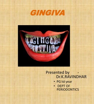
  • 1. GINGIVA Presented by - Dr.K.RAVINDHAR • PG Ist year • DEPT OF PERIODONTICS
  • 2. Contents • Introduction. • Definition. Anatomy of gingiva. 1.Macroscopic feature. a. Marginal gingiva. b. Attached gingiva. c. Interdental gingiva 2.Microscopic feature of gingiva. a. Epithelium b. Connective tissue
  • 3. • Clinical feature of gingiva a. Applied aspects • Conclusion • References
  • 4. Definition- The gingiva is the part of the oral mucosa that covers the alveolar prosses of the jaw & surronds the neck of the teeth. ~carranza Anatomically divided into : 1.Macroscopic feature of gingiva a. Marginal gingiva b. Attached gingiva. c. Interdental gingiva.
  • 6. Marginal gingiva • The marginal gingiva (unattached gingiva) is the terminal edge or border of The gingiva surrounding the teeth in collar-like fashion. • It is demarcated from the a shallow linear depression , the FREE GINGIVAL GROOVE.
  • 8. • Definition: The sulcus consists of the shallow space that is coronal to the attachment of the junctional epithelium & bounded by the tooth on one side & sulcular epithelium on the other. ~Carranza’s The coronal extent of the gingival sulcus is the gingival margin.
  • 9. • It is ‘V’ shaped. • Depth of gingival sulcus: -In ideal condition- 0 mm [Gottlieb B 1933] -Histologically- 1.8mm with variations from 0- 6mm [Orban,Kohler 1924] -Clinically- 2-3 mm.
  • 10. Development of gingival sulcus • This process takes between 1 & 2 years. ~Schroeder & Listgarten ( 1968 ). • It is formed when the tooth erupts into the oral cavity. • The junctional epithelium & REE form a broad band attached to the tooth surface from near the tip of the crown to CEJ.
  • 11. Gingival fluid ( sulculsr fluid) • It may be either transudate/exudate in manner. • It contains:  Inflammatory cells  Serum  Epithelial cells  Connective tissue components
  • 12. Diffusion of the sulcular fluid Basement membrane. Intracellular space of junctional epithelium Gingival sulcus.
  • 13. Function of sulcular fluid • Cleanse material from the sulcus. • Improve adhesion of the epithelium to the tooth. • Antimicrobial properties. • Antibody activity to defend the gingiva.
  • 15. • It is firm, resilient & tightly bound to the underlying periosteum of the alveolar bone. • It is continuous with marginal gingiva. • It extends to the relatively loose & movable alveolar mucosa & is demarcated by the mucogingival junction. • The width of the attached gingiva on the facial aspect differs in different areas of the mouth.[Bowers 1963]
  • 16. • Greatest in incisior region: - 3.5-4.5 mm in maxilla. - 3.3-3.9mm in mandible. • Narrower in posterior segments: - 1.9mm in maxilla - 1.8mm in mandible (premolar region) [Ainamo J,Loe 1966] • The width of the attached gingiva increases with age & in supraerupted teeth.
  • 18. • It is occupies the gingival embrasure, which is the interproximal space beneath the area of tooth contact. • Can be pyramidal or col shaped • The shape depends on the contact point between the two adjoining teeth & the degree of recession. • If a diastema is present, the gingiva is firmly bound over the interdental bone & form, rounded surface without interdental papillae.
  • 19. Microscopic features of gingiva • It is composed of 1.Gingival epithelium 2.Connective tissue
  • 20. 1.Gingival epithelium • Stratified squamous epithelium. Functions- • It provides a physical barrier to infection • Mechanical , chemical, water & microbial barrier • It acts as an innate defense response to gingival health & disease. ~Caffesse RG (1966)
  • 21. Features of gingival epithelium Architectural integrity Cell –cell attachments Basal lamina Keratin cytoskeleton Major cell type Keratinocyte
  • 22. • Other cell types Langerhans cells Melanocytes Markel cells
  • 23. Gingival epithelial cells • Keratinocytes • Nonkeratinocytes • Melanocyes • Langerhans cells ~Diffract CF, Toto(1985) • Markel cells
  • 24. Keratinocytes • Most of the gingiva covered with parakeratinized or nonkeratinized epithelium. • Orthokeratinized –some areas present. • These areas are progress to maturity/ differentation under different physiological / pathological condition.
  • 25. • It is activated by, 1.Proliferation 2.Differentiation
  • 26. 1.Proliferation • By mitosis in the basal layer. • Less frequently in the suprabasal layer. • Larger number of cells begin to migrate to the surface from basal layer.
  • 27. 2.Differentiation • It consists of progressions of biochemical & morphologic events that occur in the cell as they migrate from basal layer. • Morphological changes: 1. Progressive flattening of the cell. 2. Increasing prevalence of tonofilaments 3. Production of keratohyalin granules. 4. Disappearance of the nucles.
  • 28. Orthokeratinized epithelium • It is produced by complete process of keratinization. • No nucleus in stratum corneum. • Well defined nucleus in stratum granulosum. • It is present in some areas of the gingival epithelium.
  • 29. Parakeratinized epithelium • Most of the gingiva covered by parakeratinized epithelium. • The stratum corneum retains pyknotic nuclei. • The keratohylin granules are dispersed. • Not giving rise to a stratum granulosum
  • 30. Nonkeratinized epithelium • It has granulosum / corneum strata, whereas superficial cells have viable nuclei. • The epithelial cell connections are tight junctions (zonae occludens)
  • 31. Epithelial cells • Nonkeratinocytes • Melanocytes • Langerhans cells • Merkel cells
  • 32. Melanocytes • These are dendritic cells. • Located in the basal & spinous layer of the gingival epithelium. • They synthesize melanin • Also called premelanosomes / melanosomes. • Melanin granules are phagocytosed in cells of epithelium & connective tissue.
  • 34. Langerhans cells • These are dendritic cells. • Located in keratinocytes at all suprabasal levels. • They are modified monocytes. • They also act as antigen presenting cells. • Absent in junctional epithelium.
  • 35. Merkel cells • Located in deep layer of the epithelium, nerve endings. • Tactile perceptors.
  • 36. Basal lamina • The epithelium is joined to the underlying connective tissue by a basal lamina. Stern IB (1965) • It is 300 – 400 A thick • The basal lamina consists of: 1. Lamina lucida 2. Lamina densa
  • 37. 1.Lamina lucida • Hemidesmosomes of basal epithelial cells abut the lamina lucida. • Mainly composed by glycoprotein laminin.
  • 38. 2.Lamina densa • Composed of type IV collagen. • The fibrils are 750nm in length from epithelium end to connective tissue end. • PAS +ve.
  • 39. • The gingival epithelium different from the morphologically & functionally, there are;~ Karring T, Loe H (1975) 1. The oral or outer epithelium 2. Sulcular epithelium 3. Junctional epithelium
  • 40. I. Oral [outer] epithelium • Covers the crest & outer surface of marginal gingiva & surface of the attached gingiva. • Average thickness of the oral epithelium is 0.2 to 0.3mm • It is keratinized or parakeratinized or various combinations of these conditions.
  • 41. Histology of oral epithelium
  • 42. Composition of oral epithelium • The oral epithelium composed of 4 layers: 1. Stratum basale. 2. Stratum spinosum. 3. Stratum granulosum. 4. Stratum corneum
  • 43. • The degree of keratinization diminishes with age & the onset of menopause. • It is parakeratinized or orthokeratinized. • Keratinization of the oral mucosa varies in different areas (in descending order palate, gingiva, ventral aspect of the tongue,& cheek)
  • 44. 2. Sulcular epithelium • The sulcular epithelium lines the gingival epithelium. • It is thin & extends from the junctional epithelium to the crest of the gingival margin. • It is act as a semi permeable membrane.
  • 47.
  • 48. • The junctional epithelium consists of a collar like band of stratified squamous non keratinizing epithelium. • It is 3-4 layers thick in early life, but the number of layers increases with age to 10 or even 20 layers. • The junctional epithelium taper from its coronal end. • The length of the junctional epithelium ranges from 0.25-1.35mm
  • 49. • The junctional epithelium is formed by the oral epithelium & the reduced enamel epithelium, during tooth eruption. • The junctional epithelium attached to the tooth surface by internal basal lamina & it attached gingival connective tissue by external basal lamina. • The junctional epithelium attachment to the tooth by gingival fibers, which brace the marginal gingiva against the tooth surface. it is referred to as the dentogingival unit. • The internal basal lamina consists of lamina densa & lamina lucida, which hemi desmosomes are attached.
  • 50. Renewal of gingival epithelium • The mitotic rates is higher in nonkeratinized areas. • Is increased in gingivitis. • Without significant gender defferences. • The mitotic rate is increased / decreased with age.
  • 51. • The mitotic rate varies of the oral epithelium in descending order:  Buccal mucosa.  Hard palate.  Sulcular epithelium.  Junctional epithelium.  Outer surface of the marginal gingiva & attached gingiva.
  • 52. • Varies turnover time of the oral epithelium. • Palate, tongue, & cheek ~ 5-6days. • Gingiva ~10 -12days. • Junctional epithelium ~1-6days.
  • 53. Cuticular structures on the tooth 1. Cuticle 2. Coronal cementum 3. Dental cuticle
  • 54. Cuticler • A thin, acellular structure with a homogeneous matrix. • Classified into: ~Listgarten (1963 ). 1. developmental origin. 2. Acquired coatings.
  • 55. Development origin • Coatings of normally formed as part of tooth development. • They include the REE, coronal cementum, & dental cuticle.
  • 56. Coronal cementum • It deposit a thin layer of the cementum on the enamel. • A thin patches of afibrillar cementum may be seen in the cervical half of the crown.
  • 57. Dental cuticle • It consisting of a layer of homogeneous organic material. • Variable thickness [ ~0.25Um]. • It overlying the enamel surface. • It is nonmineralized. • Not always present. • Near the CEJ, it is deposited over a layer of afibrillar cementum, which in turn overlies enamel.
  • 58. Acquired origin • Coating of exogenous origin such as saliva, bacteria, calculus & surface stains
  • 59. Gingival connective tissue • The major components of the gingival connective tissue are collagen fibers , fibroblasts, vessels, nerves & matrix. • The connective tissue of the gingiva is known as the lamina propria. • it is consists of 2 layers, there are, 1. Papillary layer. 2. Reticular layer. • Connective tissue has a cellular & extra cellular compartment, composed of fibre & ground substance.
  • 60. Connective tissue fibers They are 3 types. 1. Collagen 2. Reticular 3. Elastic • Collagen type I forms the bulk of lamina propria & provide the tensile strength to gingival tissue. • Type IV collagen fibres branches between collagen type I & fibres of basement membrane & blood vessels walls.
  • 61. Ground substance • It is fills the space between fibre & cells, is amorphous,& has a high content of water. • It is composed of proteoglycans, hyaluronic acid & condroitin sulfate, & glycoproteins, fibronectin. • Laminin, another glycoprotein found in basal lamina, serves to attach it to epithelial cells.
  • 63. Cellular elements 1. Fibroblast 2. Mast cells 3. Neutrophils 4. Lymphocytes 5. Adipose cells 6. Eosinophils
  • 64. Blood supply 1. Vessels of periodontal ligament. 2. Arterioles from alveolar bone. 3. Supraperiosteal arteries.
  • 65. Nerves & Lymphatics supply • The nerve fibers arising from the periodontal ligament,& labial, buccal, palatal nerves. • Neural elements are distributed throughout the gingival tissues. • Most nerve fibers are mylinated & are closely associated with the blood vessels. • The lymphatic drainage of the gingiva into the regional lymph nodes.
  • 66. Clinical feature 1. Color 2. Physiological pigmentation [melanin] 3. Size 4. Contour 5. Shape 6. Consistency 7. Surface texture 8. Position
  • 68. Color – acute gingivitis
  • 70. size & contour of gingiva
  • 72. Surface texture - healthy gingiva
  • 74. References • Carranza`s – 11 thedition. • Orban`s- 13 th edition.