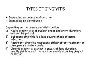
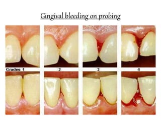
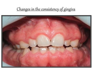
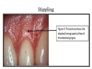
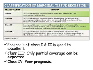
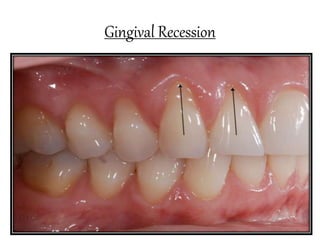
CLINICAL FEATURES OF
GINGIVITIS
PARTHA PROTIM SINGHA
B.D.S. FINAL YEAR
ROLL NO.-29
INDEX
 TYPES OF GINGIVITIS
 GINGIVA IN HEALTH AND DISEASE
 GINGIVAL BLEEDING ON PROBING
 COLOUR CHANGES IN THE GINGIVA
 CHANGES IN THE CONSISTENCY OF GINGIVA
 CHANGES IN THE SIZE OF THE GINGIVA
 SURFACE TEXTURE
 CHANGES IN THE POSITION OF THE
GINGIVA
 GINGIVAL RECESSION
 CHANGES IN GINGIVAL CONTOUR
Introduction
Inflammation of gingiva is termed as gingivitis.
The plaque microorganisms can exert its
effect on periodontium by releasing certain
products (e.g.
collagenase,hyaluronidase,protease,chondroiti
n sulfatase), which can cause damage to the
epithelial and connective tissue constituents.
The intercellular spaces between the
junctional epithelial cells are destroyed and
may permit the bacterial products or bacteria
themselves to gain access into the connective
tissue.
Normal anatomy of Gingiva
TYPES OF GINGIVITIS
 Depending on course and duration
 Depending on distribution
Depending on the course and distribution:
1) Acute gingivitis is of sudden onset and short duration;
and can be painful.
2) Subacute gingivitis is a less severe phase of acute
infection.
3) Recurrent gingivitis reappears either after treatment or
disappears spontaneously.
4) Chronic gingivitis is show in onset, of long duration,
usually painless and the most commonly occuring gingival
condition.
Depending on the distribution
 If the condition is involving a single tooth or group of
tooth, it is called localized gingivitis.
While generalized gingivitis involves
entire mouth.
 According to distribution, gingivitis could be marginal,
papillary, or diffuse.
If the inflammation is limited to the
marginal gingiva, the condition is termed as marginal
gingivitis.
In papillary gingivitis, the inflammation is
limited to the interdental papilla.
When the inlammation spreads to
attached gingiva also, it is termed as diffuse gingivitis, i.e.
involving marginal, papilla and attached gingiva.
 Papillary, marginal and diffuse gingivitis can occur as
localized or generalized conditions.
Gingival bleeding on probing
1. Significance of gingival bleeding.
2. Etiological factors responsible for
gingival bleeding.
3. Associated microscopic changes.
Significance of gingival bleeding on probing
i. It is one of the earliest visual signs of inflammation.
ii. It can appear earlier then colour changes or any
other visual signs of inflammation.
iii. It also provides an additional advantage, by being a
more objective sign that requires less subjective
estimation by the examiner.
iv. Gingival bleeding on probing also helps us to determine
whether the lesions is in an active or inactive state. In
inactive lesion, there will be little or on bleeding on
probing, whereas active lesions bleed more readily on
probing.
v. The severity and ease with bleeding can be provoked-
indicates the integrity of the inflammation.
Etiological factors responsible for gingival bleeding on
probing
Etiological factors can be divided into-
1. Local factors:-
a.Those factors that ressults in
acute bleeding.
b.Those factors that cause chronic
or recurrent bleeding.
2. Systemic factors.
Acute bleeding: It is caused due to-
1. Toothbrush trauma.
2. Impaction of sharp pieces of hard food.
3. Gingival burns from hot foods or chemicals.
4. In conditions such as acute necrotizing ulcerative
gingivitis(ANUG)
Chronic Bleeding: The most common cause are-
1. Chronic inflammation due to the presence of plaque and
calculus.
2. Mechanical trauma, e.g. from toothbrushing, tooth picks or
food impaction.
3. Biting into solids foods such as apple.
Systemic factors:- Include various systemic diseases such as
vitamin K deficiency, platelet disorders such as
thrombocytopenia purpura, other coagulation defects such as
hemophilia, leukemia and others.
Bleeding could also be as a result of excessive
administration of drugs such as salicytes and anticoagulants
such as dicumarol and heparin.
Microscopic changes associated with gingival bleeding on
probing
1. In the epithelium: Thinning and
microulcerations of the sulcular
epithelium is seen.
2. In the connective tissue: Dilation and
engorgement of the capillaries takes
place.
Gingival bleeding on probing
Colour changes in the gingiva
Colour of the gingiva is an important clinical sign of gingival
diseases. Normally, gingiva appears to be coral pink. The factors
that are responsible for this are tissue vascularity, degree of
keratinization and thickness of the epithelium. Generally, colour
of the gingiva may change to red, to bluish red to pale pink.
When there is increased vascularity or reduced epithelial
keratinization, the gingiva becomes more red. The colour
becomes pale when vascularization is reduced or epithelial
keratinization increases. Venous statis gives a bluish hue to the
gingiva.
Systemically absorbed heavy metals may also
cause gingival pigmentation, e.g. bismuth, arsenic, mercury, lead
and silver.
Abnormal melanin pigmentation of the gingiva
may be observed in conditions like Addison’s disease, peutz-
jeghers syndrome, Addison’s disease and Von Recklinghauson’s
disease.
Colour changes in the gingiva
Changes in the consistency of gingiva
Normal gingiva exhibits a firm and
resilient consistency. Factors that are
responsible are cellular and fluid
content and collagenous nature of lamina
propria.
In disease conditions, it can be
soggy and edematous or firm; and
leathery consistency.
Changes in the consistency of gingiva
Changes in the size of the gingiva
Normal size depends on the sum of the bulk
cellular and intercellular elements, and
their vascular supply.
In disease, the size is increased,
which can be termed as gingival
enlargement. The factors responsible for
this are increase in fibers and decrease in
cells as in non-inflammatory type. Whereas
in inflammatory type there will be increase
in cells and decrease in fibers.
Changes in the size of the gingiva
Surface Texture
Under normal conditions, gingiva appears
to be stippled(orange peel appearance)
due to attachment of gingival fibers to
the underlying bone. Microscopically,
alternate rounded protuberance and
depressions in the gingival layer may
rise to stippled appearance. Stippling is
absent in disease conditions. Hence, the
gingiva may appear smooth and shiny.
Stippling
Changes in the position of gingiva
Normally, the gingiva is attached to the
tooth at the cementoenamel junction.
In disease, the position can
be shifted either coronally (pseudo-
pocket) or apical to the cementoenamel
junction (gingival recession)
Gingival Recession
Defination:- Gingival recession is defined as the
exposure of the root surface by an apical shift
in the position of the gingiva.
Types:-
In gingival recession, there are two types i.e.
visible, which is clinically and hidden, which is
covered by gingiva and can only be measured
with probe. Gingival recession may also be
localized and generalized.
Classification of Gingival Recession
Two classification systems are available:-
1) According to Sullivan & Atkins:
Shallow-narrow, shallow-wide and deep-
wide.
2) According to PD Miller’s: Class-I ,Class-
II, Class III, Class IV.
Prognosis of class I & II is good to
excellent.
Class III: Only partial coverage can be
expected.
Class IV: Poor prognosis.
Etiology of gingival recession
Plaque-induced gingival inflammation is the
primary etiological factor responsible for
gingival recession; next common cause is
faulty tooth-brushing. Other secondary
factors on gingival recession are broadly
categorized as-
i. Anatomic factors
ii. Habits
iii. Iatrogenic factors
iv. Physiologic factors
Clinical significance of gingival recession
1) The exposed root surface may be
extremely sensitive.
2) Hyperemia of the pulp may result due
to gingival recession.
3) Interproximal recession creates oral
hygiene problems thereby resulting in
plaque accumulation.
4) Finally, it is aesthetically unacceptable.
Gingival Recession
Changes in gingival contour
Normally, marginal gingiva is scalloped and knife edges, whereas
interdental papilla in the anterior region is pyramidal and
posteriorly tent-shaped. The factors that maintain normal
contour are, shape of the teeth and its alignment in the arch,
location and size of the proximal contact and dimensions of the
facial and lingual gingival embrasures.
In diseased conditions, the marginal gingiva may
become rounded or rolled, whereas interdental papilla can
become blunt and flat.
Stillman’s clefts are apostrophe shaped
indentations extending from and into the gingival margin varying
distance on the facial surface.
They are two types-
1) Simple Cleft: Cleavage in a single direction.
2) Compound Cleft: Cleavage in more than one direction.
REFERENCE
Carranza's Clinical Periodontology
Essentials of Clinical Periodontology
and Periodontics-Shantipriya Reddy
Internet

clinical features of gingivitis

  • 1.
    CLINICAL FEATURES OF GINGIVITIS PARTHAPROTIM SINGHA B.D.S. FINAL YEAR ROLL NO.-29
  • 2.
    INDEX  TYPES OFGINGIVITIS  GINGIVA IN HEALTH AND DISEASE  GINGIVAL BLEEDING ON PROBING  COLOUR CHANGES IN THE GINGIVA  CHANGES IN THE CONSISTENCY OF GINGIVA  CHANGES IN THE SIZE OF THE GINGIVA  SURFACE TEXTURE  CHANGES IN THE POSITION OF THE GINGIVA  GINGIVAL RECESSION  CHANGES IN GINGIVAL CONTOUR
  • 3.
    Introduction Inflammation of gingivais termed as gingivitis. The plaque microorganisms can exert its effect on periodontium by releasing certain products (e.g. collagenase,hyaluronidase,protease,chondroiti n sulfatase), which can cause damage to the epithelial and connective tissue constituents. The intercellular spaces between the junctional epithelial cells are destroyed and may permit the bacterial products or bacteria themselves to gain access into the connective tissue.
  • 4.
  • 5.
    TYPES OF GINGIVITIS Depending on course and duration  Depending on distribution Depending on the course and distribution: 1) Acute gingivitis is of sudden onset and short duration; and can be painful. 2) Subacute gingivitis is a less severe phase of acute infection. 3) Recurrent gingivitis reappears either after treatment or disappears spontaneously. 4) Chronic gingivitis is show in onset, of long duration, usually painless and the most commonly occuring gingival condition.
  • 6.
    Depending on thedistribution  If the condition is involving a single tooth or group of tooth, it is called localized gingivitis. While generalized gingivitis involves entire mouth.  According to distribution, gingivitis could be marginal, papillary, or diffuse. If the inflammation is limited to the marginal gingiva, the condition is termed as marginal gingivitis. In papillary gingivitis, the inflammation is limited to the interdental papilla. When the inlammation spreads to attached gingiva also, it is termed as diffuse gingivitis, i.e. involving marginal, papilla and attached gingiva.  Papillary, marginal and diffuse gingivitis can occur as localized or generalized conditions.
  • 9.
    Gingival bleeding onprobing 1. Significance of gingival bleeding. 2. Etiological factors responsible for gingival bleeding. 3. Associated microscopic changes.
  • 10.
    Significance of gingivalbleeding on probing i. It is one of the earliest visual signs of inflammation. ii. It can appear earlier then colour changes or any other visual signs of inflammation. iii. It also provides an additional advantage, by being a more objective sign that requires less subjective estimation by the examiner. iv. Gingival bleeding on probing also helps us to determine whether the lesions is in an active or inactive state. In inactive lesion, there will be little or on bleeding on probing, whereas active lesions bleed more readily on probing. v. The severity and ease with bleeding can be provoked- indicates the integrity of the inflammation.
  • 11.
    Etiological factors responsiblefor gingival bleeding on probing Etiological factors can be divided into- 1. Local factors:- a.Those factors that ressults in acute bleeding. b.Those factors that cause chronic or recurrent bleeding. 2. Systemic factors.
  • 12.
    Acute bleeding: Itis caused due to- 1. Toothbrush trauma. 2. Impaction of sharp pieces of hard food. 3. Gingival burns from hot foods or chemicals. 4. In conditions such as acute necrotizing ulcerative gingivitis(ANUG) Chronic Bleeding: The most common cause are- 1. Chronic inflammation due to the presence of plaque and calculus. 2. Mechanical trauma, e.g. from toothbrushing, tooth picks or food impaction. 3. Biting into solids foods such as apple. Systemic factors:- Include various systemic diseases such as vitamin K deficiency, platelet disorders such as thrombocytopenia purpura, other coagulation defects such as hemophilia, leukemia and others. Bleeding could also be as a result of excessive administration of drugs such as salicytes and anticoagulants such as dicumarol and heparin.
  • 13.
    Microscopic changes associatedwith gingival bleeding on probing 1. In the epithelium: Thinning and microulcerations of the sulcular epithelium is seen. 2. In the connective tissue: Dilation and engorgement of the capillaries takes place.
  • 14.
  • 15.
    Colour changes inthe gingiva Colour of the gingiva is an important clinical sign of gingival diseases. Normally, gingiva appears to be coral pink. The factors that are responsible for this are tissue vascularity, degree of keratinization and thickness of the epithelium. Generally, colour of the gingiva may change to red, to bluish red to pale pink. When there is increased vascularity or reduced epithelial keratinization, the gingiva becomes more red. The colour becomes pale when vascularization is reduced or epithelial keratinization increases. Venous statis gives a bluish hue to the gingiva. Systemically absorbed heavy metals may also cause gingival pigmentation, e.g. bismuth, arsenic, mercury, lead and silver. Abnormal melanin pigmentation of the gingiva may be observed in conditions like Addison’s disease, peutz- jeghers syndrome, Addison’s disease and Von Recklinghauson’s disease.
  • 16.
    Colour changes inthe gingiva
  • 17.
    Changes in theconsistency of gingiva Normal gingiva exhibits a firm and resilient consistency. Factors that are responsible are cellular and fluid content and collagenous nature of lamina propria. In disease conditions, it can be soggy and edematous or firm; and leathery consistency.
  • 18.
    Changes in theconsistency of gingiva
  • 19.
    Changes in thesize of the gingiva Normal size depends on the sum of the bulk cellular and intercellular elements, and their vascular supply. In disease, the size is increased, which can be termed as gingival enlargement. The factors responsible for this are increase in fibers and decrease in cells as in non-inflammatory type. Whereas in inflammatory type there will be increase in cells and decrease in fibers.
  • 20.
    Changes in thesize of the gingiva
  • 21.
    Surface Texture Under normalconditions, gingiva appears to be stippled(orange peel appearance) due to attachment of gingival fibers to the underlying bone. Microscopically, alternate rounded protuberance and depressions in the gingival layer may rise to stippled appearance. Stippling is absent in disease conditions. Hence, the gingiva may appear smooth and shiny.
  • 22.
  • 23.
    Changes in theposition of gingiva Normally, the gingiva is attached to the tooth at the cementoenamel junction. In disease, the position can be shifted either coronally (pseudo- pocket) or apical to the cementoenamel junction (gingival recession)
  • 24.
    Gingival Recession Defination:- Gingivalrecession is defined as the exposure of the root surface by an apical shift in the position of the gingiva. Types:- In gingival recession, there are two types i.e. visible, which is clinically and hidden, which is covered by gingiva and can only be measured with probe. Gingival recession may also be localized and generalized.
  • 25.
    Classification of GingivalRecession Two classification systems are available:- 1) According to Sullivan & Atkins: Shallow-narrow, shallow-wide and deep- wide. 2) According to PD Miller’s: Class-I ,Class- II, Class III, Class IV.
  • 26.
    Prognosis of classI & II is good to excellent. Class III: Only partial coverage can be expected. Class IV: Poor prognosis.
  • 27.
    Etiology of gingivalrecession Plaque-induced gingival inflammation is the primary etiological factor responsible for gingival recession; next common cause is faulty tooth-brushing. Other secondary factors on gingival recession are broadly categorized as- i. Anatomic factors ii. Habits iii. Iatrogenic factors iv. Physiologic factors
  • 28.
    Clinical significance ofgingival recession 1) The exposed root surface may be extremely sensitive. 2) Hyperemia of the pulp may result due to gingival recession. 3) Interproximal recession creates oral hygiene problems thereby resulting in plaque accumulation. 4) Finally, it is aesthetically unacceptable.
  • 29.
  • 30.
    Changes in gingivalcontour Normally, marginal gingiva is scalloped and knife edges, whereas interdental papilla in the anterior region is pyramidal and posteriorly tent-shaped. The factors that maintain normal contour are, shape of the teeth and its alignment in the arch, location and size of the proximal contact and dimensions of the facial and lingual gingival embrasures. In diseased conditions, the marginal gingiva may become rounded or rolled, whereas interdental papilla can become blunt and flat. Stillman’s clefts are apostrophe shaped indentations extending from and into the gingival margin varying distance on the facial surface. They are two types- 1) Simple Cleft: Cleavage in a single direction. 2) Compound Cleft: Cleavage in more than one direction.
  • 32.
    REFERENCE Carranza's Clinical Periodontology Essentialsof Clinical Periodontology and Periodontics-Shantipriya Reddy Internet