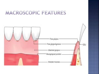
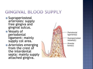
Haytham Qinawi, BDS.
 Gingiva is the part of oral mucosa that
covers the alveolar processes of the jaws
and surrounds the necks of the teeth.
 It is divided into three categories:
 1. Free gingiva
 2. Interdental gingiva
 3. Attached gingiva
 Marginal gingiva is the terminal edge of the
gingiva surrounding the teeth in a collar –
like fashion.
 Not attached to underlying periostium (free).
 Free gingival groove is positioned at a level
corresponding to the level of the CEJ, it
separates the marginal gingiva from the
attached gingiva.
 Interdental gingiva is
the part of the gingiva
which is present in the
interdental space
beneath the area of
tooth contact.
 Col is a valley like
depression which
connects the facial and
lingual papillae and
conforms to the shape
of the interproximal
contact areas.
 Attached gingiva is firm, resilient and tightly
bound to the underlying periosteum of
alveolar bone and cementum by connective
tissue fibers.
 Width of attached gingiva: It is the distance
between mucogingival junction and the
bottom of gingival sulcus/periodontal
pocket.
 Width of attached gingiva increases with age
and in supraerupted teeth.
 Significance of attached gingiva:
1. It gives support to the marginal gingiva.
2. It provides attachment or a solid base for
the movable alveolar mucosa for the action
of lips, cheeks and tongue.
3. It can withstand frictional and functional
stresses of mastication and tooth brushing.
4. It acts as a barrier for passage of
inflammation.
5. It provides resistance to tensional stresses.
 The gingival epithelium is comprised of oral
epithelium,sulcular epithelium and
junctional epithelium.
 Oral epithelium/outer epithelium: It covers
the crest and outer surface of marginal
gingiva and surface of the attached gingiva.
It is a keratinized stratified squamous
epithelium.
 Stratum Basale: cylindrical or cuboid cells
immediately adjacent to the connective
tissue and are separated from connective
tissue by a basement membrane.
 This is the germinative layer, older cells are
pushed to the next layer (Spinosum) as
keratinocytes.
 This process takes about 1 month, the
keratinocyte is then shed from the
superficial layer (Corneum).
 Stratum Spinosum: prickle cell layer in which
large polyhedral cells with short cytoplasmic
processes are present.
 Uppermost cells have granules called
keratinosomes or Odland bodies.These
granules are modified lysosomes.
 They contain a large amount of acid
phosphatase, an enzyme which is involved in
the destruction of organelle membranes.
 Cells in this layer are flattened in a plane
parallel to the gingival surface.
 These cells contain keratohyaline granules,
which is associated with keratin production.
 Keratohyaline granules are about 1 μm in
diameter, round in shape and appear in the
cytoplasm of the cell.
 Stratum Corneum: It consists of closely
packed, flattened cells that have lost nuclei
and most other organelles as they become
keratinized.
 The cells are densely packed with
tonofilaments. Clear, rounded bodies
probably representing lipid droplets appear
within the cytoplasm of the cell.
 Sulcular epithelium:
It lines the gingival
sulcus. It is a non-
keratinized,
stratified squamous
epithelium which
extends from the
coronal end of the
junctional
epithelium to the
crest of the gingival
margin.
 Junctional
epithelium (JE):
Junctional
epithelium consists
of collar like band
of stratified
squamous
nonkeratinized
epithelium. The
normal length of
junctional
epithelium is 0.25 -
1.35 mm.
 Histologically the juctional epithelium is
formed by the reduced enamel epithelium.
 Junctional epithelium is divided into three
zones: the apical, middle and coronal zone.
 The middle zone is the zone with the
maximum adhesiveness, and the coronal
zone is the most permeable of the three
zones.
 Junctional epithelium has
three surfaces: Internal
surface which faces the
tooth surface, external
surface which faces the
gingival connective tissue
and coronal surface of the
junctional epithelium
forms the base of the
sulcus.
 Junctional epithelium is
attached to the tooth
surface by means of
internal basal lamina and
to gingival connective
tissue by an external basal
lamina.
 Functions of junctional epithelium:
1. Provides attachment to the tooth.
2. Acts as an epithelial barrier against the plaque
bacteria.
3. Rapid cell division hinders bacterial
colonization and repair of damaged tissue
occurs rapidly.
4. Allows the access of GCF, inflammatory cells
and components of the immunological host
defense to the gingival margin.
5. Antimicrobial substance production.
6. Chemotaxis and cytokine secretion e.g. IL-1,
IL-6, IL-8 and TNF-α.
 Cells present in the gingival epithelium:
 1-Keratinocytes:
 90% of the total gingival cell population.
 Keratinocytes have melanosomes unlike any
other cell in the periodontium.
 Proliferation and differentiation of
keratinocytes in an apical to coronal
direction accomplishes the function of
protection.
 The microfilaments present in the
keratinocytes help in cell motility and
maintenance of the polarity.
 Cells present in the gingival epithelium:
 2-Non-Keratinocytes/Clear cells:
1. Langerhans cells (LCs): responsible for
communication with immune system by
acting as antigen – presenting cells for
lymphocytes.
2. Merkel Cells: are sensory in nature and
respond to touch.
3. Melanocytes: synthesize melanin which is
responsible for providing color to gingiva.
 Composed of 4
elements:
1. Basal cell plasma
membrane
(Hemidesmosomes
).
2. Lamina lucida: 25
to 45 nm wide.
3. Lamina densa: 40
to 60 nm with type
IV collagen.
4. Reticular layer.
 Gingival fibers:
 Collagen types: I,III,IV,V,VI. Type I predominates.
 The functions of these fibers are:
1. To stabilize the attached gingiva to the
alveolar process.
2. To stabilize the attached gingiva to the tooth.
3. Maintain the epithelial seal to the tooth.
4. Provide stability to the tooth.
5. Brace free gingiva firmly against the tooth and
adjacent attached gingiva.
6. Provide rigidity to withstand forces of
mastication.
1. Dentogingival group: provide gingival
support.
2. Alveologingival group: connects attached
gingiva to alveolar bone.
3. Circular group: Maintain contour and
position of free/marginal gingiva
4. Trans-septal fibers: Maintain relationship of
adjacent teeth, protect interproximal
bone.
5. Dentoperiosteal group: Anchors tooth to
bone, protect periodontal ligament.
6. Semicircular group.
7. Transgingival group: Secure alignment of
teeth in the arch.
8. Intergingival group: provide contour and
support for the attached gingiva.
9. Interpapillary group: Provide support for
interdental gingiva.
 Dentogingival, dentoperiosteal and
alveologingival fibers group provide the
attachment of gingiva to the tooth and to
the bony structure.
 Supraperiosteal
arterioles: supply
free gingiva and
gingival sulcus.
 Vessels of
periodontal
ligament: mainly
supply col area.
 Arterioles emerging
from the crest of
the interdental
septa: mainly supply
attached gingiva.
1. Color: described as coral pink but variations of
the normal are present.
2. Surface texture: The surface texture of free
gingiva is smooth whereas of attached gingiva
is stippled.
3. Contour: Scalloped outline normally and
straight line along teeth with relatively flat
surfaces.
4. Shape: Shape of interdental gingiva depends
upon contour of the proximal tooth surface,
location and shape of the proximal contact and
dimensions of the gingival embrasures.
5. Size: The size of the gingiva corresponds to
the sum total of the bulk of cellular and
intercellular elements and their vascular
supply.
6. Consistency: attached gingiva should be
firm, resilient and tightly bound to the
underlying hard tissues. The abundant
collagen fibers and the non-collagenous
protein combines to give gingiva, the firm
consistency.
Thank you!

Gingiva

  • 1.
  • 3.
     Gingiva isthe part of oral mucosa that covers the alveolar processes of the jaws and surrounds the necks of the teeth.  It is divided into three categories:  1. Free gingiva  2. Interdental gingiva  3. Attached gingiva
  • 4.
     Marginal gingivais the terminal edge of the gingiva surrounding the teeth in a collar – like fashion.  Not attached to underlying periostium (free).  Free gingival groove is positioned at a level corresponding to the level of the CEJ, it separates the marginal gingiva from the attached gingiva.
  • 5.
     Interdental gingivais the part of the gingiva which is present in the interdental space beneath the area of tooth contact.  Col is a valley like depression which connects the facial and lingual papillae and conforms to the shape of the interproximal contact areas.
  • 6.
     Attached gingivais firm, resilient and tightly bound to the underlying periosteum of alveolar bone and cementum by connective tissue fibers.  Width of attached gingiva: It is the distance between mucogingival junction and the bottom of gingival sulcus/periodontal pocket.  Width of attached gingiva increases with age and in supraerupted teeth.
  • 7.
     Significance ofattached gingiva: 1. It gives support to the marginal gingiva. 2. It provides attachment or a solid base for the movable alveolar mucosa for the action of lips, cheeks and tongue. 3. It can withstand frictional and functional stresses of mastication and tooth brushing. 4. It acts as a barrier for passage of inflammation. 5. It provides resistance to tensional stresses.
  • 9.
     The gingivalepithelium is comprised of oral epithelium,sulcular epithelium and junctional epithelium.  Oral epithelium/outer epithelium: It covers the crest and outer surface of marginal gingiva and surface of the attached gingiva. It is a keratinized stratified squamous epithelium.
  • 10.
     Stratum Basale:cylindrical or cuboid cells immediately adjacent to the connective tissue and are separated from connective tissue by a basement membrane.  This is the germinative layer, older cells are pushed to the next layer (Spinosum) as keratinocytes.  This process takes about 1 month, the keratinocyte is then shed from the superficial layer (Corneum).
  • 11.
     Stratum Spinosum:prickle cell layer in which large polyhedral cells with short cytoplasmic processes are present.  Uppermost cells have granules called keratinosomes or Odland bodies.These granules are modified lysosomes.  They contain a large amount of acid phosphatase, an enzyme which is involved in the destruction of organelle membranes.
  • 12.
     Cells inthis layer are flattened in a plane parallel to the gingival surface.  These cells contain keratohyaline granules, which is associated with keratin production.  Keratohyaline granules are about 1 μm in diameter, round in shape and appear in the cytoplasm of the cell.
  • 13.
     Stratum Corneum:It consists of closely packed, flattened cells that have lost nuclei and most other organelles as they become keratinized.  The cells are densely packed with tonofilaments. Clear, rounded bodies probably representing lipid droplets appear within the cytoplasm of the cell.
  • 15.
     Sulcular epithelium: Itlines the gingival sulcus. It is a non- keratinized, stratified squamous epithelium which extends from the coronal end of the junctional epithelium to the crest of the gingival margin.
  • 16.
     Junctional epithelium (JE): Junctional epitheliumconsists of collar like band of stratified squamous nonkeratinized epithelium. The normal length of junctional epithelium is 0.25 - 1.35 mm.
  • 17.
     Histologically thejuctional epithelium is formed by the reduced enamel epithelium.  Junctional epithelium is divided into three zones: the apical, middle and coronal zone.  The middle zone is the zone with the maximum adhesiveness, and the coronal zone is the most permeable of the three zones.
  • 18.
     Junctional epitheliumhas three surfaces: Internal surface which faces the tooth surface, external surface which faces the gingival connective tissue and coronal surface of the junctional epithelium forms the base of the sulcus.  Junctional epithelium is attached to the tooth surface by means of internal basal lamina and to gingival connective tissue by an external basal lamina.
  • 19.
     Functions ofjunctional epithelium: 1. Provides attachment to the tooth. 2. Acts as an epithelial barrier against the plaque bacteria. 3. Rapid cell division hinders bacterial colonization and repair of damaged tissue occurs rapidly. 4. Allows the access of GCF, inflammatory cells and components of the immunological host defense to the gingival margin. 5. Antimicrobial substance production. 6. Chemotaxis and cytokine secretion e.g. IL-1, IL-6, IL-8 and TNF-α.
  • 20.
     Cells presentin the gingival epithelium:  1-Keratinocytes:  90% of the total gingival cell population.  Keratinocytes have melanosomes unlike any other cell in the periodontium.  Proliferation and differentiation of keratinocytes in an apical to coronal direction accomplishes the function of protection.  The microfilaments present in the keratinocytes help in cell motility and maintenance of the polarity.
  • 21.
     Cells presentin the gingival epithelium:  2-Non-Keratinocytes/Clear cells: 1. Langerhans cells (LCs): responsible for communication with immune system by acting as antigen – presenting cells for lymphocytes. 2. Merkel Cells: are sensory in nature and respond to touch. 3. Melanocytes: synthesize melanin which is responsible for providing color to gingiva.
  • 22.
     Composed of4 elements: 1. Basal cell plasma membrane (Hemidesmosomes ). 2. Lamina lucida: 25 to 45 nm wide. 3. Lamina densa: 40 to 60 nm with type IV collagen. 4. Reticular layer.
  • 23.
     Gingival fibers: Collagen types: I,III,IV,V,VI. Type I predominates.  The functions of these fibers are: 1. To stabilize the attached gingiva to the alveolar process. 2. To stabilize the attached gingiva to the tooth. 3. Maintain the epithelial seal to the tooth. 4. Provide stability to the tooth. 5. Brace free gingiva firmly against the tooth and adjacent attached gingiva. 6. Provide rigidity to withstand forces of mastication.
  • 25.
    1. Dentogingival group:provide gingival support. 2. Alveologingival group: connects attached gingiva to alveolar bone. 3. Circular group: Maintain contour and position of free/marginal gingiva 4. Trans-septal fibers: Maintain relationship of adjacent teeth, protect interproximal bone. 5. Dentoperiosteal group: Anchors tooth to bone, protect periodontal ligament.
  • 26.
    6. Semicircular group. 7.Transgingival group: Secure alignment of teeth in the arch. 8. Intergingival group: provide contour and support for the attached gingiva. 9. Interpapillary group: Provide support for interdental gingiva.  Dentogingival, dentoperiosteal and alveologingival fibers group provide the attachment of gingiva to the tooth and to the bony structure.
  • 27.
     Supraperiosteal arterioles: supply freegingiva and gingival sulcus.  Vessels of periodontal ligament: mainly supply col area.  Arterioles emerging from the crest of the interdental septa: mainly supply attached gingiva.
  • 28.
    1. Color: describedas coral pink but variations of the normal are present. 2. Surface texture: The surface texture of free gingiva is smooth whereas of attached gingiva is stippled. 3. Contour: Scalloped outline normally and straight line along teeth with relatively flat surfaces. 4. Shape: Shape of interdental gingiva depends upon contour of the proximal tooth surface, location and shape of the proximal contact and dimensions of the gingival embrasures.
  • 29.
    5. Size: Thesize of the gingiva corresponds to the sum total of the bulk of cellular and intercellular elements and their vascular supply. 6. Consistency: attached gingiva should be firm, resilient and tightly bound to the underlying hard tissues. The abundant collagen fibers and the non-collagenous protein combines to give gingiva, the firm consistency.
  • 31.