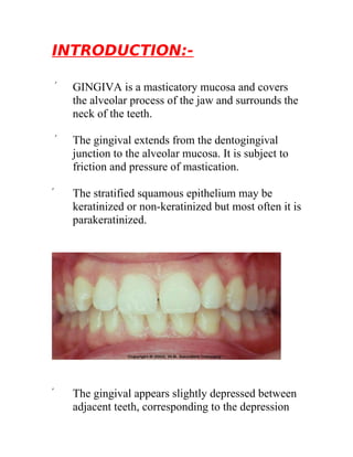
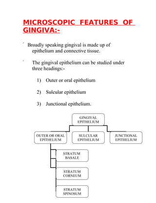
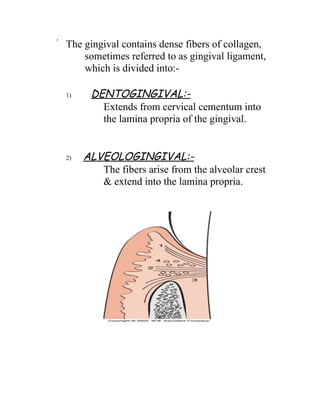
INTRODUCTION:-

    ۞   GINGIVA is a masticatory mucosa and covers
        the alveolar process of the jaw and surrounds the
        neck of the teeth.

    ۞   The gingival extends from the dentogingival
        junction to the alveolar mucosa. It is subject to
        friction and pressure of mastication.

۞       The stratified squamous epithelium may be
        keratinized or non-keratinized but most often it is
        parakeratinized.


۞       The gingival appears slightly depressed between
        adjacent teeth, corresponding to the depression
on the alveolar process between eminence of the
    sockets.

۞   The gingiva is limited on the outer surface of
    both the jaws by the mucogival junction, which
    separates it from the alveolar mucosa.

۞   The alveolar mucosa is red & contains numerous
    small vessels coursing close to the surface.

۞   The gingival is normally pink but sometimes
    have grayish tint.

۞   Gingiva is attached immovably and firmly to the
    periosteum of the alveolar bone.

۞   On the inner surface of the lower jaw a line of
    demarcation is found between the gingival and
    the mucosa on the floor of the mouth.
TYPES OF GINGIVA:-

The gingival is divided into three types:

  A)   Free or unattached or marginal gingival

  B) Gingival sulcus.

  C) Attached gingiva.
A) FREE OR UNATTACHED OR
     MARGINAL GINGIVA:-

۞        The free gingival is the terminal edge of the
         gingival which is usually about 1mm wide and
         surrounds the teeth.

۞        The free gingival forms one of the walls of the
         gingival sulcus and is separated from the
         attached gingival by a groove called free gingival
         groove.

    B)   ATTACHED GINGIVA:-

۞        It is the continuation of the free gingival and
         extends up to the alveolar mucosa.

۞        The attached gingival is separated from the
         alveolar mucosa by a mucogingival sulcus.

۞        The width :-
         →3.5-4.5 mm in the maxillary anterior region

         →3.3-3.9mm in the mandibular anterior region.

         →Posteriorly the width of the attached gingival
         is less.
→It is the least at the first premolar area being

     →1.9mm-maxilla and 1.8mm-mandible.

C)   INTERDENTAL PAPILLA:-

۞ It is the part of gingival that fills the space
        between two adjacent teeth.

۞ It is a shallow V shaped space surrounding the
        tooth.

۞ It is bounded on one side by the tooth and on the
        other side by the free gingiva.

۞ From oral or vestibular aspect, the surface of the
      interdental papilla is triangular.

۞ The depressed part of interdental papilla is called
      COL.

۞ Col is covered by thin non-keratinized
      epithelium.

۞ Elastic fibers known as OXYTALAN fibers are
      present.
MICROSCOPIC FEATURES OF
GINGIVA:-

۞ Broadly speaking gingival is made up of
    epithelium and connective tissue.

۞   The gingival epithelium can be studied under
    three headings:-

      1) Outer or oral epithelium

      2) Sulcular epithelium

      3) Junctional epithelium.

                          GINGIVAL
                         EPITHELIUM


      OUTER OR ORAL       SULCULAR          JUNCTIONAL
       EPITHELIUM        EPITHELIUM         EPITHELIUM


                  STRATUM
                   BASALE


                  STRATUM
                  CORNEUM


                  STRATUM
                  SPINOSUM
1)     OUTER OR ORAL EPITHELIUM:-

     ۞ The epithelium consists of the following layers:

       a)   Stratum basale:-      cuboidal cells.

       b)   Stratum spinosum:-   large polyhedral cells
                                    →Desmosomes

       c)   Stratum corneum:-     superficial most layer
                                  Large, wide, flat and
                                  lacking nucleus.
2)    SULCULAR EPITHELIUM:-

     ۞ Extends from the coronal area of the junctional
           epithelium to the free margin of the gingival.

     ۞ Epithelium is nonkeratinized.

     ۞ Epithelium lacks heavy ridges and papillae.

     ۞ It is not keratinized due to constant irritation of
             plaque.

3)    JUNCTIONAL EPITHELIUM:-

     ۞ Cells become cuboidal after ameloblasts have
           finished formation of enamel.

     ۞ It forms a collar around the fully erupted tooth.

     ۞ It consist of stratified squamous nonkeratinizing
            epithelium of 3-4 cell thickness having
            length of 0.25-1.35 mm.

TYPES OF GINGIVAL
LIGAMENT:-
۞ The gingival contains dense fibers of collagen,
      sometimes referred to as gingival ligament,
      which is divided into:-

  1)    DENTOGINGIVAL:-
          Extends from cervical cementum into
          the lamina propria of the gingival.


  2)   ALVEOLOGINGIVAL:-
          The fibers arise from the alveolar crest
          & extend into the lamina propria.
3)   CIRCULAR:-
          A small groups of fibers that circle the
          tooth & interlace with the other fibers.

  4)     DENTOPERIOSTEAL:-
           These fibers can be followed from the
           cementum into the periosteum of the
           alveolar crest and of the vestibular and
           oral surfaces of the alveolar bone.

۞ There are also accessory fibers that extend
      interproximally between adjacent teeth &
      are also referred to as transseptal fibers.

۞ These fibers make the interdental ligament.
DIFFERENT TYPES OF
EPITHELIAL LAYERS:-

  1)    KERATINIZATION (75%) :-

           # In which the superficial cells form
           scales of keratin & lose their nuclei.

           # A stratum granulosum is present.

  2)   PARAKERATINIZATION (15%) :-

           # In which the superficial cells retain
           pyknotic nuclei & show some signs of
           being keratinized.

           # However, the stratum granulosum is
           generally absent.

  3)   NON-KERATINIZATION (10%) :-

           # In which the surface cells are
       nucleated & show no signs of keratinization.
BlOOD SUPPLY:-
     Blood supply of gingiva is by ALVEOLAR
  ARTERY


NERVE SUPPLY:-
                               NERVE SUPPLY


   MAXILLARY                                              MANDIBULAR


                 NASOPALATINE NERVE
                                                          Inferior alveolar nerve
            (Supplies facial aspect of anterior teeth)


            POSTERIOR SUPERIOR ALVEOLAR
                             NERVE
           (Supplies facial aspect of posterior teeth)


               GREATER PALATINE NERVE
           (Supplies lingual aspect of posterior teeth)


               ANTERIOR PALATINE NERVE
            (Supplies lingual aspect of anterior teeth)
CONTENTS:-

 1) INTRODUCTION

 2) TYPES OF GINGIVA

 3) MICROSCOPIC FEATURES OF
    GINGIVA

 4) TYPES OF GINGIVAL LIGAMENT

 5) DIFFERENT TYPES OF EPITHELIAL
    LAYERS

 6) ARTERIAL SUPPLY

 7) NERVE SUPPLY

 8) REFERENCES
REFERNECES:-

۞ Oral histology and embryology    -
  ORBANS
                         S.N.
Bhaskar

۞    Oral histology and embryology -
     A.R. Ten Cate

۞    Clinical periodontology      -
     Caranza
                           Glick Man

۞    Oral pathology          -
     Shaffers

۞    Local Anaesthesia       -
     Mallamaid
 gingiva

gingiva

  • 1.
    INTRODUCTION:- ۞ GINGIVA is a masticatory mucosa and covers the alveolar process of the jaw and surrounds the neck of the teeth. ۞ The gingival extends from the dentogingival junction to the alveolar mucosa. It is subject to friction and pressure of mastication. ۞ The stratified squamous epithelium may be keratinized or non-keratinized but most often it is parakeratinized. ۞ The gingival appears slightly depressed between adjacent teeth, corresponding to the depression
  • 2.
    on the alveolarprocess between eminence of the sockets. ۞ The gingiva is limited on the outer surface of both the jaws by the mucogival junction, which separates it from the alveolar mucosa. ۞ The alveolar mucosa is red & contains numerous small vessels coursing close to the surface. ۞ The gingival is normally pink but sometimes have grayish tint. ۞ Gingiva is attached immovably and firmly to the periosteum of the alveolar bone. ۞ On the inner surface of the lower jaw a line of demarcation is found between the gingival and the mucosa on the floor of the mouth.
  • 3.
    TYPES OF GINGIVA:- Thegingival is divided into three types: A) Free or unattached or marginal gingival B) Gingival sulcus. C) Attached gingiva.
  • 4.
    A) FREE ORUNATTACHED OR MARGINAL GINGIVA:- ۞ The free gingival is the terminal edge of the gingival which is usually about 1mm wide and surrounds the teeth. ۞ The free gingival forms one of the walls of the gingival sulcus and is separated from the attached gingival by a groove called free gingival groove. B) ATTACHED GINGIVA:- ۞ It is the continuation of the free gingival and extends up to the alveolar mucosa. ۞ The attached gingival is separated from the alveolar mucosa by a mucogingival sulcus. ۞ The width :- →3.5-4.5 mm in the maxillary anterior region →3.3-3.9mm in the mandibular anterior region. →Posteriorly the width of the attached gingival is less.
  • 5.
    →It is theleast at the first premolar area being →1.9mm-maxilla and 1.8mm-mandible. C) INTERDENTAL PAPILLA:- ۞ It is the part of gingival that fills the space between two adjacent teeth. ۞ It is a shallow V shaped space surrounding the tooth. ۞ It is bounded on one side by the tooth and on the other side by the free gingiva. ۞ From oral or vestibular aspect, the surface of the interdental papilla is triangular. ۞ The depressed part of interdental papilla is called COL. ۞ Col is covered by thin non-keratinized epithelium. ۞ Elastic fibers known as OXYTALAN fibers are present.
  • 6.
    MICROSCOPIC FEATURES OF GINGIVA:- ۞Broadly speaking gingival is made up of epithelium and connective tissue. ۞ The gingival epithelium can be studied under three headings:- 1) Outer or oral epithelium 2) Sulcular epithelium 3) Junctional epithelium. GINGIVAL EPITHELIUM OUTER OR ORAL SULCULAR JUNCTIONAL EPITHELIUM EPITHELIUM EPITHELIUM STRATUM BASALE STRATUM CORNEUM STRATUM SPINOSUM
  • 7.
    1) OUTER OR ORAL EPITHELIUM:- ۞ The epithelium consists of the following layers: a) Stratum basale:- cuboidal cells. b) Stratum spinosum:- large polyhedral cells →Desmosomes c) Stratum corneum:- superficial most layer Large, wide, flat and lacking nucleus.
  • 8.
    2) SULCULAR EPITHELIUM:- ۞ Extends from the coronal area of the junctional epithelium to the free margin of the gingival. ۞ Epithelium is nonkeratinized. ۞ Epithelium lacks heavy ridges and papillae. ۞ It is not keratinized due to constant irritation of plaque. 3) JUNCTIONAL EPITHELIUM:- ۞ Cells become cuboidal after ameloblasts have finished formation of enamel. ۞ It forms a collar around the fully erupted tooth. ۞ It consist of stratified squamous nonkeratinizing epithelium of 3-4 cell thickness having length of 0.25-1.35 mm. TYPES OF GINGIVAL LIGAMENT:-
  • 9.
    ۞ The gingivalcontains dense fibers of collagen, sometimes referred to as gingival ligament, which is divided into:- 1) DENTOGINGIVAL:- Extends from cervical cementum into the lamina propria of the gingival. 2) ALVEOLOGINGIVAL:- The fibers arise from the alveolar crest & extend into the lamina propria.
  • 10.
    3) CIRCULAR:- A small groups of fibers that circle the tooth & interlace with the other fibers. 4) DENTOPERIOSTEAL:- These fibers can be followed from the cementum into the periosteum of the alveolar crest and of the vestibular and oral surfaces of the alveolar bone. ۞ There are also accessory fibers that extend interproximally between adjacent teeth & are also referred to as transseptal fibers. ۞ These fibers make the interdental ligament.
  • 11.
    DIFFERENT TYPES OF EPITHELIALLAYERS:- 1) KERATINIZATION (75%) :- # In which the superficial cells form scales of keratin & lose their nuclei. # A stratum granulosum is present. 2) PARAKERATINIZATION (15%) :- # In which the superficial cells retain pyknotic nuclei & show some signs of being keratinized. # However, the stratum granulosum is generally absent. 3) NON-KERATINIZATION (10%) :- # In which the surface cells are nucleated & show no signs of keratinization.
  • 12.
    BlOOD SUPPLY:- Blood supply of gingiva is by ALVEOLAR ARTERY NERVE SUPPLY:- NERVE SUPPLY MAXILLARY MANDIBULAR NASOPALATINE NERVE Inferior alveolar nerve (Supplies facial aspect of anterior teeth) POSTERIOR SUPERIOR ALVEOLAR NERVE (Supplies facial aspect of posterior teeth) GREATER PALATINE NERVE (Supplies lingual aspect of posterior teeth) ANTERIOR PALATINE NERVE (Supplies lingual aspect of anterior teeth)
  • 13.
    CONTENTS:- 1) INTRODUCTION 2) TYPES OF GINGIVA 3) MICROSCOPIC FEATURES OF GINGIVA 4) TYPES OF GINGIVAL LIGAMENT 5) DIFFERENT TYPES OF EPITHELIAL LAYERS 6) ARTERIAL SUPPLY 7) NERVE SUPPLY 8) REFERENCES
  • 14.
    REFERNECES:- ۞ Oral histologyand embryology - ORBANS S.N. Bhaskar ۞ Oral histology and embryology - A.R. Ten Cate ۞ Clinical periodontology - Caranza Glick Man ۞ Oral pathology - Shaffers ۞ Local Anaesthesia - Mallamaid